Introduction
There are only about a dozen lichen species known that produce conidia in hyphomycetous structures. Only a few of them, viz. Cheiromycina globosa Aptroot & Schiefelbein (Reference Aptroot and Schiefelbein2003), Blarneya hibernica D. Hawksw., Coppins & P. James, Sclerococcum griseisporodochium Etayo, and Micarea adnata Coppins (Smith et al. Reference Smith, Aptroot, Coppins, Fletcher, Gilbert, James and Wolseley2009), and anamorphs of Tylophoron (Tibell Reference Tibell1991), form conidia in sporodochia. Whether some Milospium spp. are further examples remains under dispute (Aptroot & Sipman Reference Aptroot and Sipman2001); we believe these also to be lichenized hyphomycetes with sporodochia.
While identifying corticolous sterile crusts from the tropics that had accumulated in our collections over the years, we came across several specimens which share the following characters: thallus effuse, whitish, somewhat byssoid, with myrmecioid algae, with discrete, white or variously coloured soralium-like mounds, corresponding to the definition of sporodochia, that contain only hyphae (no algae) and are covered with a few or many relatively large, hyaline to pale brown, mostly unidistoseptate ellipsoidal conidia that originate by arthric conidiogenesis.
There is no doubt that the conidia and sporodochia belong to the lichen thallus (not to a parasite), as their correlation is perfect. Very probably this group of lichens has escaped attention thus far because sterile tropical crustose lichens are not much studied, and the sporodochia can be easily mistaken for soralia. We were unable to find any hyphomycetous form genus that fits these species.
The similarity in conidium production and shape, in thallus structure and sporodochium shape indicates a close relationship between the four undescribed species; in the absence of ascocarps their relationship with other lichen groups is unknown. Therefore we describe them here as new species and place them all together in a new genus of unknown affinity.
Materials and Methods
The study is mostly based on material collected by the authors and deposited in ABL or B. The morphology of all specimens was studied using an Olympus SZX7 stereomicroscope and an Olympus BX50 compound microscope with differential interference contrast optics and photomicrographs taken with an attached Nikon Coolpix 995 digital camera. Chemistry was investigated by TLC (TDA and HEF).
Taxonomy
Sporodochiolichen Aptroot & Sipman gen. nov
MycoBank No: 561143
Thallus crustaceus, continuus, subbyssoideus, sine hypothallo, algis myrmecioideis, sporodochiis instructis, conidia arthrica, 0–2-distoseptata, hyalina ad pallide fuscescentia, ascomata ignota.
Typus: Sporodochiolichen lecanoricus Aptroot & Sipman, see below.
Thallus corticolous, continuous, whitish or grey to greenish grey or ochraceous, dull, somewhat byssoid, consisting of thick-walled, rather wide, branched hyphae, in places with needle-like crystals, with myrmecioid algae, without hypothallus.
Conidiomata sporodochia, numerous, mostlyorbicular, hemispherical to applanate. Conidiaoriginating by septation of the conidiogenous hyphae (arthric), few to numerous and then amassing on top of the sporodochia, hyaline to pale brown, smooth or papillate, 0–2-distoseptate, predominantly 1-septate, ellipsoidal, septation distoseptate. Ascomata unknown.
Chemistry. Lecanoric or norstictic acid and/or yellow or pink pigments present.
Sporodochiolichen flavus Aptroot & Sipman sp. nov
MycoBank No: 561144
Sporodochiis flavis, conidiis hyalinis, praecipue unidistoseptatis, 5–8 × 1·5–3·5 µm, laevibus, sine acidis lichenicis, pigmentum flavum ignotum continens.
Typus: Papua New Guinea, Madang Prov., Mouth of Boroi river near Bogia, alt. 1 m, on Rhizophora in mangrove forest, 21 July 1992, A. Aptroot 30426 (ABL—holotypus).
Thallus grey, thin, dull, covering an area of up to 5 cm diam. Algal cells, 3–4 µm diam.
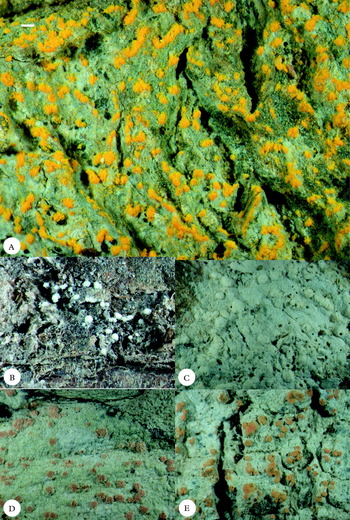
Sporodochiolichen, habitus of thallus and sporodochia. A, S. flavus (holotype); B, S. lecanoricus (holotype); C, S. lecanoricus (Aptroot 10806); D, S. papillatus (holotype); E, S. pigmentatus (holotype). Scale = 1·0 mm.

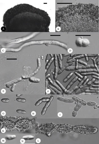
Sporodochiolichen, internal structures. A & B S. lecanoricus (Sipman 48660); A, section through sporodochium; B, conidial mass; C & D, S. flavus (holotype); C, thallus hyphae; D, conidia; E–I, S. lecanoricus; E, conidia (holotype); F, conidia (Sipman 48660); G-I, conidia in various optical sections showing distosepatation (Sipman 444313); K & L, S. papillatus, conidia (holotype); M & N, S. pigmentatus, conidia (holotype). Scales: A & B = 50 µm; C–M (E–M all on same scale) = 10 µm.

Sporodochia starting as punctiform dots c. 0·2 mm diam., soon confluent to form larger, irregular, often partly linear, patterns overlaying the thallus, golden yellow. Conidia hyaline, smooth, ellipsoidal, generally 1-distoseptate, 5–8 × 2·5–3·5 µm, often remaining long attached to each other, eventually some simple and then cubical.
Chemistry. Yellow pigment, KOH+ violet, not dissolving, Rf 5 in TDA.
Additional specimens examined. Papua New Guinea: Madang: Gogol valley, Tgubi logging site, on bark in virgin lowland rainforest, 1992, A. Aptroot 33011 (ABL); Jais Aben, c. 10 km NW of Madang, on Terminalia in coastal Cocos plantation, 1995, A. Aptroot 38239 (ABL).
Sporodochiolichen lecanoricus Aptroot & Sipman sp. nov
MycoBank No: 561145
Sporodochiis albis, hemisphaericis, conidiis hyalinis ad pallide flavorufis, unidistoseptatis vel aseptatis, 4–12 × 3–4 µm, laevibus, sine pigmento, acidum lecanoricum continens.
Typus: Papua New Guinea, Eastern Highlands Prov., Mount Gahavisuka Provincial Park, 11 km N of Goroka, alt. 2300–2450 m, on bark in mountain forest, 11 August 1992, A. Aptroot 31060 (ABL—holotypus).
Thallus whitish to ochraceous, dull, nearly absent to rather thick, somewhat byssoid, covering areas up to 5 cm diam. Algal cells, 3–4 µm diam.
Sporodochia mostly hemispherical, 0·2–0·6 mm diam., pure white to whitish. Conidia hyaline to pale yellowish brown, smooth, ellipsoidal, partly simple and then 4–8 × 3–4 µm, partly 1-distoseptate and then 6–12 × 3–4 µm.
Chemistry. Lecanoric acid; sporodochia and often also thallus C+ red.
Remarks. This is the species with the largest known distribution, and also with the widest morphological variation. There is variation in the amount of thallus and average sporodochium size, both of which seem correlated to bark type, and also to some extent in the pigmentation of the conidia, although this may well be age-related.
Additional specimens examined. Thailand: Chiang Mai: Doi Suthep Pui National Park, in forest relict near temple, 2002, H. J. M. Sipman 48660 (B).—Ecuador: Manabi: Puerto Lopez, in coastal Cereus-savannah, 1982, A. Aptroot 10806 (ABL).—Costa Rica: Cartago: La Esperanza, Rio Macho Forest Reserve, in primary mountain forest, 1999, H. J. M. Sipman 44313 (B).—Venezuela: Merida: Monte Zerpa, 1998, K. Wiegleb 125 (B).
Sporodochiolichen papillatus Aptroot & Sipman sp. nov
MycoBank No: 561146
Sporodochiis punctiformibus, pallide violaceis, conidiis hyalinis, plerumque aseptatis, 15–28 × 10–12 µm, dense papillatis, papillis ad 0·1 × 0·5 µm, sine pigmento, acidum norsticticum continens.
Typus: Papua New Guinea, Madang Prov., Awar near Bogia, alt. 1 m, on Cocos in coastal plantation, 19 July 1992, A. Aptroot 30162 (ABL—holotypus).
Thallus whitish to pale ochraceous, dull, rather thin, somewhat byssoid, covering areas up to 5 cm diam. Algal cells, 3–4 µm diam.
Sporodochia punctiform, 0·4–1·0 mm diam.,arising from the thallus and initially surrounded by a flimsy, whitish thalline rim, partly confluent to form larger patterns, pink. Conidia hyaline, strongly papillate, ellipsoidal, generally non-septate, 15–28 × 10–12 µm, papillae c. 0·1 × 0·5 µm.
Chemistry. Norstictic acid repeatedly detected with TLC in thallus and sporodochia, but apparently in low concentration, because all structures are KOH−.
Sporodochiolichen pigmentatus Aptroot & Sipman sp. nov
MycoBank No: 561147
Sporodochiis praecipue hemisphaericis, pallide violaceis, conidiis hyalinis, unidistoseptatis, 10–12 × 2·5–3·0 µm, laevibus, acidum lecanoricum et pigmentum violaceum ignotum continens.
Typus: Papua New Guinea, Madang Prov, foothills of Finisterre Range, along road Madang-Lae, km 30, alt. 230 m, on bark in virgin lowland rainforest, 14 August 1992, A. Aptroot 31779 (ABL—holotypus).
Thallus whitish to pale ochraceous, dull, rather thin, somewhat byssoid, covering areas up to 5 cm diam. Algae 3–4 µm diam.
Sporodochia mostly hemispherical, 0·2–0·6 mm diam., pink. Conidia hyaline, smooth, ellipsoidal to often clavate with one end pointed, generally 1-distoseptate, 10–12 × 2·5–3·0 µm.
Chemistry. Lecanoric acid, C+ red, and a pink pigment, KOH+ purple to rusty red, dissolving, Rf 4 in TDA.
Key to the species of Sporodochiolichen
1 Sporodochia yellow, KOH+ violet … S. flavus
Sporodochia whitish to pink, KOH+ purple to rusty red or usually
negative … 2
2(1) Sporodochia whitish, C+ red … S. lecanoricus
Sporodochia pink, C+ red or negative … 3
3(2) Conidia strongly papillate; all parts C− and KOH− … S. papillatus
Conidia smooth; sporodochia C+ red, KOH+ purple to rusty red … … S. pigmentatus
Discussion
At present, the affinity of the new genus to any described lichen or hyphomycete group is unclear. The general aspect of the thalli is arthonialean, and most hyphomycetous lichens are of that affinity, but the presence of myrmecioid algae is aberrant. Micarea is the only lichen group with known hyphomycetous anamorphs that often contains myrmecioid algae. However, the thallus of Sporodochiolichen does not resemble any Micarea at all (nor any other genus with myrmecioid algae). Further insights may arise when DNA from fresh material has been sequenced. We decided to describe the group even when affinities are unknown, in order to attract attention to its existence. After all, the affinity of many more described lichens is still unknown.