Introduction
Despite the wide use of porous bioceramics as scaffolds, the design and optimization of scaffolds for bone tissue engineering remain an inexact science. Criteria which must be considered in the design of biomaterials include the provision of appropriate biodegradability, high bioactivity, appreciable mechanical strength, and adequate pore interconnectivity with pore sizes large enough to allow continuous tissue ingrowth and sufficient fluid diffusivity.[ Reference Hollister 1 ] The compressive strength of cortical bone, primarily in the shaft of long bones, is usually in the range of 100–150 MPa in the direction parallel to the axis of orientation. There is a general consensus that porous scaffolds should be highly open macroporous structures (>40%–60%) to favor rapid diffusion or the flow of cell nutrients to allow cell migration.[ Reference Henkel, Woodruff, Epari, Steck, Glatt, Dickinson, Choong, Schuetz and Hutmacher 2 ] Macropore sizes are suggested to be in the range of 150–500 µm for bone regeneration.[ Reference Karageorgiou and Kaplan 3 , Reference Kasten, Beyen, Niemeyer, Luginbühl, Bohner and Richter 4 ] These mechanical and structural requirements are heavily influenced by the pore sizes, pores morphology, pores interconnectivity in scaffolds, and in particular, the material itself.[ Reference Hollister 1 , Reference Karageorgiou and Kaplan 3 , Reference Jone, Arns, Hutmacher, Milthorpe, Sheppard and Knackstedt 5 ] Particularly for large bone defects in thin-wall skull and load-bearing limb surgery, the regeneration of new bone tissue remains a challenge.
It is known that the chemical composition of the scaffolds is crucial for osteoconduction and biodegradation of materials. It has been widely reported that releasing specific elements (ions) from bioactive materials, e.g., bioactive phosphate and silicate biomaterials, can induce positive effects in their surrounding biological environment.[ Reference Hoppe, Guldal and Boccaccini 6 ] Ca-phosphate (CaP) ceramics such as hydroxyapatite (HA), β-tricalcium phosphate (β-TCP), and biphasic calcium phosphate (BCP) of mixtures of HA/β-TCP, have been widely used in bone replacement due to their chemical similarity to bone mineral. In vitro and in vivo studies have reported that CaPs, no matter in which form (bulk, coating, or porous) or phase (crystalline or amorphous) it is used, it consistently supports the attachment, proliferation, and differentiation of osteoblasts. However, the clinical investigations have demonstrated that the porous BCP and HA are virtually inert, remaining in body for as long as 3–7 years post-implantation.[ Reference Weiss, Layrolle, Clergeau, Enckel, Pilet, Amouriq, Daculsi and Giumelli 7 , Reference Marcacci, Kon, Moukhachev, Lavroukov, Kutepov, Quarto, Mastrogiacomo and Cancedda 8 ] This should make BCP and HA less favored as scaffold material for bone tissue engineering.
Over the past decade, special and increasing attention has been paid to Ca-silicate (CSi) bioceramics for bone repair due to their exceptional bioactivity.[ Reference Wu and Chan 9 ] However, the pure wollastonite scaffold undergoes pore-structure collapsing through rapid biodegradation, which is unfavorably for its strength stability.[ Reference Xu, Lin, Wang, Chang, Wang, Lu and Ning 10 ] Some researchers have investigated a series of studies involving the stoichiometric Ca–Mg silicate compounds with Mg content increased from 3.61 wt% for bredigite [Ca7Mg(SiO4)4, 7.39 wt% for merwinite (Ca3Mg(SiO4)2], 8.92 wt% for akermanite (Ca2MgSi2O7), 11.22 wt% for diopside (CaMgSi2O6), and to 15.53 wt% for monticellite (CaMgSiO4).[ Reference Diba, Goudouri, Tapia and Boccaccini 11 ] Mg is very important in living body, and it plays a key role in bone mineralization and indirectly influences bone remodeling. Besides its application to modify physical, thermal, and biological properties of Ca–Mg silicate bioceramics, it also makes MgO a very interesting component of bioactive glasses (BGs) for medical applications.[ Reference Diba, Tapia and Boccaccin 12 ] However, deficiency in mechanical strength of such porous BG and bioceramic materials may limit their applications in thin-wall bone and segmental bone defect conditions.
On the other hand, the formation of a fully interconnected porous structure is the primary objective in bone scaffold fabrication. A variety of techniques, including thermal bonding of microsphere template,[ Reference Descamps, Duhoo, Monchau, Lu, Hardouin, Hornez and Leriche 13 ] freeze drying,[ Reference Deville, Saiz, Nalla and Tomsia 14 ] and three-dimensional (3D) printing[ Reference Fu, Saiz and Tomsia 15 ] have been used to fabricate bioceramic scaffolds with isotropic, anisotropic, and periodic pore structure, respectively. Ice-template process has encouraged to prepare porous scaffolds with hierarchical porosity and unusually high compressive strength (20–60 MPa for a porosity of 40%–70%), whereas the too small pore size (usually <150 µm) and a low degree of interconnection among the pores restricts its applications in bone scaffolds.[ Reference Deville, Saiz and Tomsia 16 ] The 3D printing is an advanced additive manufacturing technique which facilitates in building up complex constructs with periodic pores and adjustable geometrical parameters. However, there are few examples that fully leverage the potential of 3D printing to prepare strong porous bioceramics to match with the mechanical conditions in load-bearing bone, mainly due to raw material unavailability.[ Reference Butscher, Bohner, Hofmann, Gauckler and Müller 17 ]
Here we reported a new dilute Mg-doping β phase of wollastonite (β-CSi; Mg-substituting Ca by 0–17 mol%; corresponding to 0–3.5 wt% of Mg content in β-CSi) that enables the direct ink writing of porous scaffolds with adequate open porosity (50%–60%). Using this unique Mg-doped β-CSi (thereafter denote as CSi–Mgx, x = 0–17) and one- or two-step sintering approaches [see the Methods section in the Supplementary Material (SM)], the new bioceramic scaffolds exhibited ultrahigh compressive strength (>120 MPa) that is an order of magnitude higher than the pure CSi and other Ca–Mg silicate porous bioceramics. It is reasonable to consider that such new bioactive scaffolds are particularly beneficial for osteogenic cell activity, and are potentially perfect for enhancing large segmental bone and thin-wall bone defect repair.
Primary characterization of bioceramic powders and scaffolds
A series of dilute Mg-doped wollastonite ceramic powders (CSi–Mgx) were prepared with Mg-substituting Ca by 0%, 6%, 10%, 14%, and 17% in molar percent. According to inductively coupled plasma (ICP) measurement, the Mg content in these crystalline products was 0%, 1.21%, 2.11%, 2.95%, and 3.67% in weight, respectively.
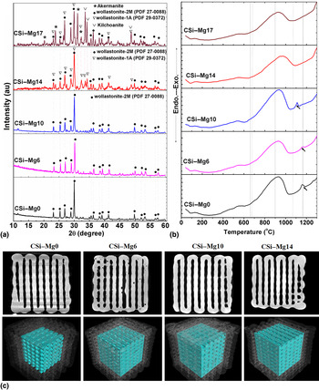
Figure 1(a) shows the X-ray diffraction (XRD) patterns of the ceramic powders for five of the compositions in this series. The pattern of pure wollastonite (i.e., CSi–Mg0) was shown for comparison. At very low Mg-substituting Ca levels (≤14%), the predominant phase was wollastonite, even though the CSi–Mg14 produced two types of wollastonite phase, including wollastonite-2M (PDF# 27-0088) and wollastonite-1A (PDF# 29-0372). With increasing Mg-substituting-Ca percent ratio up to 17% (CSi–Mg17), the other hybrid phases including akermanite (Ca2MgSi2O7; PDF# 35-0592) and kilchoanite (Ca3Si2O7; PDF# 29-0370), became clearly detectable, suggesting an evolution in composition from mono- to bi-phase wollastonite, and further toward the composites of at least three compounds of Ca–Mg silicate systems. Moreover, all the ball-milled bioceramic powders showed a superfine particle size below 1.5 µm (see Fig. S1, SM).
Characterization of the CSi–Mgx powder after calcining at 950 °C and bioceramic scaffolds after sintered at 1150 °C, respectively. (a) XRD patterns of the CSi–Mgx powders. (b) DTA curves of the CSi–Mgx powders. (c) 2D and 3D microstructures of the porous ceramics by μCT reconstruction.

The bioceramic scaffolds with filament distance of ~220 µm were patterned by extruded the CSi–Mgx inks through 450 µm nozzle. According to the quantitative analyses for the scaffold structure parameters, it was displayed that the strut thickness, pore size, and porosity slightly decreased while increasing Mg-substituting-Ca percent ratio (0%–17%) and sintering temperature from 1100 to 1200 °C (see Fig. S2, SM). Specifically, CSi–Mg6 and CSi–Mg10 (1150 °C) exhibited higher pore size (185–220 µm) and open porosity (40%–46%) than those of the CSi–Mg17 (1150 °C). These quantitative analyses suggest that, the more is the Mg-substituting-Ca percent ratio, the higher is the shrinkage of the bioceramic scaffolds during the sintering process. Generally, depending on the initial green body density, this is typically associated with high and anisotropic shrinkage for the 3D-printed BG scaffolds.[ Reference Bergmann, Lindner, Zhang, Koczur, Kirsten, Telle and Fischer 18 ] The large or complex parts may deform significantly as a result of gravity, surface tension, intrinsic strain or temperature, and density gradients. To overcome this problem, glass–ceramic composite structures may be considered, where the glass fraction and, hence, the extent of viscous sintering is reduced, and where a secondary stiff phase is used to geometrically stabilize the green body during sintering, to reduce overall shrinkage.[ Reference Winkel, Meszaros, Reinsch, Müller, Travitzky, Fey, Greil and Wondracze 19 ]
Figure 1(b) shows the differential thermal analysis (DTA) curves for the bioceramic powders. It is showed the crystallization temperature (T c, exothermic effect) in the range of ~700 °C. The onset of T c shifted toward lower temperature with increasing Mg substitution ratio from 0% to 10% in the powders. As the low heat process proceeded, only a very limited weight loss (<3%) occurred at 30–600 °C on thermogravimetric analysis (TG) curves (not shown) for all samples, which could be attributed to physically absorbed water.
The internal 2D and 3D visualizations of the bioceramic scaffolds were characterized by μCT. As shown in Fig. 1(c), the CSi–Mgx (x = 0–14) scaffolds had a uniform structure with well regular pore morphology. The constructs retained their shape with no noticeable deformation of the filaments after sintering. There were no closed pores present in the internal porous network, which was in good agreement with the scanning electric microscopy (SEM) observation below.
Additionally, it was seen from the optical images for the stop-view appearance of the CSi–Mgx scaffolds that the sintering process only improved the densification of the filament while avoiding deformation of the pore structure (Fig. S3, SM). In comparison with the CSi–Mg0 scaffold, the crossing filaments in the CSi–Mgx (x = 6–17) were fused together from 1100 to 1200 °C, suggesting the dilute Mg doping in wollastonite may be highly effective for improving the sintering property of its porous bioceramic.
Effect of sintering temperature on mechanical strength of bioceramic scaffolds
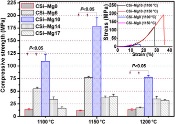
The effect of the sintering temperature on the compressive strength of the porous scaffolds is shown in Fig. 2. For five groups of scaffolds, the compressive strength (in the direction parallel to the pore channel) was increased from CSi–Mg0 to CSi–Mg10, and then decreased to CSi–Mg17. The strength (110–178 MPa) of the CSi–Mg10 (1100 and 1150 °C) was an order of magnitude higher than those (11–15 MPa) of CSi–Mg0 (1100 and 1150 °C), though the porosity (open pores) of the later was only higher by 2%–4% than those of the Mg-doped ceramics (see Fig. S2, SM). This significant difference in mechanical strength is also demonstrated from the representative curves for the compressive stress versus strain for the CSi–Mg0 and CSi–Mg10 scaffolds tested at the same deformation rate (Fig. 2, inset). Both groups of scaffolds showed similar “elastic” responses, as well as a similar rapid failure mode. However, the response of the ceramic scaffolds showed substantial difference in strain for failure (<18% versus 25%–35%). However, the strength of CSi–Mg14 and CSi–Mg17 scaffolds (1100 and 1150 °C) only showed that it was equal to threefold of the CSi–Mg0 (1100 and 1150 °C). Although the porosity (open pores) of the CSi–Mgx scaffolds was decreased with a higher temperature of sintering (see Fig. S2, SM), the strength of the CSi–Mgx (1200 °C) in compression was not more than 40 MPa, except for the CSi–Mg10. These experimental results suggest that a higher sintering temperature (e.g., 1200 °C) may enhance the shrinkage of CSi–Mgx, but not substantially contribute to their compressive strength.
Effects of sintering temperature on the compressive strength of the small-pore CSi–Mgx scaffolds. Inset shows the representative stress–strain curves of the compression test.

Microstructure of bioceramic scaffolds
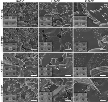
The reason why some CSi–Mgx scaffolds sintered at 1100–1200 °C possess exceptionally high compressive strength and this specific property may be related to the microstructure of the ceramic strut in the scaffolds. We carried out the SEM analysis using the facture surface of the ceramic scaffolds. As shown in Fig. 3, there were no significant changes in the microstructure along the length of the ceramic ink filaments. So each fracture surface was, in general, representative of the whole construct. However, instead of nearly rectangular pores in the CSi–Mg0 (1100–1200 °C) scaffolds, Mg dilute doping resulted in rectangular-to-round corner transformation around the pore with increasing sintering temperature (Fig. 3, inset), implying the substantially Mg-assisted viscous-flow sintering in the CSi ceramics. Moreover, it indicated a transgranular type of fracture for the CSi–Mg0 (1100 °C) and CSi–Mg6 (1100 °C), but both transgranular and intergranular types of fracture can be observed for the CSi–Mg10 (1100 °C). At low temperature, i.e., 1100 °C, the shape of particles in CSi–Mg0 and CSi–Mg6 almost remained unchanged. At 1150 °C, the micrographs of these ceramic particles show softening while beginning to bond to each other, and here the pores were the connective open-pores. With increasing Mg-substituting-Ca percent ratio up to 10% (CSi–Mg10), it appeared that the open-pores in the ceramic struts disappeared when sintering at 1150 °C, and the closed pores were also hardly noticeable, which is thought to contribute the increased mechanical strength.[ Reference Fu, Saiz, Rahaman and Tomsia 20 ] However, it is noticed that all of porous CSi–Mgx (1200 °C) resulted in significant grain growth, which would be disadvantageous for scaffold's strength.
Fracture surface microstructures of the small-pore CSi–Mgx scaffolds after one-step sintering at 1100–1200 °C for 240 min, respectively. Insets represent the low magnified SEM images of the porous scaffolds (side view). The white and black arrows respectively show the closed and open pores in the ceramic struts.

Effect of pore size on mechanical strength of bioceramic scaffolds
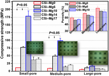
It is well known that, for bone tissue engineering, the scaffolds with larger pore size, and high porosity and interconnectivity are required to facilitate the neovascularization and new bone tissue ingrowth.[ Reference Henkel, Woodruff, Epari, Steck, Glatt, Dickinson, Choong, Schuetz and Hutmacher 2 – Reference Kasten, Beyen, Niemeyer, Luginbühl, Bohner and Richter 4 ] To increase the overall pore size and porosity, the CSi–Mgx scaffolds with initial filament distance up to 350 and 500 µm were fabricated and produced medium- or large-pore scaffolds with top-view pore dimensions of ~320–340 and 430–460 µm after sintering at 1150 °C. Also, the side-view pore size (Fig. S4, SM) and the average porosity (Fig. 4, inset) of the ceramic scaffolds increased with increasing the filament distance, but decreased with the increasing Mg substitution ratio. The compressive strengths of CSi–Mgx scaffolds with different pore sizes were measured and compared (Fig. 4). For the three groups of as-sintered scaffolds, the compressive strength was decreased with the increasing macropore dimension. Generally, the CSi–Mg10 displayed a highest strength and the CSi–Mg6 obtained the second-highest strength in each group. The strengths (50–100 MPa) of CSi–Mg6 and CSi–Mg10 with the medium-pore structure were 5–10 times higher than that of the medium-pore CSi–Mg0.
Relationship between pore size and compressive strength of CSi–Mgx scaffolds sintered at 1150 °C for 3 h. Inset showing the porosity of the scaffolds with different pore size levels. Optical images (side view) showing the porous structure of the sintered scaffolds.

Effect of heating schedules on mechanical strength of bioceramic scaffolds
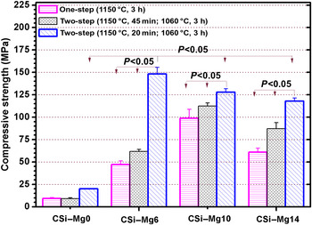
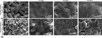
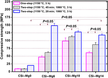
It is well known that two-step sintering can be used to control the grain growth of modern technical ceramics during the final-stage sintering process.[ Reference Chen and Wan 21 ] In this regard, we also used two-step sintering as new heating schedule to explore the possibility of enhancing mechanical strength on the medium-pore scaffolds. In fact, a shorter (from 4 to 3 h) one-step sintering treatment at 1150 °C for the CSi–Mgx ceramics may not substantially enhance the compressive strength, except for CSi–Mg14 (Fig. 5 versus Fig. 4). However, the medium-pore CSi–Mgx (x = 6–14) scaffolds undergoing two-step sintering treatment, especially shortening of sintering time (from 45 to 20 min) at the first step temperature of 1150 °C, exhibited ultrahigh strength in comparison with the conventional secondary phase-reinforced CSi-based scaffolds.[ Reference Shuai, Gao, Feng and Peng 22 , Reference Wu, Fan, Zhou, Luo, Gelinsky, Chang and Xiao 23 ] For instance, the CSi–Mg6 group, which was initially heated at 1150 °C for 20 min, and then held at 1060 °C for 3 h, showed threefold higher strength (~148.2 ± 7.3 MPa) than this material after one-step sintering at 1150 °C for 3 h (Fig. 5) and graphene-reinforced CSi scaffolds.[ Reference Shuai, Gao, Feng and Peng 22 ] Meanwhile, the CSi–Mg14 scaffolds according to this heat schedule also showed appreciable strength (~120 MPa), similar to that of the human cortical bone. Fortunately, the porosity of two-step sintering treatment for CSi–Mgx (x = 6–14) scaffolds still reached 49%–53%, with negligible decrease in porosity belonging to open pores in the scaffolds. It is confirmed from the SEM micrographs that a two-step sintering method readily suppressed the CSi–Mgx grain growth and enhanced the densification of pore struts (Fig. S5, SM). After undergoing an acid-heat-etching treatment, the grains in one-step sintered CSi–Mgx scaffolds [Fig. 6(a)] all showed a significant growth, except that the pure wollastonite ceramic showed a small grain but less densification structure. In contrast, the smaller grains were retained and the fracture surface displayed a high degree of densification in the two-step sintered CSi–Mgx scaffolds [Fig. 6(b)]. These findings demonstrate that two-step sintering is an efficient approach to control the grain growth and enhance mechanical strength of the dilute Mg-doped CSi scaffolds.
Relationship between heating schedule and compressive strength of medium-pore CSi–Mgx scaffolds.
SEM micrographs of the acid-heat-etched fracture surface of the pore strut in the medium-pore CSi–Mgx scaffolds after undergoing one-step sintering at 1150 °C for 3 h (a) and two-step sintering at 1150 °C for 20 min and then at 1060 °C for 3 h (b), respectively.

Comparison of CSi–Mgx and Ca–Mg silicate bioceramic scaffolds
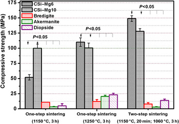
To clarify the potential influence of dilute and high doping of Mg on the mechanical strength of CaO–SiO2 system, the medium-pore akermanite, bredigite, and diopside porous scaffolds were also fabricated using ceramic ink writing technique. These porous bioceramics and CSi–Mgx scaffolds were treated using one-step (1150 or 1250 °C, 3 h) or two-step sintering (1150 °C, 20 min; 1060 °C, 3 h), respectively. The quantitative measurement for physical properties was confirmed that the CSi–Mgx scaffolds showed lower porosity by 5%–8% than the stoichiometric Ca–Mg silicate scaffolds (Fig. S6, SM). This was partly attributed to the significant shrinkage of the pore struts of the CSi–Mgx scaffolds. This bigger shrinkage might result from viscous sintering and contribute to the densification in pore struts of CSi–Mg6 and CSi–Mg10, which was observed in silicate glass-ceramics.[ Reference Ryu, Lee, Kim, Hong, Kim, Lee, Lee, Chang, Lee and Chung 24 ] However, the average pore size of the medium-pore scaffolds of CSi–Mgx still remained above 300 µm (Fig. S6, SM). As it can be seen from Fig. 7, the Ca–Mg silicate scaffolds showed significantly different compressive strengths when sintering at the same temperature condition. Meanwhile, the heating schedule also heavily influenced the strengths of the porous bioceramics. Increasing sintering temperature from 1150 to 1250 °C showed the benefits of enhancing strength of diopside and akermanite porous bioceramics, but this contributed to just a little for the bredigite. Meanwhile, the compressive strengths of all stoichoimetric compound porous scaffolds were less than 25 MPa, similar to the data by using the conventional pore-making methods,[ Reference Wu, Chang, Zhai, Ni and Wang 25 – Reference Wu, Chang, Zhai and Ni 27 ] but much higher than that of the inorganic–organic hybrid scaffolds.[ Reference Chen, Watkins-Curry, Smoak, Hogan, Deese, McCandless, Chan and Hayes 28 ] In contrast, the two-step sintering has important implications for suppressing grain growth of the CSi–Mgx, which is more beneficial for improving their strength in porous scaffolds, even though the one-step high heating schedule (1250 °C, 3 h) may enhance the CSi–Mgx's strength after undergoing β-to-α-phase transition.
Comparison of compressive strength of medium-pore CSi–Mgx and calcium–magnesium silicates ceramic scaffolds sintered at different heat schedule conditions.

Discussion
In this study, we found that the dilute Mg doping can increase the densification of pore struts in wollastonite scaffolds and significantly reinforce its mechanical strength when one- or two-step sintering at 1150 °C. The compressive strength of the 3D-printed CSi–Mg6 and CSi–Mg10 scaffolds is nearly ten times higher than the pure CSi scaffolds. The dilute Mg-doped CSi porous scaffolds, moreover, has certain inherent advantages over other Ca–Mg silicate scaffolds, viz. mechanical strength and sintering properties. Thus, this study provides a new strategy that the dilute Mg doping into single-phase bioceramic is beneficial in fabricating ultrahigh strength porous scaffolds.
Sintering at high temperature is widely used to form porous bioceramics which have a high melting point. However, this method requires high temperature and long duration for heating in order to bond the junctions between ground material particles. In general, the prerequisite for optimizing the fabrication of porous bioceramics is to understand the heat treatment conditions of bioceramic particles and the interaction between heat treatment and grain growth, which controls the material bioactivity, resorbability, and mechanical strength. This raises the question as to what composition design might be the best for the balance between mechanical and physicochemical properties of Ca/Ca–Mg silicate bioceramic scaffolds.
It is of special note that the introduction of MgO into Ca–SiO2-based BGs and bioceramic systems is usually expected to enhance their bioactivity, and Mg ions may increase the osteoblastic activity and bone remodeling in vitro and in vivo.[ Reference Diba, Goudouri, Tapia and Boccaccini 11 , Reference Diba, Tapia and Boccaccin 12 ] In our studies, it is interesting to see that dilute Mg doping may enhance the densification of CSi grains in pore struts. This contribution in CSi–Mgx is possibly attributed to the Mg ion substitution for Ca ion in the crystal lattice. In our previous studies, we found that a new B2O3-added, 45S5-derived BG which exhibits low melting temperature (<1000 °C) and may reinforce the porous CSi porous scaffolds via a low-temperature co-firing process[ Reference Xie, Zhang, Yang, Yang, Wang, Zhang, Xu, Shao, He, Fu and Gou 29 , Reference Shao, Yang, He, Fu, Liu, Zhang, Yang, Gao and Gou 30 ]; unfortunately, the possible mismatch's coefficient of thermal expansion for the biphasic composites leads to poor densification in the sintered BGs at higher temperature (>1080 °C). The present study confirms that pressureless sintering of CSi–Mg6 and CSi–Mg10 scaffolds at 1150 °C readily maintains considerably high porosity (~50%) and keeps appreciable compressive strength (>80 MPa). This can be confirmed by the SEM observation, in which the CSi–Mg6 and CSi–Mg10 sintered at 1150 °C showed more dense fracture microstructures compared with the pure CSi (Fig. 3).
It appears from images of the fracture microstructure in CSi–Mgx that with increasing Mg content, open-pores in the pore struts disappear, whereas closed pores increase and the pores show regular spherical shapes (Fig. 3). By increasing sintering temperature up to 1150 °C, the grain growth of the CSi–Mgx leads to a pore decrease and as a result, the sintering densification is achieved. Microstructural changes of CSi–Mgx samples with temperature are different to those of pure CSi scaffolds. Based on the results presented in Fig. 3, it can be concluded that an optimum temperature at 1150 °C for the maximum densification exists for CSi–Mg6 and CSi–Mg10. Accordingly, the pressureless sintering of the 3D-printed CSi–Mgx scaffolds readily produce an enhanced densification at such temperature level in comparison with the other Ca–Mg silicate bioceramic scaffolds.
Sintering, as an important stage in ceramics processing, has also undergone significant modifications and some novel sintering technologies and routes have been widely introduced. For example, using pressure sintering such as hot (isostatic) pressure, ultrahigh pressure, and spark plasma sintering are reported as successful approaches to control grain growth during extremely high rates of heating process.[ Reference Champio 31 ] However, the application of pressure and also high rates of heating would need special equipment and it is particularly disadvantageous for macropore maintenance in ceramic scaffolds. It is accepted that at the final stage of conventional sintering, the density of the free surfaces diminishes and the thermal processes are associated with slower kinetics via diffusion along the grain boundaries and in the bulk. Especially, the relative low transition temperature (<1250 °C) from pure β-to-α-phase transformation of CSi is disadvantageous for the densification of pure β-CSi ceramic by a pressureless sintering method. Therefore, the preservation of the microstructure characteristic and mechanical strength in single-phase β-CSi macroporous ceramics represents a significant challenging target due to the inevitable and accelerated grain growth during conventional sintering processes.[ Reference Long, Chen, Bai, Chang and Lin 32 ]
The sintering under different two-step sintering regimes may be used to investigate the effect of sintering parameters on the densification behavior and grain growth suppression, which eventually contribute to the improvement of mechanical strength.[ Reference Nadernezhad, Moztarzadeh, Hafezi and Barzegar-Bafrooei 33 ] In this study, our investigation indicates that the dilute Mg-doping approach is of benefit for improving the sinterability of single-phase β-CSi porous bioceramics and obtaining ultrahigh compressive strength by a simple two-step sintering (for CSi–Mg6), without applied pressure (Fig. 5). The feasibility of densification without grain growth relies on the suppression of grain-boundary migration, while keeping grain-boundary diffusion active (Fig. 6). In general, interface kinetics in very fine-grained polycrystals is limited due to difficulties in maintaining source and sinks to accommodate point defects.[ Reference Land, Martin, Potapenko, Palmore and De Yoreo 34 ] However, this effect may diminish at larger grain size above 100 nm, allowing the kinetic window to extend to lower temperature. For examples, Chen and Wang's investigation demonstrated that MgO doping has a window kinetic shifting at a lower temperature in Y2O3 nanocrystalline ceramic.[ Reference Champio 31 ] Accordingly, the control of CSi–Mgx grain growth can be achieved using a two-step sintering method, in which the high-temperature heating followed by structural freezing via rapid cooling to a constant temperature levels stop the grain growth but not the densification. Speaking totally, this is probably due to the two following reasons: (1) the sinterability of CSi can be substantially modified by Mg dilute doping, and (2) the grain growth of CSi–Mgx is drastically suppressed during the final stage of sintering.[ Reference Chen and Wan 21 ]
Conclusion
In summary, the dilute Mg-doped β-CSi bioceramic scaffolds with ultrahigh strength is successfully developed under conventional pressureless sintering condition. Compressive strengths of the porous scaffolds have shown 120–180 MPa and have an order of magnitude higher than Ca–Mg silicate porous bioceramics upon compression tests. The heating schedule has been found to play an important role in suppressing grain growth and improving densification of pore struts. Successful completion of these investigations will enable us to manufacture bioceramic scaffolds for a variety of repair applications directly by 3D printing technique. Also, these findings might be a breakthrough in 3D-printed high-strength bioceramic scaffolds for large and/or geometrically complex craniomaxillofacial bone and load-bearing segmental bone defect repair areas.
Supplementary Material
For supplementary material for this article, please visit http://dx.doi.org/10.1557/mrc.2015.74
Acknowledgments
This work was supported by the Zhejiang Provincial Natural Science Foundation of China (grant no. LZ14E020001), the National Natural Science Foundation of China (grant numbers 81271956 and 51221004), Science and Technology Department of Zhejiang Province Foundation (grant numbers 2015C33119 and 2014C33202), and the National “Twelfth Five-Year” Plan for Science & Technology Support of China (grant no. 2012BAI08B01).