Cardiovascular malformations constitute the single most important cause of death in infants and young children with Down's syndrome.1–4 Quite understandably, therefore, several studies, but mostly from Europe and America, have focused on the cardiovascular defects associated with this trisomy, particularly on their prevalence and spectrum.1, 5–9 The published rates of prevalence have varied widely, from under 20 to over 60%, 1, 2, 7, 9–12 and considerable differences also exist in the reported relative frequency of the individual defects.
Comparable studies in Arab children are lacking. To our knowledge, only one previous report specifically addressed the question of associated cardiac defects in Arab children with Down's syndrome,13 and that lone report did not provide any clear data on the relative frequency of the specific defects. Thus, knowledge about the congenital cardiac malformations to be found in Arab children with Down's syndrome is still very fuzzy. We hope with this report, addressing the prevalence and spectrum of cardiovascular malformations in Omani Arab children with trisomy-21, to remedy that situation.
Patients and methods
Patients for this study, all Omani Arabs, were recruited from two secondary-level health facilities in the Sultanate of Oman; namely, Buraimi Hospital, examining the period from January, 1996, to December, 1998, and Nizwa Regional Referral Hospital, taking in the period January, 1999, to December, 2004. They were a subset of the subjects in a wider clinical prospective study of congenital cardiac disease in Arab children.
A uniform methodology was followed in both hospitals. The patients were enrolled from three sources: the maternity wards, the Special Care Baby Units, and a paediatric cardiology clinic conducted by one of us (OJ). The diagnosis of Down's syndrome was based initially on phenotypical features and confirmed later by karyotyping. Prior to the onset of the study in each centre, and at appropriate intervals subsequently, the doctors in the peripheral health institutions were contacted and urged to refer all children with Down's syndrome phenotype to the designated paediatric cardiology clinic, whether or not cardiac disease was suspected.
All the patients had a detailed cardiac evaluation, including cross-sectional, Doppler, and colour-imaging echocardiography, performed by the same paediatric cardiologist (OJ). The echocardiographic studies were performed in the standard fashion, using cardiac ultrasonography machines (Hewlett Packard Sonos 1800) fitted with 5.0 and 7.5 megahertz transducers. The studies were video-taped, and the records reviewed subsequently in order to maximize accuracy of the diagnoses. Neonates whose initial echocardiography was performed in the first week of life had the procedure repeated at the second- or sixth-week postnatal follow-up clinic, barring default. Overall, each of the subjects in this study had echocardiography on at least two occasions.
Criterions for exclusion
We excluded from analysis data from preterm babies with lone patent arterial duct, babies detected to have a lone duct in the first week of life but who, for whatever reason, could not be restudied to confirm continuing patency of the duct; neonates with small defects, measuring less than 4 mm, in the oval fossa detected in the first week of life but not reconfirmed subsequently, infants of diabetic mothers, and patients with trivial tricuspid or mitral valvar regurgitation considered to be a normal echocardiographic finding.
Results
After applying the criterions for exclusion stated above, we assembled data from 110 patients for analysis. Of these 110 patients, 26, 16 males and 10 females, were studied in the first hospital and the remaining 84, 51 males and 33 females, in the second hospital. The age range for the two groups were from 1 day to 10 years, with a median of 6.5 months, and 1 day to 11 years, with a median of 2 months, respectively. For the groups combined, the range was from 1 day to 11 years, with a median of 2 months. Thus, both groups of patients were demographically similar. Furthermore, as stated above, a uniform methodology was followed in both hospitals, and the cardiac evaluation in all the patients was performed by the same paediatric cardiologist. We felt justified, therefore, in combining the data for analysis.
Cardiovascular malformations were identified in 63 (57%) of the 110 patients. The prevalence in the first group was 62% (16/26), and in the second group, 56% (47/84). These rates are not significantly different (χ2 equal to 0.25; P greater than 0.05). Neither are the prevalences in neonates (55%), older infants (64%), and patients aged more than 12 months (50%) (Table 1). But cardiac malformations occurred significantly more frequently in the female subjects. Of the 43 female subjects in both groups, 30 (70%) had cardiovascular defects, compared with 33 (49%) of the 67 male subjects. This difference is significant (χ2 equal to 4.50, P less than 0.02).
Table 1. Prevalence of cardiac disease in three age groups

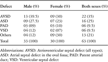
Table 2 shows the main defects identified in the 63 subjects with cardiovascular malformations. Atrioventricular septal defects, including the so-called “ostium primum” defect, were the most prevalent, followed by atrial septal defects confined to the oval fossa. These two malformations were detected in 38 (60%) of the 63 patients with cardiac disease. Patency of the arterial duct, and perimembranous ventricular septal defect, each occurred as lone defects in six patients. Thus, defects of cardiac septation and patency of the arterial duct were present as lone defects in 50 (79%) of the 63 patients.
Table 2. Relative frequency and sex distribution of the main defects in 63 affected children

Of the 63 patients, 13 (21%) each had two or more cardiovascular defects. These are shown in Table 3, with the main defect listed first. Of the 13 patients, 11 had defects which permitted left-to-right shunts and excessive pulmonary blood flow. In all, therefore, 61 (97%) of the 63 affected children had defects which allowed left-to-right shunts. The remaining two patients had tetralogy of Fallot.
Table 3. Miscellaneous cardiovascular defects identified in 13 patients
When analysed in terms of the relative frequencies of the specific malformations, the preponderance of defects which permitted left-to-right shunts was again obvious (Figure). Atrioventricular septal defect accounted for 24 (32%) of the 76 defects detected in the affected children. Of the 24 cases, half were detected in neonatal life, 10 in older infants, and only two in children older than 12 months. Defects within the oval fossa accounted for 22 (29%) of the 76 defects, patency of the arterial duct for 13 (17%), and perimembranous ventricular septal defect for 11 (14%). Thus, defects characterized by left-to-right shunts accounted for 70 (92%) of the 76 malformations. Mitral regurgitation (two cases), tetralogy of Fallot (two cases), mild coarctation of the aorta (one case), and right-sided heart (one case) completed the tally. Of these 6 lesions, 4 coexisted with defects that allowed left-to-right shunts.

Figure. Frequency distribution of 76 cardiovascular defects identified in 63 children with Down's syndrome.
Discussion
Cardiovascular malformations were detected in almost three-fifths of the Omani Arab children with Down's syndrome who formed the subjects of this study. The prevalence in neonates, at 55%, and in the older children recruited from the paediatric cardiology clinics, at 59%, were similar; and both figures are in tandem with the prevalence of three-fifths reported from a study of 90 children in the university hospital in Muscat, the capital city of Oman.13 Furthermore, the rates from this study, and that from Muscat, fall within the range of two to three-fifths, which has been reported from most of the studies from several other countries.5, 6, 8, 14–16
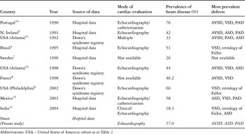
Lower and higher rates have also been reported occasionally, as shown in Table 4. Most of the low rates were reported from studies2, 3, 7, 11, 12, 17 in which the cardiac evaluation was, judging by current practice, arguably inadequate. This raises the possibility of under-detection. Indeed, proof of this was apparent in the study by Khoury and Erickson,11 in which improved ascertainment resulted in a remarkable increase in the reported prevalence, from around one-fifth in the early 1970s to over half in the late 1980s, all in the same surveillance programme in Atlanta, United States of America. At the other extreme, Pinto et al.,10 from Portugal, reported an exceptionally high prevalence of three-quarters. There was, however, an obvious selection bias in that study, because the subjects comprised children with Down's syndrome who were referred to a paediatric cardiology clinic because of suspected cardiac disease.
Table 4. Some recent publications on cardiovascular malformations associated with Down's syndrome

We sought to eliminate selection bias in our clinic-derived patients by urging the doctors in our practice area to refer all children with features suggestive of Down's syndrome, whether or not cardiac disease was suspected. We believe that they complied with our bidding because, as stated above, the prevalence of heart disease in our consecutively recruited newborn patients, at 55%, and the older, clinic-derived patients, at 59%, are similar. These figures, coupled with the previous report from Muscat,13 indicate that the likelihood of associated cardiovascular defects in Omani Arab children with Down's syndrome is high, at from half to two-thirds. The data further indicate that the likelihood is higher in female than in male patients, at 70% versus 49%. The reason for this female predilection, which has also been noted by some other workers,2, 10 is unknown.
In the absence of comparable data from other Arab countries, we are unable to determine with certainty the reasons for the high prevalence of congenital cardiac malformations in our patients. But a high rate of ascertainment appears to be one main, and perhaps the major, reason. We almost certainly achieved a very high rate of ascertainment owing to our methodology, which included echocardiographic evaluation of all children with Down's syndrome, whether or not cardiac disease was suspected.
Next, we wish to address the issue relating to the spectrum of cardiovascular defects associated with Down's syndrome. In our patients, atrioventricular septal defect was the most common malformation, accounting for one-third of associated defects. Next, in descending order, were atrial septal defects within the oval fossa, at one-third, patency of the arterial duct, at just over one-sixth, and perimembranous ventricular septal defect, at just under one-sixth. Altogether, defects which are characterized by increased pulmonary blood flow and the potential for pulmonary hypertension accounted for over nine-tenths of the malformations. Isomerism, and stenotic lesions were very uncommon, and we did not detect any case of discordant ventriculo-arterial connections.
Just like it is in this present report, atrioventricular septal defect has been the most prevalent defect in most publications (Table 4). But dissenting reports also do exist.12, 14–16, 18 Among the most recent in this latter category is the publication by de Rubens Figueroa et al.,15 who reported that the most prevalent defects in Mexican children with trisomy-21 were atrial septal defects, within the oval fossa, ventricular septal defect, and patency of the arterial duct, in that order. These three defects accounted for nine-tenths of the malformations in the children they studied. Atrioventricular septal defect ranked fourth, having been identified in only 14 (9%) of the 160 patients in the study. Much earlier, in a study of Chinese children with Down's syndrome, Lo et al.19 detected atrioventricular septal defect in only 15% of 149 patients; and atrial septal defect in an even smaller proportion, 13%. Ventricular septal defect was the most prevalent, at 44%. Similarly, in a reaction to the paper by de Rubens Figueroa et al.,15 Digilio20 wrote that ventricular septal defect was the most prevalent cardiovascular malformation in Chinese and Japanese children with trisomy-21.
Children with Down's syndrome are predisposed to alveolar hypoventilation, hypoxaemia, and pulmonary hypertension by several adverse respiratory factors, including recurrent pulmonary infections, reduced ventilatory excursion due to muscle hypotonia, chronic upper airway obstruction, obstructive sleep apnoea, and alveolar hypoplasia.1 Hence, the coexistence of any cardiac malformation that permits a left-to-right shunt will increase the proclivity for pulmonary hypertension. Therein lies the additional danger of cardiac defects associated with Down's syndrome. The case for early detection and prompt, appropriate treatment can hardly be stronger.
Another reason for the continuing interest in the cardiac malformations associated with Down's syndrome is the uncertainty about their pathogenesis. The defects have been variously attributed to a postulated increased adhesiveness of the trisomic cells during morphogenesis,21 to an interaction between the trisomic genome and environmental factors,22, 23 and to the effects of genes located in a putative Down's syndrome critical region.24–27 Recently, Barlow et al.28 suggested that the defects might result from the effects of a gene, which they called the “Down's syndrome cell adhesion molecule”, which is unrelated to the critical region.
These theories are, no doubt, fascinating. A careful reading of the subject, nonetheless, suggests that the prevalence and spectrum of the defects reported from different centres probably reflect the effects of several factors, including genetic influence, the source of the patients, their age at the time of the study, and the mode of cardiac evaluation. Environmental factors may play a role in high-altitude locations like Mexico.15
Without doubt, the methodology used could profoundly influence the prevalence and pattern reported from any given centre. For example, an echocardiographic study of trisomic neonates in the first week of life will likely yield a falsely high incidence of patency of the arterial duct and atrial septal defects within the oval fossa if appropriately timed repeat evaluations are not performed. Conversely a study of older institutionalized children will under-report two categories of defects. These are the ones that are liable to resolve spontaneously with time, such as atrial septal defects within the oval fossa and patency of the arterial duct, and those that are apt to be fatal in infancy if untreated, such as atrioventricular septal defect. Hence, data from different centres should be interpreted discerningly, with due attention to the variable confounding factors.
In conclusion, we wish to reiterate some of the major practice implications of cardiac malformations encountered in the setting of Down's syndrome. First, any physician tasked with the evaluation of a child with the Down's syndrome phenotype will do well to remember that the likelihood of associated cardiovascular malformations is high, at up to three-fifths. For an Omani Arab child, this is the likelihood. Second, the most prevalent defects are atrioventricular septal defect, atrial septal defects within the oval fossa, perimembranous ventricular septal defect, and patency of the arterial duct. Third, and this is common knowledge, those with atrioventricular septal defects are particularly liable to develop severe pulmonary vascular disease in early infancy. Any indicated surgical intervention, therefore, should be appropriately timed. Finally, it is also worth remembering that, in neonates with trisomy-21, clinical evaluation, including chest radiography and electrocardiography, may fail to reveal an associated congenital cardiac malformation.5, 9, 29 Echocardiography is essential. This, indeed, is one of the main thrusts of the recommendations of the American Academy of Pediatrics for the care of children with Down's syndrome.30
Acknowledgements
We thank the paediatric residents in Buraimi and Nizwa hospitals, and most especially Dr S M George, for assisting with the detection of cases, and the authorities in Nizwa Hospital and Dhakiliya Regional Health Directorate for administrative support.