Introduction
The blood-feeding nematode Haemonchus contortus inhabits the abomasum (stomach) of both domestic and wild small ruminants during its adult stage. Recognized as one of the most pathogenic parasitic nematodes, H. contortus is responsible for severe health deterioration in affected animals and causes substantial economic losses in the global livestock industry (Ma et al Reference Ma, Jiang, Fan, Hao, Li, Zhang, Li, Wang and Luo2025; Rodríguez-Vivas et al Reference Rodríguez-Vivas, Grisi, Pérez-de León, Silva, Torres-Acosta, Fragoso, Romero, Rosario, Saldierna and García2017). Infected animals exhibit various clinical signs, including apathy, weight loss, anaemia, malnutrition, and weakened immune defences (Cruz-Tamayo et al Reference Cruz-Tamayo, López-Arellano, Gonzalez-Garduno, Torres-Hernandez, De la Mora-Valle, Becerril-Perez and Huchin-Cab2021), which can ultimately result in death (Jesse et al Reference Jesse, Chung, Abba, Lila, Aishah, Affandi and Hambali2019; Kandil et al Reference Kandil, Hany, Elsawy and Fadlallah2025). Traditional control of haemonchosis and other nematode infections relies primarily on chemical anthelmintic drugs to reduce parasite burden; however, this approach can lead to undesirable outcomes, such as the emergence of anthelmintic-resistant gene mutations (Reyes-Guerrero et al Reference Reyes-Guerrero, Higuera-Piedrahita, Maza-Lopez, Mendoza-de-Gives, Camas-Pereyra and López-Arellano2024). In addition, drug residues can remain in animal tissues, including milk, meat, and other derivatives intended for human consumption (Dai et al Reference Dai, Zhou, Zhai, Li, Dai, Wang and Zhang2025). Furthermore, animals excrete these drug residues through their faeces and urine, introducing them into the soil and posing a threat to beneficial soil microbiota (Belete et al Reference Belete, Adugna and Yirsa2025). Over the past several decades, alternative methods for controlling these parasitic diseases have been investigated. Examples include the use of plants and plant metabolites with nematocidal activity (Boyko and Brygadyrenko Reference Boyko and Brygadyrenko2025), copper particles that kill adult nematodes in the abomasum of animals (Johnson et al Reference Johnson, Mackey, DeWees and Papajeski2018; Vatta et al Reference Vatta, Waller, Githiori and Medley2012), as well as vaccines and immunogens (Sajovitz-Grohmann et al Reference Sajovitz-Grohmann, Adduci, Werling, Wiedermann, Wortha, Prole and Lichtmannsperger2025).
More recently, the study of natural enemies of nematodes has attracted the attention of researchers seeking potential biological control agents. Nematophagous fungi are a group of organisms that, over thousands of years, have adapted to live in the soil primarily as saprophytes. However, when stimulated by proteins from the nematode cuticle known as ‘nemins’ (Pramer and Stoll Reference Pramer and Stoll1959), these fungi shift from a saprophytic lifestyle to a predatory one. Nemins act as signals, prompting the fungi to differentiate their mycelia into specialized trapping devices designed to attract, capture, destroy, and feed on nematode tissues (Faria et al Reference Faria, dos Santos Fonseca, de Araújo, de Carvalho, Albuquerque and de Souza Perinotto2025; Mendoza-de Gives Reference Mendoza-de Gives2022).
The species Dactylellina candida and Arthrobotrys musiformis belong to the Orbiliales, a group of micro-fungi that, in their sexual phase, act as predators of nematodes of agricultural and livestock importance (Pérez-Anzúrez et al Reference Pérez-Anzúrez, Olmedo-Juárez, von-Son de Fernex, Alonso-Díaz, Delgado-Núñez, López-Arellano, González-Cortázarv, Zamilpa, Ocampo-Gutiérrez and Paz-Silva2022; Wu et al Reference Wu, Kim and Zhou2012). This study aimed to evaluate the effects of adding crude extracts from three different nematodes – H. contortus, Turbatryx aceti, and Panagrellus redivivus – as potential elicitors of nematocidal activity in liquid culture filtrates of Dactylellina candida and Arthrobotrys musiformis.
Materials and methods
Location
This study was conducted at the Laboratory of Helminthology, National Center for Disciplinary Research in Animal Health and Safety (CENID-SAI), part of the National Institute for Research in Forestry, Agriculture and Livestock (INIFAP), located in Jiutepec City, Morelos, Mexico.
Biological material
Obtaining strains of nematophagous fungi
Soil samples were collected from the protected natural area ‘Chapultepec Ecological Park’, located in Cuernavaca, Morelos (Figure 1). Covering 12.844 hectares at an elevation between 1,412 and 1,466 meters above sea level, this park features an intermediate subhumid climate. It is notable for its diverse wild flora and fauna, effective moisture retention, and regulation of rainfall, which aid in aquifer replenishment (Arriola-Ponsin et al Reference Arriola-Ponsin, Orozco-Lugo and Franch-Pardo2022). Additional soil samples were randomly gathered from the Ecotouristic Park ‘El Hoyanco’, in Coatlán del Río, Morelos (Figure 1). This park lies at an elevation between 1,200 and 1,400 meters, has a warm subhumid climate with summer rainfall, and is recognized for its rich biological diversity. It also plays a vital role in conserving the local ecosystem (Álvarez-Vargas et al Reference Álvarez-Vargas, Alia-Tejacal, López-Martínez, Acosta-Durán, Andrade-Rodríguez, Colinas-León, Delgado-Escobar and Villegas-Torres2006).
Map of the Mexican Republic, highlighting the locations of the ‘Chapultepec Ecoturistic Park’ in Cuernavaca, State of Morelos, and the ‘El Hoyanco’ Ecoturistic Park in Coatlán del Río, State of Morelos.

Nematode
A strain of the free-living nematode Panagrellus redivivus, cultivated in an oatmeal medium, was commercially acquired from a pet store in Jiutepec, Morelos, where it is sold as fish food. To prepare the culture medium, approximately 20 g of oat flakes and 200 mL of distilled water were combined in a beaker and mixed until homogeneous. The mixture was then microwaved for 5 minutes, with pauses every 30 seconds. After allowing the medium to cool, it was transferred to a sterile glass container, and an inoculum containing an undetermined number of P. redivivus specimens was introduced. The nematode culture was incubated at 27°C for 7 days (De Lara et al Reference De Lara, Castro, Castro and Castro2007). To harvest the nematodes, the sieving technique was employed: a sample of the oatmeal culture was placed in a beaker with distilled water and washed through three sieves of different mesh sizes. Finally, nematodes were resuspended in sterile distilled water and recovered using the Baermann funnel technique.
Obtaining Haemonchus contortus infective larvae. To obtain infective larvae of Haemonchus contortus (L3), a Pelibuey lamb was orally inoculated with an aqueous suspension containing 350 infective larvae of H. contortus per kilogram of body weight. After 21 days, faecal samples were collected directly from the rectum once the animal tested positive for nematode eggs using the McMaster technique (Daş et al Reference Daş, Klauser, Stehr, Tuchscherer and Metges2020). Fresh faeces were ground and mixed with small pieces of polyurethane foam to promote oxygenation, then incubated at room temperature (18–28°C) for 7 days (Liébano Reference Liébano2004). Infective larvae were subsequently recovered using the Baermann Funnel technique for 24 hours (Hernández et al Reference Hernández, Prats and Ruvalcaba1998), followed by cleaning via differential centrifugation with 40% sucrose gradients and several washes with sterile water to remove residual material. The larvae were then unsheathed by treatment with a 0.187% sodium hypochlorite solution for 3–5 minutes, followed by washing with water to remove the sheaths (Cedillo-Borda et al Reference Cedillo-Borda, López-Arellano and Reyes-Guerrero2020). The clean larvae were resuspended in sterile distilled water and immediately used for an in vitro larval mortality assay. It is important to note that the management of the parasite egg-donor lamb was conducted under strict animal welfare standards, avoiding unnecessary animal suffering, and following well-established best practices at INIFAP. The Norma Oficial Mexicana (Official Rule number) NOM-052-ZOO-1995 (https://www.gob.mx/senasicaas), as well as the Ley Federal de Sanidad Animal (Federal Law for Animal Health) DOF 07-06-2012 (http://www.diputados.gob.mx/LeyesBiblio/ref/lfsa.htm), were strictly followed. All procedures performed in this study adhered to the ethical standards of INIFAP.
Obtaining a Turbatrix aceti population. A strain of the free-living nematode T. aceti was obtained from the Helminthology Laboratory of INIFAP and cultivated in apple cider vinegar medium. To prepare the culture medium, five 1 cm2 pieces of apple were added to a mixture of 250 mL distilled water, 250 mL apple cider vinegar, and 20 g table sugar. This mixture was transferred to a sterile glass container, and an inoculum containing an unspecified number of T. aceti specimens was introduced. The culture was incubated at 27°C for 14 days. After incubation, the vinegar medium containing the nematodes was transferred to a volumetric flask. A small cotton plug was inserted, and 5 mL of distilled water was added, prompting the larvae to migrate to the water surface (Fenwick and Mohamed Reference Fenwick and Mohammed1967).
Nematophagous fungus isolation procedure
Five soil samples of approximately 50 g each were collected from a garden with Pachystachys lutea in Chapultepec, and another five 50 g soil samples were collected from El Hoyanco. Twenty Petri dishes containing bacteriological agar (20 g/L) were prepared for the plate sprinkling technique. For this method, about 0.5 g of soil was sprinkled onto each agar plate and incubated for seven days at room temperature (Barron Reference Barron1977). Fungal growth was monitored daily under a microscope. To stimulate the development of predatory fungal structures, several drops of an aqueous suspension containing approximately 1 × 10³ free-living nematodes (Panagrellus redivivus) were also added to the plate surface.
Traditional taxonomic identification by morphological procedures
In a microculture plate, two slides were stacked with a 0.5 cm square of potato dextrose agar (PDA) medium placed between them. The fungus was inoculated at the four corners of the agar block using a bacteriological loop, and a coverslip was placed on top. To maintain optimal humidity during the 7-day incubation at 27°C, 50 μL of sterile distilled water was added to each quadrant of the plate. After incubation, when fungal growth became visible, the coverslip was carefully transferred to a slide containing a drop of sterile distilled water. Fungal structures were then observed using a Leica DM6 microscope (Leica, Wetzlar, Germany) equipped with LAS V4.9 software. Twenty-five characteristic structures – including conidiophores, conidia, and trapping organs – were photographed and measured for taxonomic comparison.
Molecular identification
Genomic DNA was extracted using the Wizard® Genomic DNA Purification Kit (PROMEGA, USA) and quantified with an IMPLEN NanoPhotometer NP80. PCR amplification of the ITS region (White et al Reference White, Bruns, Lee, Taylor, Innis, Gelfand, Sninsky and White1990) utilized ITS4 (5′-GGAAGTAAAAGTCGTAACAAGG-3′) and ITS5 (5′-TCCTCCGCTTATTGATATGC-3′) primers. Each 20 μL PCR reaction included 100 ng genomic DNA, GoTaq® Green Master Mix, primers, and nuclease-free water. The thermal cycling protocol comprised an initial denaturation at 94°C for 3 minutes; 35 cycles of denaturation (94°C, 60 s), annealing (42°C, 90 s), and extension (72°C, 90 s); and a final extension at 72°C for 90 seconds. The PCR products were analysed using 1.5% agarose gel electrophoresis at 70 V for 70 minutes, and the bands were visualized with a gel documentation system. Sequencing of the amplified DNA region was performed at the Institute of Biotechnology of the National Autonomous University in Cuernavaca, with an Applied Biosystems Sequencer (Ocampo-Gutiérrez et al Reference Ocampo-Gutiérrez, Hernández-Velázquez, Aguilar-Marcelino, Cardoso-Taketa, Zamilpa, Lopez-Arellano and Mendoza-de-Gives2021).
Phylogenetic analysis
DNA sequences obtained in FASTA format were first compared to those available in the NCBI database using BLAST. The closest matching sequences were selected and aligned using the CLUSTAL 2.0 algorithm within MEGA11 software. Based on the Akaike Information Criterion (AIC) calculated in the Command Prompt, the optimal substitution model was identified as K3P+I+G4. Bayesian inference analysis was then performed to construct the phylogenetic tree with the BEAST package for Windows. The resulting phylogenetic tree was visualized and edited using FigTree software.
Fungal predatory activity against Haemonchus contortus infective larvae
The infective larvae of Haemonchus contortus were activated by placing the assay tube containing the aqueous larval suspension near a lamp. The larvae were then washed three times by centrifugation with 40% sucrose at 800 xg for 3–5 minutes (ELMI CM-6MT-8 Skyline Centrifuge, Latvia), followed by rinsing with distilled water to remove residual sucrose. Finally, the larvae were suspended in sterile distilled water. Clean larvae in the aqueous suspension were counted by examining and quantifying the number of larvae in ten 10-μL aliquots on a microscope slide using a light microscope at 50× magnification (Zeizz ICS 25 standard microscope, Jena, Germany). The total number of larvae in suspension was estimated by calculating the mean number of larvae in the aliquots and extrapolating this value to the 3 mL final volume of the assay tube (Olmedo-Juárez et al Reference Olmedo-Juárez, Rojo-Rubio, Zamilpa, Mendoza de Gives, Arece-García, López-Arellano and von Son-de Fernex2017).
Thirty plastic Petri dishes (60 × 15 mm diameter) containing bacteriological agar were prepared. Ten plates were inoculated with a sample from the surface of a 14-day-old Dactylellina candida culture using a bacteriological loop, while another ten plates were inoculated with Arthrobotrys musiformis from a culture of the same age. The remaining ten plates served as a control group without any fungi. All plates were incubated for 7 days at room temperature (18–25°C). Afterwards, 5 mL of an aqueous suspension containing 300 active H. contortus infective larvae was added to each plate, and incubation continued for another 7 days at the same temperature. Following incubation, the agar from each plate was removed and inverted (with the agar side facing down) onto a Baermann funnel, where it remained for 24 hours. This allowed the non-captured larvae to migrate towards the base of the tubes. The recovered larvae were then counted using the same procedure described above.
The larvae reduction percentage attributed to the predatory fungal effect was determined following the ABBOTT Formula:

Where:
MRLCG = Mean of recovered larvae in the control group
MRLTG =Mean of Recovered larvae in the treated group
Nematode crude extracts obtaining
Crude extracts from three different nematode species – H. contortus (L3), P. redivivus, and T. aceti – were prepared to evaluate their ability to enhance the nematocidal activity of fungi. Preparation followed the method of Mendoza-de Gives et al (Reference Mendoza-de Gives, Rodríguez-Labastida, Olmedo-Juárez, Gamboa-Angulo and Reyes-Estebanez2022). In summary, a 1 mL aqueous suspension containing 150 × 103 nematodes of each species (H. contortus, P. redivivus, or T. aceti) was homogenized in PBS buffer (pH 7.4, 1X) with 0.01% Triton. The nematodes were disrupted five times for 45 seconds each using a Bead Bug microtube homogenizer (Benchmark Scientific, New Jersey, USA). Tissue disintegration was confirmed microscopically at 10x magnification. The homogenate was collected in 1.5 mL Eppendorf tubes and centrifuged for 15 minutes at 4°C at 14,000 rpm using a Thermo Fisher centrifuge (model 75007203, Osterode am Harz, Germany). The resulting supernatant was stored at −20°C until use.
Quantification of total proteins from the nematode crude extracts
Protein quantification of NCEs was carried out using the Qubit Protein Assay Kit (Zavan et al. Reference Zavan, Fang, Johnston, Whitchurch, Greening, Hill and Kaparakis-Liaskos2023). This approach employs a Qubit fluorometer and specific dye kits that emit fluorescence upon binding to proteins, allowing for direct protein measurement without the need for standard curves. Protein concentrations are reported in mg/mL. For each sample, 199 μL of buffer solution (provided in the kit) was mixed with 1 μL of fluorophore. Subsequently, 2 μL of this mixture was replaced with 2 μL of the NCE. The presence of protein bands was verified using 5% and 12% polyacrylamide gels (Jaramillo-Tlalapango et al Reference Jaramillo-Tlalapango, Mendoza-de Gives, Higuera-Piedrahita, Ocampo-Gutiérrez, López-Arellano, Pérez-Anzúrez and González-Cortázar2023).
Liquid cultures of nematophagous fungi
Preparation and inoculation of liquid cultures
Fungi were cultured in SPDB using sterile 125 mL flasks. Each flask contained 30 mL of SPDB, inoculated with three 5 mm discs of bacteriological agar embedded with the respective fungus after 15 days of plate growth. Following inoculation, cultures were incubated at 25 ± 2°C under a 12-hour light/12-hour dark photoperiod.
Obtaining filtrates from liquid cultures of nematophagous fungi
At the end of the incubation period, the mycelium was separated from the liquid medium by filtration using Whatman filter papers with pore sizes of 25 μm and 11 μm. The collected mycelium was air-dried at room temperature. The filtrates were sterilized using a filtration unit equipped with two sequential membranes of 1.5 μm and 0.22 μm pore sizes to prevent contamination. The sterilized filtrates were then transferred into 50 mL Falcon tubes, concentrated with a rotary evaporator, frozen, and finally lyophilized.
Nematocidal activity of fungal liquid culture filtrates against Haemonchus contortus infective larvae
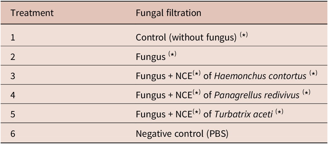
The lyophilized filtrate (LF) was diluted in PBS to a final concentration of 200 mg/mL. Assays were conducted in 96-well microtiter plates, with each well receiving 50 μL of LF and 50 μL of PBS containing 100 H. contortus infective larvae. Before the assay, the larvae were washed with sucrose and unsheathed by incubating them in 186 μL of sodium hypochlorite in 6 mL of water for 3 minutes. The treatments evaluated included LFs at concentrations of 25, 50, and 100 mg/mL, as well as PBS as a negative control. Each treatment was performed in quadruplicate (n = 4 wells). The plates were incubated for 72 hours at 28°C (Table 1).
Experimental design for evaluating the interaction between Haemonchus contortus infective larvae and filtrates from two nematophagous fungi: Dactylellina candida and Arthrobotrys musiformis at three different concentrations (100, 50, and 25 mg/mL); after growing in Sweet Potato Dextrose Broth

Table 1. Long description
The table consists of two columns: Treatment and Fungal filtration.
* Treatment 1: Control without fungus.
* Treatment 2: Fungus.
* Treatment 3: Fungus plus N C E of Haemonchus contortus.
* Treatment 4: Fungus plus N C E of Panagrellus redivivus.
* Treatment 5: Fungus plus N C E of Turbatrix aceti.
* Treatment 6: Negative control P B S.
A footer specifies that N equals 4 and N C E stands for Nematode Crude Extract. The note indicates the entire experiment was repeated in triplicate.
N = 4; (*) = NCE = Nematode Crude Extract. Note: The whole experiment was repeated in triplicate.
Subsequently, the readings of each of the wells in the microtiter plates were taken.
To quantify the number of live and dead larvae, 10 aliquots of 10 μL from each well were taken and placed on a glass slide for observation under a microscope (5×). To identify live and dead larvae, larval movement was observed. Larvae that showed no movement were stimulated by touching them with a metal needle, and those that remained motionless after the stimulus were considered dead.
The mortality percentage was determined in each case using the following formula:

Statistical analysis of the predatory and nematicidal activity of nematophagous fungi
The predatory activity (PA) data for both fungi against the nematode H. contortus were compared using mean and standard deviation calculations for each group. Meanwhile, larval mortality assay data from the liquid filtrates of both fungi were examined with a completely randomized design and analysed via multivariate methods. In this analysis, the mortality means of the different treatments served as the dependent variable. Tukey’s test was then applied to generate confidence intervals and identify significant differences between treatment means.
Results
Morphological identification of the isolated strains
After seven days of incubation, examination of bacteriological agar plates sprinkled with soil from the sampling site revealed structures indicative of nematophagous fungi. This observation led to the identification of two distinct species. Measurements of taxonomically important structures for both isolates are presented in Table 2.
Characteristics of isolated nematophagous fungi Dactylellina candida and Arthrobotrys musiformis, including average measurements of taxonomically relevant structures

Table 2. Long description
The table consists of three columns: Characteristic, Isolate D c, and Isolate A m.
* Conidium shape: Isolate D c is fusiform, elongated, broadened at the medial septum, and narrowed at the basal septum. Isolate A m is ellipsoidal to ovoid, slightly curved, and rounded at the broader distal end.
* Trapping organ: Isolate D c uses simple rings and adhesive knobs. Isolate A m uses three-dimensional adhesive networks.
* Chlamydospores: Absent in Isolate D c. Present and globose in Isolate A m.
* Septa: Isolate D c has 1 to 4 throughout the conidium. Isolate A m has 1 below the midpoint of the conidium.
* Conidium length in micrometers: Isolate D c averages 38.21 with a range of 31.2 to 48.5. Isolate A m averages 28.05 with a range of 26.22 to 38.13.
* Conidium width in micrometers: Isolate D c averages 10.2 with a range of 9.6 to 10.7. Isolate A m averages 8.26 with a range of 8.68 to 12.5.
* Conidiophore length in micrometers: Isolate D c averages 172.22 with a range of 130.12 to 213.25. Isolate A m averages 213.2 with a range of 130.44 to 282.88.
The strain identified as isolate ‘Dc’ exhibited hyaline, erect conidiophores with an apical region where conidia formed in clusters of 1 to 5. These conidia were fusiform, containing 1 to 4 septa, and appeared broadened at the medial septum while tapering at the basal end. Furthermore, two distinct types of capture organs were observed: simple rings and adhesive knobs similar to those of Dactylellina candida (Figure 2).
Microscopic photographs illustrating the fungal structures of Dactylellina candida: (A) Appearance of conidia; (B) Germination of conidia; (C) Formation of adhesive knobs; (D) Erect conidiophores; (E) Simple, non-constricting rings.

The other isolate displayed septate, branched hyphae and hyaline, septate, erect, unbranched conidiophores, which formed candelabra structures. From these, hyaline, elongated, ellipsoidal, and slightly curved conidia – characteristic of A. musiformis – emerged. Furthermore, when nematodes of the species P. redivivus were introduced to Petri dishes containing this fungus, the fungus captured them using intricate three-dimensional networks (Figure 3).
Microscopic photographs of the typical taxonomic characteristics of the nematophagous fungus Arthrobotrys musiformis. (A) Conidial clusters; (B) Three-dimensional hyphal network; (C) Erect conidiophore; (D) Candelabra; (E) Chlamydospore.

Figure 3. Long description
A multi-panel microscopic study consisting of five panels labeled A through E.
Panel A, top left, shows clusters of elongated, multi-cellular Conidia attached to hyphal branches. A scale bar indicates 20 micrometers.
Panel B, bottom left, displays a Three-dimensional adhesive net formed by looping hyphal structures. A scale bar indicates 20 micrometers.
Panel C, center, features a long, vertical Conidiophore stalk. At the top, an arrow points to the Apex candelabrelloid, which supports two oval Conidia. A scale bar at the bottom indicates 50 micrometers.
Panel D, top right, provides a close-up of the branching Apex candelabrelloid structure at the tip of a hypha. A scale bar indicates 20 micrometers.
Panel E, bottom right, shows a single, thick-walled, circular Chlamydospore integrated into a hyphal strand. A scale bar indicates 20 micrometers.
After comparing the morphological characteristics and measurements of conidia and conidiophores with those reported in the morphological taxonomic keys, we assumed that these isolates belong to the species Dactylellina candida and Arthrobotrys musiformis.
The information resulting from the observation and measurement of the main morphological structures of taxonomic interest for each of the two isolated strains is summarized in Table 2.
Molecular identification
DNA sequences obtained from the PCR products of both isolates, supplied by the Institute of Biotechnology, were analysed for homology using BLAST on the NCBI database. Tables 3 and 4 present the similarity percentages for the top five sequences identified in NCBI.
Coverage and similarity percentages for Dactylellina candida in relation to strains reported in NCBI corresponding to the partial sequence of the ITS1, 5.8S, and ITS2 regions

Table 3. Long description
The table consists of four columns: Strain, Query cover percent, Similarity percent, and N C B I database access number.
* Row 1: Dactylium candidum, 98 percent query cover, 99.28 percent similarity, access number M H 1 7 9 7 2 1 dot 1.
* Row 2: Dactylellina miltoniae, 99 percent query cover, 98.58 percent similarity, access number N R underscore 1 8 5 4 3 2 dot 1.
* Row 3: Dactylellina species, 99 percent query cover, 98.22 percent similarity, access number O R 0 1 5 8 7 2 dot 1.
* Row 4: Dactylella lysipaga, 96 percent query cover, 99.08 percent similarity, access number F J 3 8 0 9 4 4 dot 1.
* Row 5: Dactylellina haptotyla, 99 percent query cover, 93.81 percent similarity, access number M H 8 5 6 5 8 8 dot 1.
Coverage and similarity percentages for Arthrobotrys musiformis in relation to strains reported in NCBI corresponding to the partial sequence of the ITS1, 5.8S, and ITS2 regions

Table 4. Long description
The table consists of four columns: Strain, Query cover percent, Similarity percent, and N C B I database access number. There are five rows of data, all listing the strain as Arthrobotrys musiformis with a Query cover of 100 percent and a Similarity of 99.46 percent. The N C B I database access numbers for each row are as follows:
* Row 1: O L 4 5 4 9 3 1 dot 1
* Row 2: P P 3 3 3 2 0 6 dot 1
* Row 3: O L 4 5 4 9 2 9 dot 1
* Row 4: M H 8 5 5 8 4 2 dot 1
* Row 5: K P 8 5 9 6 2 4 dot 1
Evaluation of the predatory activity of nematophagous fungi against Haemonchus contortus
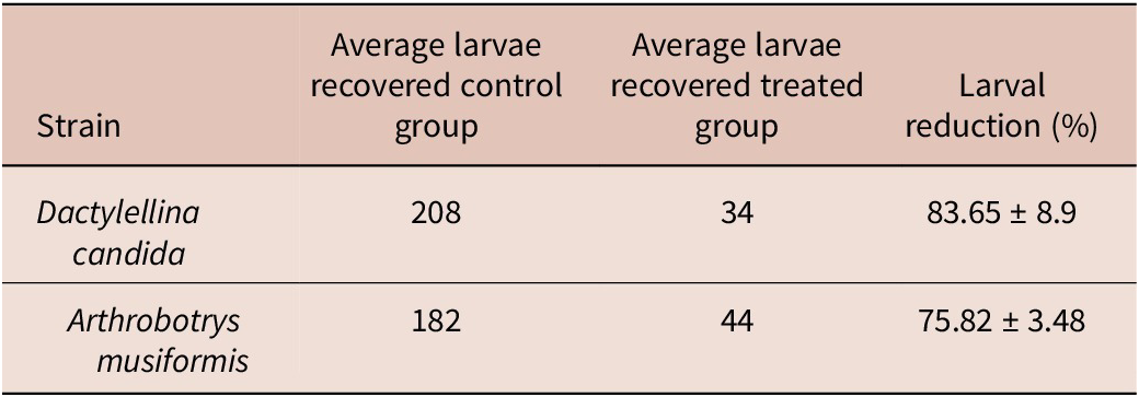
Table 5 presents the mean numbers of H. contortus (L3) larvae recovered from both fungal and non-fungal plate groups following confrontation, along with the corresponding predation percentages.
Percentages of predatory activity of nematophagous fungal strains after 7 days of confrontation against Haemonchus contortus

Table 5. Long description
The table consists of four columns: Strain, Average larvae recovered control group, Average larvae recovered treated group, and Larval reduction percentage.
* Row 1: Dactylellina candida. Control group: 208. Treated group: 34. Larval reduction: 83.65 plus or minus 8.9 percent.
* Row 2: Arthrobotrys musiformis. Control group: 182. Treated group: 44. Larval reduction: 75.82 plus or minus 3.48 percent.
Figure 4 presents microscopic images of L3 H. contortus larvae captured by the trapping organs of D. candida and A. musiformis isolates.
Microscopic view of infective Haemonchus contortus (L3) larvae trapped by adhesive knobs of the nematophagous fungus Dactylellina candida (A) and by the three-dimensional adhesive networks of Arthrobotrys musiformis (B).

Evaluation of the nematicidal activity of liquid culture filtrates of nematophagous fungi against Haemonchus contortus
Tables 6 and 7 present the larval mortality results caused by liquid filtrates from both nematophagous fungi. These filtrates were supplemented with NCE from three distinct nematode taxonomic groups and cultured in SPDB. Mortality was evaluated at three concentrations after 72 hours of exposure to H. contortus.
Percentages of mortality caused by liquid culture filtrates of Dactylellina candida after 72 hours of confrontation against Haemonchus contortus

Table 6. Long description
The table is organized into three main concentration sections plus a control. Columns are labeled Treatment, D L forward slash T L (Dead Larvae over Total Larvae), and Mortality percentage with standard deviation.
* Control: P B S treatment shows 2 forward slash 66 D L forward slash T L and 3.31 plus or minus 2.58 percent mortality.
* 25 milligrams per milliliter section:
- S P D B: 2 forward slash 92, 2.2 plus or minus 1.57 percent.
- Dactylellina candida without inducer: 20 forward slash 97, 20.6 plus or minus 12.25 percent.
- Dactylellina candida plus N C E Haemonchus contortus: 28 forward slash 92, 29.89 plus or minus 24.89 percent.
- Dactylellina candida plus N C E Panagrellus redivivus: 8 forward slash 77, 10.77 plus or minus 6.31 percent.
- Dactylellina candida plus N C E Turbatrix aceti: 14 forward slash 82, 17.77 plus or minus 24.79 percent.
* 50 milligrams per milliliter section:
- S P D B: 3 forward slash 91, 3.57 plus or minus 2 percent.
- Dactylellina candida without inducer: 34 forward slash 98, 36.4 plus or minus 24.89 percent.
- Dactylellina candida plus N C E Haemonchus contortus: 40 forward slash 99, 40.47 plus or minus 25.03 percent.
- Dactylellina candida plus N C E Panagrellus redivivus: 14 forward slash 95, 14.69 plus or minus 8.94 percent.
- Dactylellina candida plus N C E Turbatrix aceti: 15 forward slash 90, 16.17 plus or minus 15.62 percent.
* 100 milligrams per milliliter section:
- S P D B: 5 forward slash 98, 5.55 plus or minus 4.3 percent.
- Dactylellina candida without inducer: 62 forward slash 99, 63.38 plus or minus 22.4 percent.
- Dactylellina candida plus N C E Haemonchus contortus: 77 forward slash 89, 86.61 plus or minus 7.85 percent.
- Dactylellina candida plus N C E Panagrellus redivivus: 22 forward slash 94, 22.81 plus or minus 11.67 percent.
- Dactylellina candida plus N C E Turbatrix aceti: 37 forward slash 82, 45.33 plus or minus 32.04 percent.
(±) standard deviation. (p < 0.05). * Means with different letters at the same concentration are significantly different. Experiment conducted in triplicate. NCE = Neatode Crude Extract; DL = Dead Larvae; TL = Total Larvae.
Percentages of mortality caused by liquid culture filtrates of Arthrobotrys musiformis after 72 hours of confrontation against Haemonchus contortus

Table 7. Long description
The table consists of three columns: Treatment, Dead larvae forward slash Total larvae, and Mortality percentage.
At the top, the P B S control shows 1 forward slash 70 dead larvae with 1.87 plus or minus 1.53 percent mortality.
Under the 25 milligrams per milliliter concentration:
- S P D B: 3 forward slash 81 larvae, 3.16 plus or minus 2.62 percent.
- Arthrobotrys musiformis without inducer: 7 forward slash 84 larvae, 9.72 plus or minus 5.23 percent.
- Arthrobotrys musiformis plus N C E Haemonchus contortus: 9 forward slash 76 larvae, 11.25 plus or minus 3.42 percent.
- Arthrobotrys musiformis plus N C E Panagrellus redivivus: 5 forward slash 68 larvae, 7.97 plus or minus 5.51 percent.
- Arthrobotrys musiformis plus N C E Turbatrix aceti: 4 forward slash 75 larvae, 5.77 plus or minus 4.05 percent.
Under the 50 milligrams per milliliter concentration:
- S P D B: 4 forward slash 76 larvae, 5.14 plus or minus 2.81 percent.
- Arthrobotrys musiformis without inducer: 15 forward slash 76 larvae, 18.32 plus or minus 5.96 percent.
- Arthrobotrys musiformis plus N C E Haemonchus contortus: 21 forward slash 85 larvae, 25.01 plus or minus 6.74 percent.
- Arthrobotrys musiformis plus N C E Panagrellus redivivus: 7 forward slash 78 larvae, 8.49 plus or minus 3.49 percent.
- Arthrobotrys musiformis plus N C E Turbatrix aceti: 5 forward slash 67 larvae, 8.53 plus or minus 4.4 percent.
Under the 100 milligrams per milliliter concentration:
- S P D B: 5 forward slash 87 larvae, 5.75 plus or minus 1.15 percent.
- Arthrobotrys musiformis without inducer: 39 forward slash 105 larvae, 37.14 plus or minus 3 percent.
- Arthrobotrys musiformis plus N C E Haemonchus contortus: 46 forward slash 99 larvae, 47.49 plus or minus 1.46 percent.
- Arthrobotrys musiformis plus N C E Panagrellus redivivus: 15 forward slash 86 larvae, 17.61 plus or minus 3.36 percent.
- Arthrobotrys musiformis plus N C E Turbatrix aceti: 13 forward slash 75 larvae, 17.33 plus or minus 3.23 percent.
N C E stands for Nematode Crude Extract. Mortality values include standard deviation.
(±) standard deviation. (p < 0.05). * Means with different letters at the same concentration are significantly different. Experiment conducted in triplicate. NCE = Nematode Crude Extract.
Figure 5 presents photographs of H. contortus infective larvae after 72 hours of exposure to liquid culture filtrates from Dactylellina candida and Arthrobotrys musiformis, both supplemented with H. contortus crude extract – the treatment that resulted in the highest mortality rate among all filtrates tested. Some larvae exhibited deformations on the cuticular surface and loss of structural integrity in intestinal cells, while others displayed a smooth cuticular surface with notable constrictions along the larval body.
Microscopic images (40×) of Haemonchus contortus infective larvae exposed to liquid culture filtrates of the two nematophagous fungal strains supplemented with Haemonchus contortus crude extract L3 larvae. (A) Live (normal) L3 larva; (B) Damage caused by Dactylellina candida culture filtrate (100 mg/mL) supplemented with Haemonchus contortus crude extract; (C) Damage caused by Arthrobotrys musiformis culture filtrate (100 mg/mL) supplemented with Haemonchus contortus crude extract.

Figure 5. Long description
A three-panel microscopic display at 40 times magnification. Each panel includes a 100 mu m scale bar at the bottom right.
Panel A shows a live, normal L 3 larva. The organism is coiled in a smooth, continuous S-shape with a clear, well-defined cuticle and visible internal structures. The body is uniform in thickness and appears translucent.
Panel B shows an L 3 larva exposed to Dactylellina candida culture filtrate. The larva is tightly coiled and exhibits significant morphological damage. The cuticle appears irregular and distorted, with visible vacuolization and degradation of internal tissues compared to the healthy specimen.
Panel C shows an L 3 larva exposed to Arthrobotrys musiformis culture filtrate. The larva is coiled in a circular loop. The body shows signs of thinning and structural breakdown, with a less distinct internal organization and a granular appearance throughout the length of the organism.
Discussion
Morphological identification
The microorganism isolated from ‘Ecoturistic Park El Hoyanco’ exhibited similarities to species of the genus Dactylellina. Several species within this genus possess overlapping morphological traits, which can cause identification challenges and necessitate a detailed structural analysis (Hyde et al Reference Hyde, Swe, Zhang, Zhang and Hyde2014). Table 8 presents a comparative overview of the key taxonomic features, such as conidia type, conidiophores, septation in conidia, and the various trapping organs found in Dactylellina candida, Dactylellina lysipaga, and Dactylellina drechsleri.
Main taxonomic characteristics of three similar species of the genus Dactylellina

Table 8. Long description
The table compares taxonomic characteristics across three columns.
Row 1: Conidia measurements in micrometers.
- Dactylellina candida: Seven spindle-shaped conidia with 2 to 5 septa, measuring 27.5 to 57.5 by 7.5 to 12.5.
- Dactylellina lysipaga: Six spindle-shaped conidia with 2 to 4 septa, measuring 27.5 to 85 by 9 to 17.5.
- Dactylellina drechsleri: Six spindle-shaped conidia with 2 to 4 septa, measuring 29.5 to 48.7 by 10.7 to 15.4.
Row 2: Conidiophores in micrometers.
- Dactylellina candida: Three long, septate stalks with tips ranging from simple forks to multi-pronged crowns, measuring 100 to 335.
- Dactylellina lysipaga: Three vertical stalks, two with rounded tips and one with a Y-shaped apex, measuring 125 to 760.
- Dactylellina drechsleri: Three vertical stalks with one Y-shaped apex, measuring 93 to 184.
Row 3: Capture organs.
- Dactylellina candida: Line drawings of simple rings consisting of three-celled circular traps on short stalks and adhesive buttons.
- Dactylellina lysipaga: Line drawings showing both simple rings and adhesive buttons, which are spherical structures on short stalks branching from the hypha.
- Dactylellina drechsleri: Line drawings of adhesive buttons branching from a segmented hyphal filament.
Although all three of the aforementioned species produce adhesive knobs, only D. candida and D. lysipaga also form simple rings; D. drechsleri does not. The conidia of D. candida are fusiform and elongated, whereas those of D. lysipaga are slightly larger and range from obovoid to fusiform. Based on the taxonomic identification guides by De Hoog and Van-Oorschot (Reference De Hoog and Van-Oorschot1985), Rubner (Reference Rubner1996), and Hyde et al (Reference Hyde, Swe, Zhang, Zhang and Hyde2014), this organism has been classified as Dactylellina candida in traditional taxonomy. However, the taxonomy of this species has been controversial. Initially, Nees von Esenbeck named the fungus Dactylium candidum in Reference Nees von Esenbeck1817, and in Reference Saccardo1886 Saccardo renamed it Dactylaria candida. Since then, it has undergone several nomenclatural changes, including reassignment to Monacrosporum by Gams (Reference Gams1999). Most recently, Li et al (Reference Li, Hyde, Jeewon, Cai, Vijaykrishna and Zhang2005) proposed the current classification as Dactylellina candidum. With regard to the other fungus isolated from the ‘Chapultepec Ecoturistic park’, due to its general taxonomic characteristics, including conidia, conidiophores, and trapping organs, it was initially identified as belonging to the genus Arthrobotrys. A detailed analysis of its taxonomically relevant structures revealed that this fungus shares similar characteristics with three species of the genus Arthrobotrys: Arthrobotrys musiformis, A. javanica, and A. eryuanensis (Table 9).
Key taxonomic features distinguishing three closely related species of the genus Arthrobotrys

Table 9. Long description
The table compares taxonomic features across three columns.
Row 1: Conidia measurements in mu m.
- A. musiformis: Six elongated, two-celled conidia with a septum below the middle, measuring 20 to 47.5 times 7 to 12.5.
- A. javanica: Six pear-shaped conidia with a septum near the middle, measuring 20 to 37.5 times 7.5 to 10.
- A. eryuanensis: Seven pear-shaped spores including Macro (18 to 44.5 times 5 to 11.5) and Micro (7.5 to 28 times 4 to 11) varieties.
Row 2: Conidiophores in mu m.
- A. musiformis: Three vertical stalks with septa and branched apices, height 104 to 640.
- A. javanica: Three vertical stalks with branched apical structures, height 220 to 400.
- A. eryuanensis: Three stalks with lobed apical structures, height 110 to 308.
Row 3: Capture organs.
- All three species (A. musiformis, A. javanica, and A. eryuanensis) feature line drawings of three-dimensional adhesive networks consisting of interconnected circular and curved hyphal loops.
Row 4: Chlamydospores.
- A. musiformis: Globose or ellipsoidal, yellow.
- A. javanica: Globose, yellow.
- A. eryuanensis: Cylindrical in chain, hyaline.
Although all three species possess candelabrum-like apices, only A. eryuanensis has conidiophores that may be branched – a feature absent in both A. musiformis and A. javanica. Notably, A. eryuanensis is also unique among the three species in producing two types of conidia: macro- and microconidia (Hyde et al Reference Hyde, Swe, Zhang, Zhang and Hyde2014). Discrepancies exist in the reported lengths of conidiophores: Drechsler (Reference Drechsler1937) reported that those of A. musiformis measured 104–640 μm, while Rifai and Cooke (Reference Rifai and Cooke1966) documented conidiophores of A. javanica at 220–400 μm. By contrast, De Hoog and Van-Oorschot (Reference De Hoog and Van-Oorschot1985) reported maximum lengths of up to 1000 μm for A. javanica and 900 μm for A. musiformis. In the present study, the conidiophores from the isolate collected at the ‘Chapultepec Ecoturistic Park’ measured 130.44–282.88 μm, a range more consistent with A. musiformis than with A. javanica, according to the descriptions by Drechsler (Reference Drechsler1937) and Rifai and Cooke (Reference Rifai and Cooke1966).
Based on the results and a comparison with the available literature for these species, the organism was classified as A. musiformis. To further validate the morphological identification of both fungi, a molecular diagnosis was conducted using Polymerase Chain Reaction, as described and discussed below.
Molecular identification
The DNA sequence of the D. candida isolate was analysed and compared to sequences of other isolates previously reported in the NCBI database. The analysis showed high coverage (98%) and similarity (99.28%) with fungi of the species Dactylium candidum. These findings provide strong scientific support for the taxonomic identification, confirming that the strain isolated from ‘El Hoyanco Ecoturistic Park’ in Coatlán del Río, Morelos, belongs to Dactylellina candida. Conversely, the isolate from ‘Chapultepec Ecological Park’ in Cuernavaca, Morelos, which was morphologically identified as A. musiformis, demonstrated 100% coverage and 99.46% similarity with A. musiformis fungi when genotyped using the NCBI database. These results corroborate the previously described morphological diagnosis and validate the classification of this strain from Cuernavaca, Morelos, as A. musiformis.
Evaluation of the predatory activity of nematophagous fungi against Haemonchus contortus
Both nematophagous fungi species demonstrated strong PA, with predation rates nearing 80%. However, a comprehensive literature review indicated that reports on the biological activity of Dactylellina candida against other organisms are scarce. Dowsett and Reid (Reference Dowsett and Reid1976) found that Dactylaria (Dactylellina) candida could capture juvenile stages of the plant-parasitic nematode Aphelenchus avenae and parasitize nematode eggs using adhesive trapping knobs. Similarly, Wu et al (Reference Wu, Kim and Zhou2012) observed that Dactylellina candidum was capable of capturing and destroying free-living nematodes of the genus Rhabditis sp. In contrast, Zouhar et al (Reference Zouhar, Douda, Novotny, Novakova and Mazakova2010) assessed the virulence of several nematophagous fungi strains, including D. candida, and reported low virulence for this species, possibly resulting from prolonged laboratory maintenance of the isolate.
Multiple studies have highlighted the PA of A. musiformis against H. contortus. For example, Pérez-Anzúrez et al (Reference Pérez-Anzúrez, Olmedo-Juárez, von-Son de Fernex, Alonso-Díaz, Delgado-Núñez, López-Arellano, González-Cortázarv, Zamilpa, Ocampo-Gutiérrez and Paz-Silva2022) found that a strain from Cuautla, Morelos, reduced larval populations by 71.54%. Similarly, Jaramillo-Tlalapango et al (Reference Jaramillo-Tlalapango, Mendoza-de Gives, Higuera-Piedrahita, Ocampo-Gutiérrez, López-Arellano, Pérez-Anzúrez and González-Cortázar2023) and Colinas-Picazo et al (Reference Colinas-Picazo, Mendoza-de Gives, Pérez-Anzúrez, Gutiérrez-Medina, Bautista-García, Delgado-Núñez and Olmedo-Juárez2024) reported predatory activities of 74.9% and 70.95%, respectively, for strains isolated in Cuernavaca, Morelos. These results are consistent with the present study, where the strain from the ‘Chapultepec Ecological Park’ also showed significant predatory effects against H. contortus.
Assessment of nematicidal effects of liquid culture filtrates from nematophagous fungi on Haemonchus contortus
Larval mortality resulting from the liquid culture filtrates of both nematophagous fungi, tested at three different concentrations, showed a significant effect at the highest concentration when supplemented with H. contortus crude extract. This treatment achieved nearly 87% mortality for D. candida, outperforming the other treatments. An extensive literature review found no previous reports on the nematicidal activity of D. candida liquid culture filtrates, suggesting this may be the first publication on the subject. Conversely, a study found that liquid culture filtrates of A. musiformis grown in potato dextrose broth produced a nematicidal effect of 49.84% against H. contortus after 72 hours of exposure (Jaramillo-Tlalapango et al Reference Jaramillo-Tlalapango, Mendoza-de Gives, Higuera-Piedrahita, Ocampo-Gutiérrez, López-Arellano, Pérez-Anzúrez and González-Cortázar2023). This percentage is slightly higher than our findings with A. musiformis in the absence of NCE, which showed a 37.14% effect. In another publication by our group (Mendoza-de Gives et al Reference Mendoza-de Gives, Rodríguez-Labastida, Olmedo-Juárez, Gamboa-Angulo and Reyes-Estebanez2022), we evaluated the nematicidal activity of four nematophagous fungi, including A. musiformis, considering the presence or absence of crude protein extract from H. contortus in the liquid culture medium. In that study, when nematophagous fungi were cultured with H. contortus extract for 48 hours, A. musiformis demonstrated a lethal activity of 82.2%, whereas in the absence of the extract, mortality decreased to 41.2%.
These findings demonstrate that crude extracts from infective larvae of H. contortus serve as effective elicitors of nematicidal compounds (Mendoza-de Gives et al Reference Mendoza-de Gives, Rodríguez-Labastida, Olmedo-Juárez, Gamboa-Angulo and Reyes-Estebanez2022). In this study, comparable outcomes were observed when NCE of H. contortus was added to A. musiformis cultures. Notably, this is the first report of the nematode extract acting as an elicitor of nematicidal compounds in D. candida, particularly at the highest filtrate concentration tested (100 mg/mL). The nematicidal effect of D. candida filtrates alone reached 63.38%, while supplementation with crude extract from H. contortus L3 larvae significantly increased mortality to 86.61% (p < 0.05).
Conclusions
The results of this study indicate that both fungi have significant PA against one of the most pathogenic parasitic nematodes, H. contortus, and may be suitable candidates for in vivo biocontrol assays. Regarding the liquid culture filtrates, it was demonstrated one more time that crude extracts from infective larvae of H. contortus are effective elicitors of nematicidal compounds. Importantly, this is the first report describing the nematode extract as an elicitor of nematicidal compounds in D. candida, particularly at the highest tested filtrate concentration (100 mg/mL). The nematicidal effect of D. candida filtrates alone reached 63.38%, but supplementation with crude extract from H. contortus L3 larvae significantly increased mortality to 86.61% (p < 0.05).
The practical application of these findings in controlling H. contortus in sheep flocks will depend on several factors, including the gastrointestinal microbiome, pH, temperature, and other conditions that may limit the effectiveness of fungal filtrates. Therefore, it is essential to conduct in vivo assays to validate the benefits of this technology in practical settings.
Acknowledgements
This study was conducted as part of the thesis project of Miss Ana Jatziri Tórres-Armendaris to fulfil the requirements for a bachelor’s degree in Biotechnology Engineering at the Universidad Politécnica del Estado de Morelos (UPEMOR), Mexico, under the supervision of Dr. Pedro Mendoza-de Gives and MSc Enrique Gutiérrez Medina.
Financial support
This study utilised reagents and consumables purchased with funding from CONAHCYT (SECIHTI), Proyecto Ciencias de Frontera-2023, CF-2023-I-2309.
Competing interests
The authors declare there is no conflict of interest.
Ethical standard
Not applicable.