Introduction
Polycystic ovarian syndrome (PCOS) is a chronic endocrinopathy that is manifested in 2.2–27% of women of pre-menopausal age, and may start between 15 to 44 years old (Knochenhauer et al., Reference Knochenhauer, Key, Kahsar-Miller, Waggoner, Boots and Azziz1998). This syndrome is represented by ovulatory dysfunction, hirsutism, acne and menstrual disorder, found either as oligomenorrhea or amenorrhea (Teede et al., Reference Teede, Deeks and Moran2010). Other associated disorders are for example endometrial cancer, as well as 40% of the cases of female infertility (Dahlgren et al., Reference Dahlgren, Friberg, Johansson, Lindström, Odén, Samsioe and Janson1991). The commonly used name ‘PCOS’ is derived from the observation of multiple ovarian cysts during ultrasonography; these are premature follicles arrested at the primordial stage (Azziz, Reference Azziz2006). Scientists believe the syndrome is due to excessive androgen hormone expression, genetic susceptibility, along with environmental influences (Rebar et al., Reference Rebar, Judd, Yen, Rakoff, Vandenberg and Naftolin1976; Strauss, Reference Strauss2003). The genetic base of this disease was deduced from familial clustering of the cases studied, as 3–35% of adult female individuals with PCOS had mothers or sisters (∼2%) with the same syndrome. Most adolescent female individuals with polycystic ovarian morphology (PCOM) had either an asymptomatic mother or a father with metabolic syndromes (Sam et al., Reference Sam, Legro, Bentley-Lewis and Dunaif2005; Leibel et al., Reference Leibel, Baumann, Kocherginsky and Rosenfield2006; Coviello et al., Reference Coviello, Sam, Legro and Dunaif2009). The diagnosis of the syndrome is based on three sets of criteria for the accurate determination given by the National Institute of Health (NIH) in the 1990 international conference on PCOS, the Rotterdam criteria of 2003 and Androgen and Excess PCOS Society (AE-PCOS) in 2006 (Amato et al., Reference Amato, Galluzzo, Finocchiaro, Criscimanna and Giordano2008).
The WNT signal transduction pathway is a classical evolutionary pathway that regulates aspects of cell proliferation, migration and cell fate determination in the tissue and early embryonic development. The WNT protein is associated with a family of lipid modified glycoproteins, which help the attachment of WNT protein to Wntless proteins and transports them to the plasma membrane for secretion (Cadigan and Nusse, Reference Cadigan and Nusse1997). Activation of co-receptors LRP-5/6 follows and a signal is sent to Dishevelled (Dsh) protein in the cytoplasm (Habas and Dawid, Reference Habas and Dawid2005). Activation of Dishevelled protein leads to the inhibition of GSK-3β, which consequently leads to the destruction of the multiprotein complex. This stabilizes the intracellular accumulation of β-catenin (CTNNB1) in the cytoplasm. Next, β-catenin translocates to the nucleus, where it acts as a transcriptional co-activator of TCF/LEF to trigger WNT-target genes. However, if WNT ligands are absent, β-catenin is phosphorylated by a cytoplasmic multiprotein complex of Axin, APC, GSK3β and CK1α. The phosphorylated β-catenin is recognized by E3 ubiquitin and targeted to proteasomal degradation, causing a reduction in the cytoplasmic β-catenin level (Moon et al., Reference Moon, Bowerman, Boutros and Perrimon2002).
In the present study we aimed to measure the expression levels of WNT/β-catenin signal transduction pathway genes (AXIN2, FZD4, TCF4, WNT3, WNT4, WNT5A, WNT7A, WNT 1, APC, GSK3B and β-catenin) in the ovaries of PCOS women and compare these with expression levels in control healthy women, as these genes might substantially effect the development of ovaries and oogenesis, as well as PCOS. The AXIN2 gene acts as a scaffold for β-catenin stabilization in the WNT signalling pathway (Mai et al., Reference Mai, Qian, Yokomizo, Smith and Liu1999). This gene and its identical isoform AXIN1 are important components of the cytoplasmic complex that targets β-catenin degradation in the cell, if the ligand is absent (Gluecksohn-Schoenheimer, Reference Gluecksohn-Schoenheimer1949; Zeng et al., Reference Zeng, Fagotto, Zhang, Hsu, Vasicek, Perry, Lee, Tilghman, Gumbiner and Costantini1997). The WNT1, WNT3, WNT4, WNT5A and WNT7A genes are members of a large WNT family that have a significant role in embryonic development and adult tissue homeostasis (Yamaguchi et al., Reference Yamaguchi, Bradley, McMahon and Jones1999; Nishita et al., Reference Nishita, Enomoto, Yamagata and Minami2010). TCF4 is a transcriptional factor in humans that is encoded by TCF7L2 (Castrop et al., Reference Castrop, van Norren and Clevers1992) and plays a part in activation of WNT-targeted genes, pro-glucagon regulation through the WNT signalling pathway and metabolic glucose balancing in liver cells instead of pancreatic B-cells (Jin and Liu, Reference Jin and Liu2008; Facchinello et al., Reference Facchinello, Tarifeño-Saldivia, Grisan, Schiavone, Peron, Mongera, Ek, Schmitner, Meyer, Peers, Tiso and Argenton2017). The FZD4 gene encodes a 537 amino acid receptor that is a member of the frizzled family receptors on the plasma membrane, controlling cell polarity and proliferation in embryonic development (Peifer, Reference Peifer1999). Abnormal activation of this gene has been associated with cell malfunction and exudative vitreoretinopathy, which is an inherited form of retinal degradation (Toomes et al., Reference Toomes, Bottomley, Jackson, Towns, Scott, Mackey, Craig, Jiang, Yang, Trembath, Woodruff, Gregory-Evans, Gregory-Evans, Parker, Black, Downey, Zhang and Inglehearn2004; Robitaille et al., Reference Robitaille, Zheng, Wallace, Beis, Tatlidil, Yang, Sheidow, Siebert, Levin, Lam, Arthur, Lyons, Jaakkola, Tsilou, Williams, Weaver, Shields and Guernsey2011; Milhem et al., Reference Milhem, Ben-Salem, Al-Gazali and Ali2014).
The investigation of the expression levels of the genes involved in this canonical pathway enabled us to further understand the factors leading to PCOS symptoms in patients and would be also important for the development of therapy options in the future.
Materials and methods
Sample collection
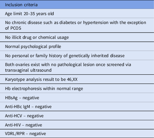
In this case–control study, in total, 13 metaphase II (MII) stage oocyte samples were collected from non-obese and young women who were admitted to the Near East University Hospital (NEUH) IVF Center, after the approval of the Near East University Scientific Review Board (registration numbers: YDU/2020/800-1120 and YDU/2020/79-1056) (Table 1). Seven of these samples were from PCOS patients and were categorized as the PCOS group and the other six were from healthy individuals and categorized as the control group.
Inclusion criteria for the 13 study participants

RNA extraction, cDNA synthesis and gene expression analysis
Total RNA extraction was performed using Norgen’s RNA/DNA Purification Kit (Norgen, Canada) following the manufacturer’s guidelines. cDNA synthesis was performed using the TruScript First Strand cDNA Synthesis Kit (Norgen, Canada) following the manufacturer’s guidelines.
Expression levels of 11 genes (AXIN2, FZD4, TCF4, WNT3, WNT4, WNT5A, WNT7A, WNT 1, APC, GSK3B and β-catenin) were analyzed using gene-specific primers and the Light Cycler® 480 SYBR Green I Master system (Roche, UK) and their qPCR reaction mix, according to the manufacturer’s guidelines.
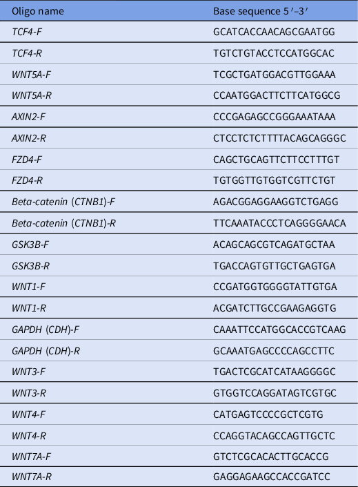
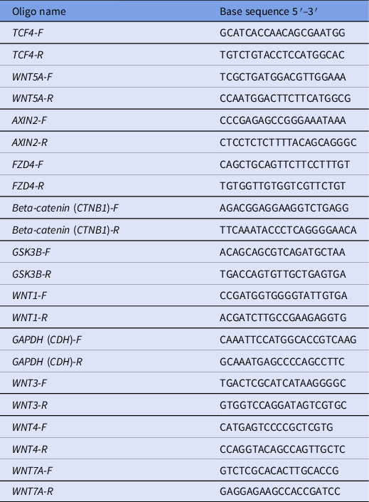
Gene-specific primers were designed to flank exon–exon junctions using the Primer3 tool (Table 2).
Primer sequences of the investigated genes
Statistical analysis
Statistical analysis was performed using analysis of variance (ANOVA) (GraphPad Prism 8.4.3) and Student’s t-test to find the correlation between each gene and PCOS. P-values were used to compare the results between the genes, P-values <0.05 were considered as significant, while those >0.05 were considered non-significant.
Gene expression levels were calculated using the comparative ΔΔCt method (Tulay et al., Reference Tulay, Naja, Cascales-Roman, Doshi, Serhal and SenGupta2015). The Rotor-Gene Q Software (Qiagen, Germany) system was used to evaluate the Ct values. For each gene expression profile analysis, GAPDH was used as a housekeeping gene for normalization. The ΔCt values were calculated as described previously (Tulay et al., Reference Tulay, Naja, Cascales-Roman, Doshi, Serhal and SenGupta2015).
Results
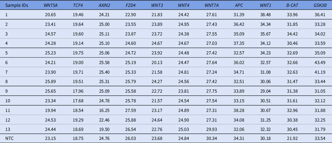
In the present study we assessed the expression levels of WNT/β-catenin signalling pathway genes (AXIN2, FZD4, TCF4, WNT3, WNT4, WNT5A, WNT7A, WNT1, APC, GSK3B and β-catenin) in human oocyte samples obtained from female donors with or without PCOS (Table 3). MII stage oocytes were collected from female oocyte donors applying at the Near East University Hospital IVF Center. All participated donors were of similar age and body mass index (BMI). AXIN2, FZD4, TCF4, WNT3, WNT4, WNT5A, WNT7A genes were not expressed or showed extremely low expression profiles in seven human MII oocytes from patients with PCOS, as well as six human MII oocytes from the healthy donors. APC, GSK3B, WNT1 and β-catenin were expressed in all oocytes including PCOS and control groups.
Expression levels of the 11 genes in all 13 samples

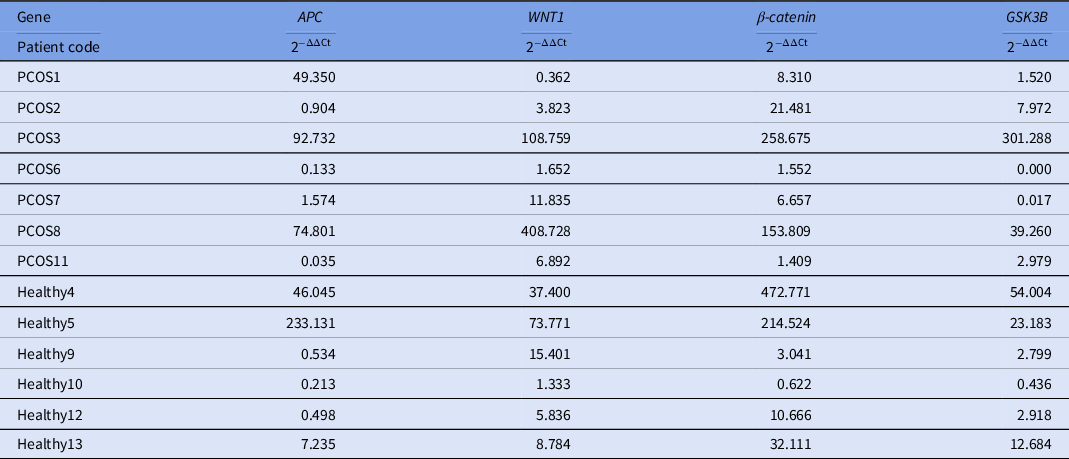
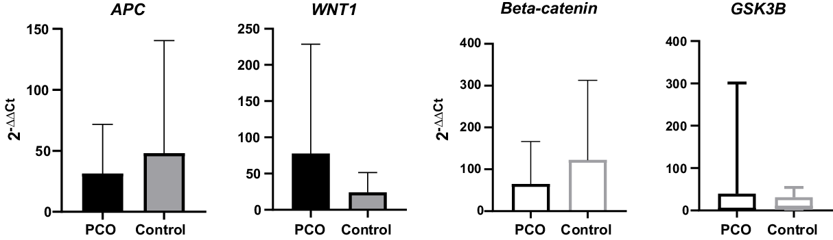
Student’s t-test with 2−ΔΔCt analysis was used for the statistical investigation and aimed to detect the presence of significant values between APC, GSK3B, WNT1 and β-catenin genes and PCOS (Table 4). There were no significant differences for the β-catenin gene and the APC gene (P = 0.4995 and P = 0.6744, respectively); while the expression profiles of WNT1 and GSK3B genes both indicated a significant difference in the oocytes obtained from the patients with PCOS (P = 0.0018 and P = 0.0019, respectively) (Figure 1).
Results for 2−ΔΔCt using Student’s t-test

Graphs for APC, WNT1, GSK3B and β-catenin using Student’s t-test for 2−ΔΔCt values.

Discussion
For the past few decades, PCOS has remained a major health challenge and infertility trigger in women. The complete patho-physiological effects of the syndrome are still unclear. Nonetheless, scientists believe it is due to androgen excess and environmental effects associated with genetic inheritance of the disease. Depending on the diagnostic criteria, prevalence statistics vary between populations. A study conducted by Amato et al. (Reference Amato, Galluzzo, Finocchiaro, Criscimanna and Giordano2008) indicated that PCOS prevalence diagnosed by NIH criteria was ∼51%, 83% for Rotterdam and 70% by AE-PCOS criteria. When these were all combined the prevalence only reached 49%. The inclusion of WNT in normal ovarian activity is not too surprising given the diversity of physiological systems regulated by the family. However, the bulk of data collected on the WNT ligand and its involvement in oocyte development is based on embryo investigations. The number of studies that have addressed WNT ligand expression in adult oocytes is quite small. The purpose of the current research was to compare the expression levels of 11 WNT signalling pathway genes in oocytes obtained from the PCOS group and the healthy group.
AXIN2 is considered as a negative regulator of the WNT signalling pathway as it restricts the action of β-catenin and leads to complete shutdown of TCF gene transduction (Jho et al., Reference Jho, Zhang, Domon, Joo, Freund and Costantini2002). In a study investigating the role of the APC2 gene in the WNT signal transduction pathway conducted by Mohamed et al. (Reference Mohamed, Hay, Reed, Smalley and Clarke2019) they revealed that ovaries with concealed APC2 expression exhibited higher levels of AXIN2 when compared with normal ovaries. In addition, a study by Leung et al. (Reference Leung, Kolligs, Wu, Zhai, Kuick, Hanash, Cho and Fearon2002) conducted on patients with PCOS who had ovarian carcinoma revealed that the carcinogenic ovary with de-regulated β-catenin had higher AXIN2 expression compared with normal ovaries. Abnormal expression of the WNT5A gene has been associated with lung and hepatic fibrosis (Iozzo et al., Reference Iozzo, Eichstetter and Danielson1995; Xiong et al., Reference Xiong, Hu, Jian, Wang, Jiang, Li and He2012). Recently, scientists described proinflammation as a progression factor of PCOS pathogenesis. A recent study by Zhao et al. (Reference Zhao, Zhang, Huang, Yu, Li, Li, Liu, Liu and Qiao2015) showed that the ovaries and granulosa cells in patients with PCOS showed signs of proinflammation and oxidative stress as the expression level of WNT5A gene was increased. In another study by Wu et al. (Reference Wu, Wang, Xu, Liu, Bi, Wang and Zhang2017), investigating the expression levels of Wnt family genes in the granulosa cells of patients with polycystic ovaries and in healthy ovary patient, WNT1, WNT3 and WNT4 were more highly expressed in PCOS ovaries than in healthy ovaries, while WNT5A exhibited no significant difference whatsoever. Lower mRNA expression levels of WNT7a, WNT8a and WNT3 ligands were detected in patients with PCOS with endometrial dysfunction (Mehdinejadiani et al., Reference Mehdinejadiani, Amidi, Mehdizadeh, Barati, Safdarian, Aflatoonian, Alyasin, Aghahosseini, Pazhohan, Mohammad Zadeh Kazorgah and Sobhani2018). Furthermore, Sanchez et al. (Reference Sanchez, Viganò, Quattrone, Pagliardini, Papaleo, Candiani and Panina-Bordignon2014) found lower levels of WNT1 and higher levels of WNT4 and WNT5 as independent components of β-catenin found in the luteinized granulosa cells of patients with PCOS. Vainio et al. (Reference Vainio, Heikkilä, Kispert, Chin and McMahon1999) reported that the inactivation of WNT4 in female mice resulted in a partial female to male reversal and oocyte depletion at birth. However, male mice with mutant WNT4 had normal gonadal growth at birth. In a subsequent study, the role of WNT4 was demonstrated in sorting adrenal and gonadal cells at the fetal stage of development, as well as in cell migration. Expression of the WNT7a gene as a WNT family member has been reported as a required component in the development of female reproductive tract. A study by Kobayashi and Behringer (Reference Kobayashi and Behringer2003) found that the WNT7a gene is expressed in the Müllerian duct during embryonic development, and after birth is only present in the oviductal and uterine epithelium. They also reported that WNT7a mutant mice were infertile due to impaired endometrial uterine glands. Just as WNT protein standard expression has been of significant importance in normal ovulation and folliculogenesis, there are frizzled receptors that carry out the same process. Studies observing mutant mice with deletion of the FZD4 gene resulted in mice suffering from oesophagus and auditory dysfunctions (Wang et al., Reference Wang, Huso, Cahill, Ryugo and Nathans2001). A study by Gupta et al. (Reference Gupta, Folger, Rajput, Lv, Yao, Ireland and Smith2014) on rodent ovaries demonstrated that luteinizing hormone (LH) overexpression promoted FZD1 and FZD4 increased levels. Conversely, a study on adult female mice granulosa cells with germ line deletion of the FZD4 gene showed normal ovulation and production of fertilized oocytes, however mice were still sterile as a result of failed embryo implantation. This outcome was due to abnormal corpus luteum formation and progesterone reduction (Hsieh et al., Reference Hsieh, Boerboom, Shimada, Lo, Parlow, Luhmann, Berger and Richards2005). Last, for the TCF4 gene, scientists were undecided on the basis of this gene’s association with the PCOS pathogenicity. Some researchers have noted the correlation of this gene with the syndrome; for example Christopoulos et al. (Reference Christopoulos, Mastorakos, Gazouli, Panidis, Deligeoroglou, Katsikis, Papadias, Diamandi-Kandarakis and Creatsas2008) who observed polymorphism of the TCF4 gene in the Greek population, while others (Xu et al., Reference Xu, Che, Cao, Wu, Sun, Liang, Sun, Ke, Yi and Wang2010; Ben-Salem et al., Reference Ben-Salem, Ajina, Suissi, Daher, Almawi and Mahjoub2014) denied the association due to absence of polymorphisms in Chinese, Korean and Tunisian populations, respectively. In a recent study by Prabhu et al. (Reference Prabhu, Sekar and Abilash2018) polymerase chain reaction–restriction fragment length polymorphism (PCR-RFLP) was utilized to validate that TCF4 gene polymorphisms were not associated with PCOS pathology. Glycogen synthase kinase (GSK) is a multifactorial serine/threonine kinase enzyme that is encoded by the GKS3 gene; this gene plays a crucial role in the WNT/β-catenin signalling pathway and is a mediator of glycogen synthesis alongside insulin signalling (Doble and Woodgett, Reference Doble and Woodgett2003). The GSK3 gene has two distinct isoforms in WNT signalling: GSK3α regulates the transcriptional activity of CTNNB1 (Asuni et al., Reference Asuni, Hooper, Reynolds, Lovestone, Anderton and Killick2006), however GSK3β regulates glucose metabolism in the muscle and acts as a negative regulator of the WNT signalling pathway by forming a complex with AXIN1 and APC for proteasomal degradation of CTNNB1. Wu et al. (Reference Wu, Zhou, Liu, Pöllänen, Sallinen, Mäkinen and Erkkola2003) reported lower levels of glycogen synthesis in cultured ovarian granulosa cells of women with PCOS as a result of insulin resistance caused by GSK3 deficiency. The APC gene is an antagonist regulatory component of the WNT/β-catenin pathway and promotes the degradation of β-catenin protein through influencing the recruitment of β-catenin protein to other Axin destruction proteins such as β-catenin/GSK3. This facilitates subsequent ubiquitination by ubiquitin ligase (β-Trcp) and degradation by proteasomes (Coleman, Reference Coleman, Coleman and Tsongalis2018). To date, there have been no studies that demonstrated the role of APC in normal ovarian development. However, there have been reports on its role in ovarian tumours.
There is no known expression of AXIN2, WNT5A, FZD4, TCF4, WNT3, WNT4, WNT5A and WNT7A genes in the oocytes of the PCOS group nor in the healthy group. This might count as a limitation of this study as the total extracted RNA from oocytes that passed purity and quality controls was less than 2 ng/μl. Another limitation was the small sample size of the utilized oocytes. Therefore, confirmation of these finding is needed in further studies using wider data range.
Conversely, a significant increase in the expression was observed for both GSK3β and WNT1 gene levels in the WNT/β-catenin signalling pathway in oocytes form all seven patients with PCOS when compared with oocytes from the control group (P = 0.0019 and P = 0.0018, respectively). By contrast, no significant difference was detected in the expression of both APC and β-catenin genes in oocytes from both the PCOS and the control groups.
In conclusion, PCOS remains the most predisposed disorder among women of reproductive age with severe consequences such as infertility. With the contemporary treatment prototypes hardly addressing the direct bases of the disease, much effort is needed to reduce reproduction infertility. Overall, the results of this study displayed the absence of expression of AXIN2, FZD4, TCF4, WNT5A, WNT3, WNT4 and WNT7A genes in ovaries in women with PCOS and in healthy women. In addition, APC and β-catenin expression levels were similar in the oocytes of both the patients and controls. Conversely, WNT1 and GSK3β showed increased expression in the oocytes of patients with PCOS, therefore suggesting an association between aberrant expression of WNT1 and GSK3β and the pathogenesis of PCOS. This finding of aberrant gene expressions may be the connection for both pathogenesis and a pointer towards of the unsolved mystery of PCOS.
Funding
This project was funded by the institutional scientific research project unit (BAP) with SAG-2017-2-029 application number.
Conflicts of interest
The authors declare no conflict of interest.