Introduction
Some taxa of thoracican barnacles are known to be endemic to chemosynthetic hydrothermal vents and seeps on the deep-sea floor (Chan & Chang, Reference Chan and Chang2018). Most of these endemic species occur in the Pacific Ocean; only two stalked barnacles, Vulcanolepas scotiaensis Buckeridge & Linse, 2013 (East Scotia Ridge) and Neolepas marisindica Watanabe et al., Reference Watanabe, Chen, Marie, Takai, Fujikura and Chan2018 (Indian Ocean), are known outside of the Pacific. Seventeen extant species belonging to eight genera have been described, and are separated into two molecular phylogenetic lineages descended from different common ancestors settled in hydrothermal vent ecosystems by two independent events: the major clade including brachylepadomorphs, neoverrucids and neolepadids, and the balanomorphan Eochionelasmus clade (Herrera et al., Reference Herrera, Watanabe and Shank2015; Chan, Reference Chan, Kado, Mimura and Endo2018).
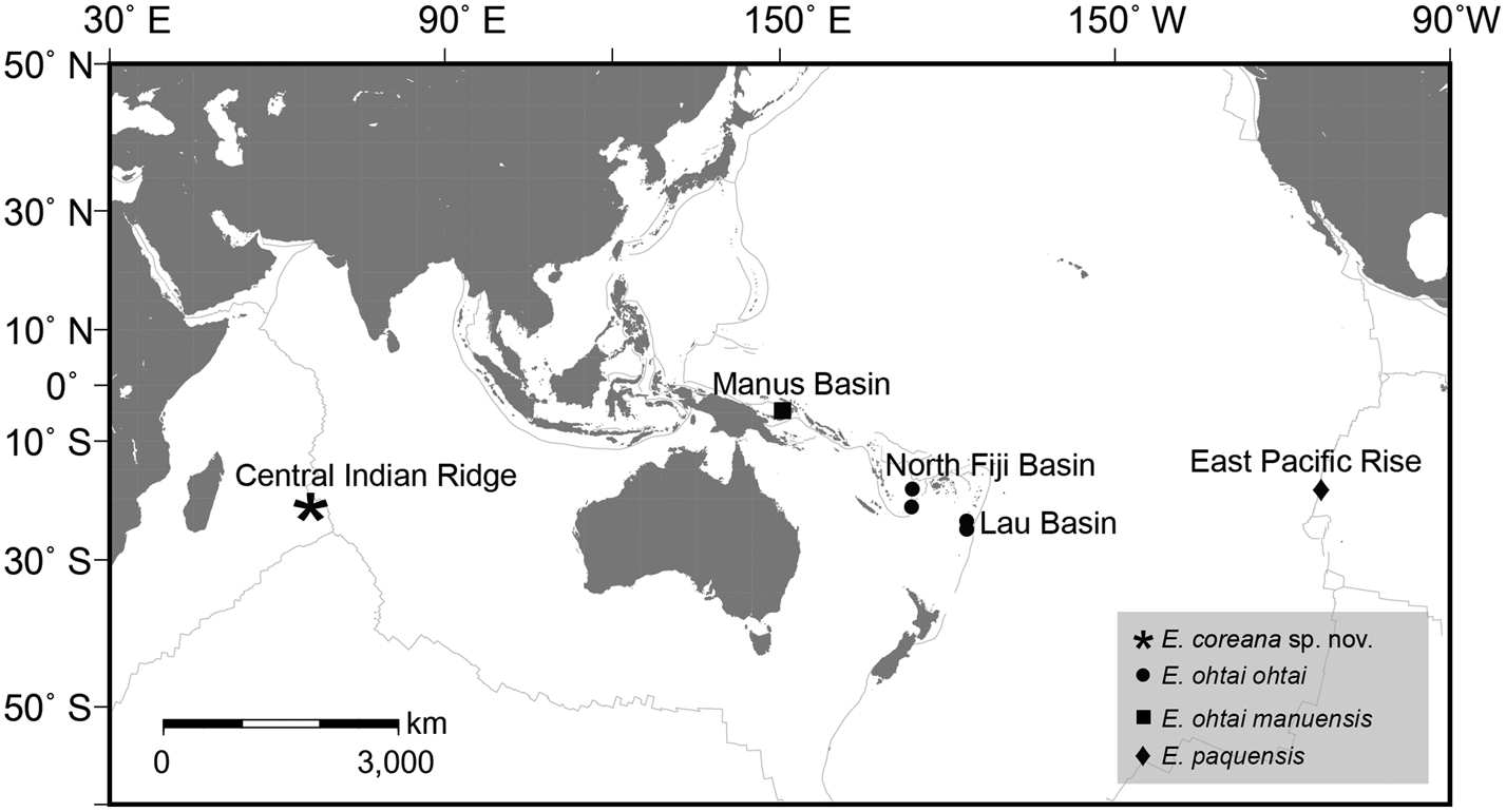
Among the extant descendants of hydrothermal vent barnacles, the Eochionelasmus clade includes only the genus Eochionelasmus Yamaguchi, 1990, which is characterized by six principal wall plates and multiple whorls of basal imbricating plates (Yamaguchi & Newman, Reference Yamaguchi and Newman1990, Reference Yamaguchi and Newman1997a, Reference Yamaguchi and Newman1997b). Two Eochionelasmus species, E. ohtai Yamaguchi, 1990 and E. paquensis Yamaguchi, 1997, have been recorded in different vent fields, the South-west Pacific Ocean and East Pacific Rise, respectively (Yamaguchi & Newman, Reference Yamaguchi and Newman1997a, Reference Yamaguchi and Newman1997b; Figure 1). Eochionelasmus ohtai, which is distributed widely in hydrothermal vent fields of the South-west Pacific Ocean, comprises two subspecies, E. ohtai ohtai for the North Fiji-Lau populations and E. ohtai manusensis for the Manus population, based on distinct differences in the ontogenetic development of the imbricating plates (Yamaguchi & Newman, Reference Yamaguchi and Newman1997a, Reference Yamaguchi and Newman1997b). Phylogenetic and population genetic analyses have been conducted to elucidate their taxonomic position and dispersal capacity (Plouviez et al., Reference Plouviez, Schultz, McGinnis, Minshall, Rudder and van Dover2013; Herrera et al., Reference Herrera, Watanabe and Shank2015; Kim et al., Reference Kim, Lee, Kim and Ju2018); however, DNA sequences of E. paquensis are unavailable in public databases.
Distribution map of Eochionelasmus species based on Yamaguchi & Newman (Reference Yamaguchi and Newman1990, Reference Yamaguchi and Newman1997a, Reference Yamaguchi and Newman1997b) and this study.

The Indian Ocean ridges have an intermediate spreading rate (50–60 mm year−1) compared with those of the East Pacific Rise and Mid-Atlantic Rise, and the organisms living there remain poorly studied (Beedessee et al., Reference Beedessee, Watanabe, Ogura, Nemoto, Yahagi, Nakagawa, Nakamura, Takai, Koonjul and Marie2013). The stalked neolepadid Neolepas marisindica Watanabe et al., Reference Watanabe, Chen, Marie, Takai, Fujikura and Chan2018 is currently the only known vent-endemic barnacle in the Indian Ocean and is distributed widely among five hydrothermal vent fields of the Indian Oceanic ridges: Karei, Solitaire, Onnuri, Longqi and Site 21 (Watanabe et al., Reference Watanabe, Chen, Marie, Takai, Fujikura and Chan2018; Ryu et al., Reference Ryu, Woo and Lee2019).
In this study, we describe a new species of the chionelasmatid Eochionelasmus found in the Solitaire hydrothermal vent field of the Central Indian Ridge; its CO1 barcodes for DNA taxonomy have been deposited in GenBank. We also discuss the phylogenetic position of the new species with known congener sequences.
Materials and methods
Vent barnacle sampling
Eochionelasmus specimens were collected at depths of >2600 m from the Solitaire hydrothermal vent field on the Central Indian Ridge (Figure 1) using a TV-guided grab sampler equipped by the research vessel ‘ISABU’ during a vent ecosystem study in the Indian mid-ocean ridge by the Korean Institute of Oceanographic Science and Technology. The specimens were immediately frozen and kept at − 80 °C for morphological and molecular studies.
Morphological examination
Each specimen was examined and dissected under stereomicroscopes. Soft parts including the mouth parts, cirri and penis were photographed using a compound microscope (Zeiss Axioplan compound microscope installed with objective APO40 × and APO20 × lenses; Germany). Morphological terminology generally follows Yamaguchi & Newman (Reference Yamaguchi and Newman1997a, Reference Yamaguchi and Newman1997b) for descriptions of the imbricating plates and arthropodal characters, and Chan et al. (Reference Chan, Hoeg and Garm2008) for setal classification. The dissected holotype and the dissected paratype have been deposited in the Korea National Institute for Biological Resources (Incheon, Korea, NIBR) and the Biodiversity Research Museum, Academia Sinica, Taiwan (ASIZCR), respectively.
Phylogenetic analysis
A small amount of muscle tissue was dissected from the Eochionelasmus specimens for DNA extraction. Total genomic DNA was extracted using a QIAamp DNA Mini Kit (Qiagen, Hilden, Germany). The partial sequences of three mitochondrial (12S, 16S rDNA and CO1) and two nuclear (18S rDNA and histone 3) genes were determined using the universal primers (Folmer et al., Reference Folmer, Black, Hoeh, Lutz and Vrijenhoek1994; Chan et al., Reference Chan, Corbari, Rodriguez and Tsang2017). PCR amplification was performed in a total volume of 50 μl containing 1 μl of genomic DNA, 4 μl of dNTP mixture (2.5 mM each), 1 μl of each primer (10 pmol), 5 μl of 10 × ExTaq Buffer (Mg2+ plus), and 1.25 U of Takara Ex Taq DNA Polymerase (Takara Biotechnology Co., Tokyo, Japan) under the following conditions: initial denaturation at 94 °C for 2 min, followed by 40 cycles of denaturation (15 s at 95 °C), primer annealing (30 s at 50 °C in the first 10 cycles and 30 s at 55 °C in the last 30 cycles), and extension (2 min at 72 °C), with a final extension step at 72 °C for 5 min. Then, 5 μl of each PCR product was run on a 1.0% agarose gel and visualized under UV light. Finally, Sanger sequencing was performed at Macrogen Service (Macrogen, Seoul, Korea) using the ABI PRISM 3730XL Analyzer (Applied Biosystems, Foster City, CA, USA) with a BigDye(R) Terminator v3.1 Cycle Sequencing Kit (Applied Biosystems).
The newly determined sequences from this study were trimmed and annotated, and then aligned with the other balanomorph genera retrieved from GenBank using T-Coffee (Notredame et al., Reference Notredame, Higgins and Heringa2000). To construct a phylogenetic tree of balanomorph barnacles, individual 12S rRNA, 16S rDNA, 18S rDNA and histone 3 gene alignments were concatenated to form a single multiple-sequence alignment with DAMBE5 (Xia, Reference Xia2013). The best-fitting model of nucleotide substitution was then determined using the Akaike information criterion in jModelTest 2.1.7 (Darriba et al., Reference Darriba, Taboada, Doallo and Posada2012); the model GTR + I + G was selected as the best evolution model. Maximum likelihood (ML) and Bayesian inference (BI) tests were performed using RAxML version 8.2.11 (Stamatakis, Reference Stamatakis2014) and MrBayes 3.2.6 (Ronquist et al., Reference Ronquist, Teslenko, Van Der Mark, Ayres, Darling, Höhna, Suchard and Huelsenbeck2012), respectively, implemented in Geneious Prime with the gene partition option. Confidence in the resulting balanomorph relationships was assessed based on the bootstrap proportion (BP) with 200 replications for the ML model. For BI analysis, four Markov chain Monte Carlo chains were run for 1000,000 generations and sampled every 200 generations. Bayesian posterior probability (BPP) values were estimated after the initial 500 (10%) trees were discarded as burn-in.
Results
Systematics
Superorder THORACICA Darwin, 1854
Order SESSILIA Lamarck, 1818
Suborder BALANOMORPHA Pilsbry, 1916
Superfamily CHIONELASMATOIDEA Buckeridge, Reference Buckeridge1983
Family CHIONELASMATIDAE Buckeridge, Reference Buckeridge1983
Genus Eochionelasmus Yamaguchi, 1990
Eochionelasmus coreana sp. nov.
(Figures 2–10)
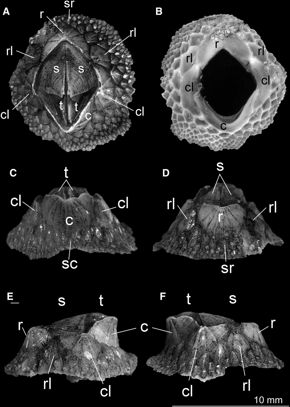
External shell of Eochionelasmus coreana sp. nov. (holotype). (A) Top view. (B) Basal view showing the sheath is formed by the combination of rl, paired cl and c. rl is located outside the sheath. (C) Carinal view. (D) Rostral view. (E) Left view. (F) Right view. R, rostrum; c, carina; cl, carinal latus; rl, rostral latus; s, scutum; t, tergum; sc, subcarina; sr, subrostrum.

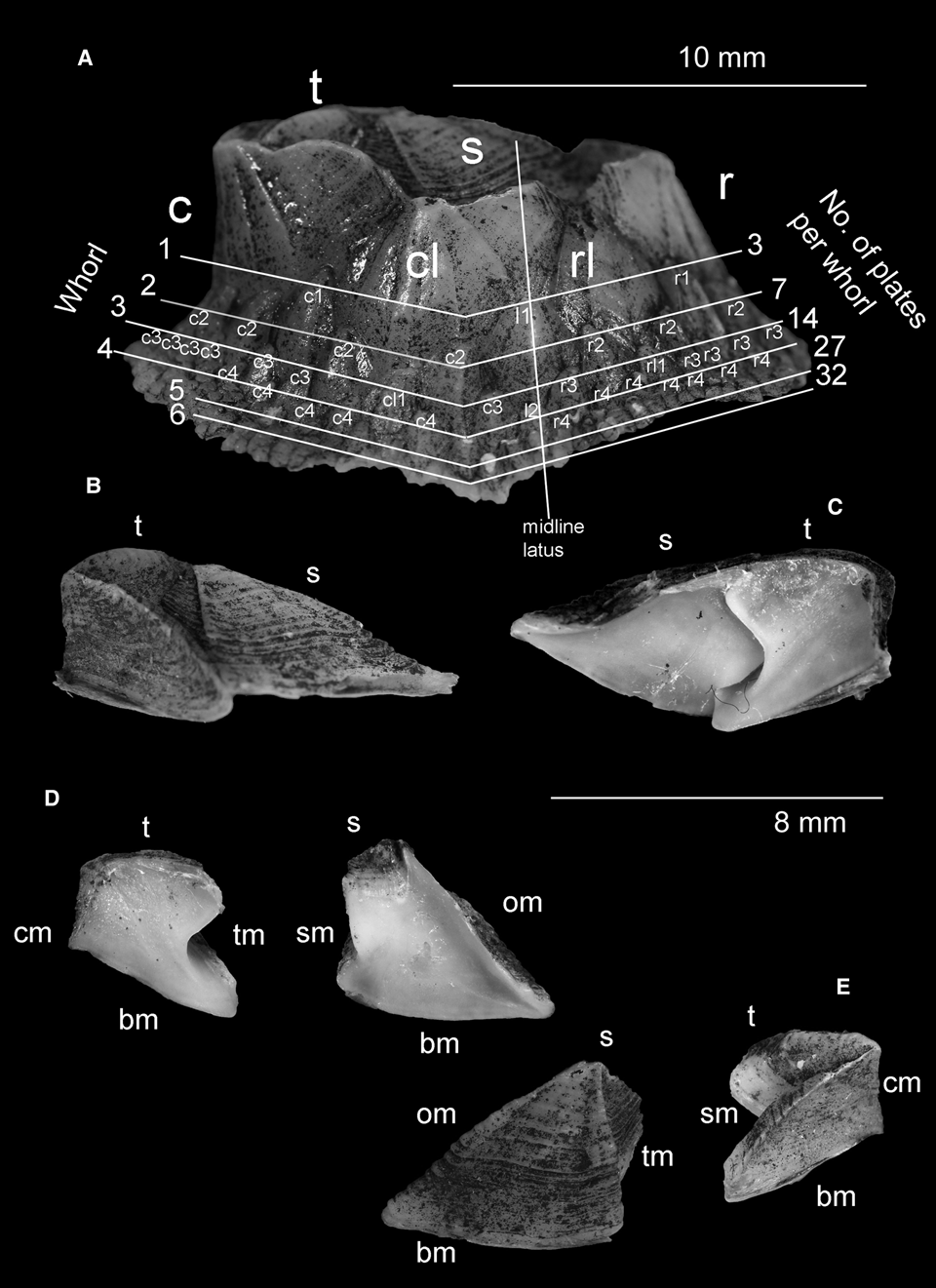
(A) Side view of Eochionelasmus coreana sp. nov. (holotype), showing the number of whorls of imbricating plates and number of imbricating plates per row. Naming of imbricating plates follows the system in Yamaguchi & Newman (Reference Yamaguchi and Newman1997a, Reference Yamaguchi and Newman1997b). Imbricating plates on the carinal side from the latera are abbreviated as c. Imbricating plates on the rostral side from the latera are abbreviated as r. The numeral of the imbricating plates indicates the number of whorls from their first appearance. cl1 indicates the first cl that appears directly under cl. rl1 indicates the first rl that appears directly under cl. l is the latera. Note that not all the imbricating plates are labelled for clarity reasons. (B) Outer side of inter-locked scutum and tergum. (C) Inner side of inter-locked scutum and tergum. (D) Inner side of tergum and scutum. (E) Outside of scutum and tergum. cm, carinal margin; om, occludent margin; bm, basal margin; tm, tergal margin; sm, scutal margin; t, tergum; s, scutum.

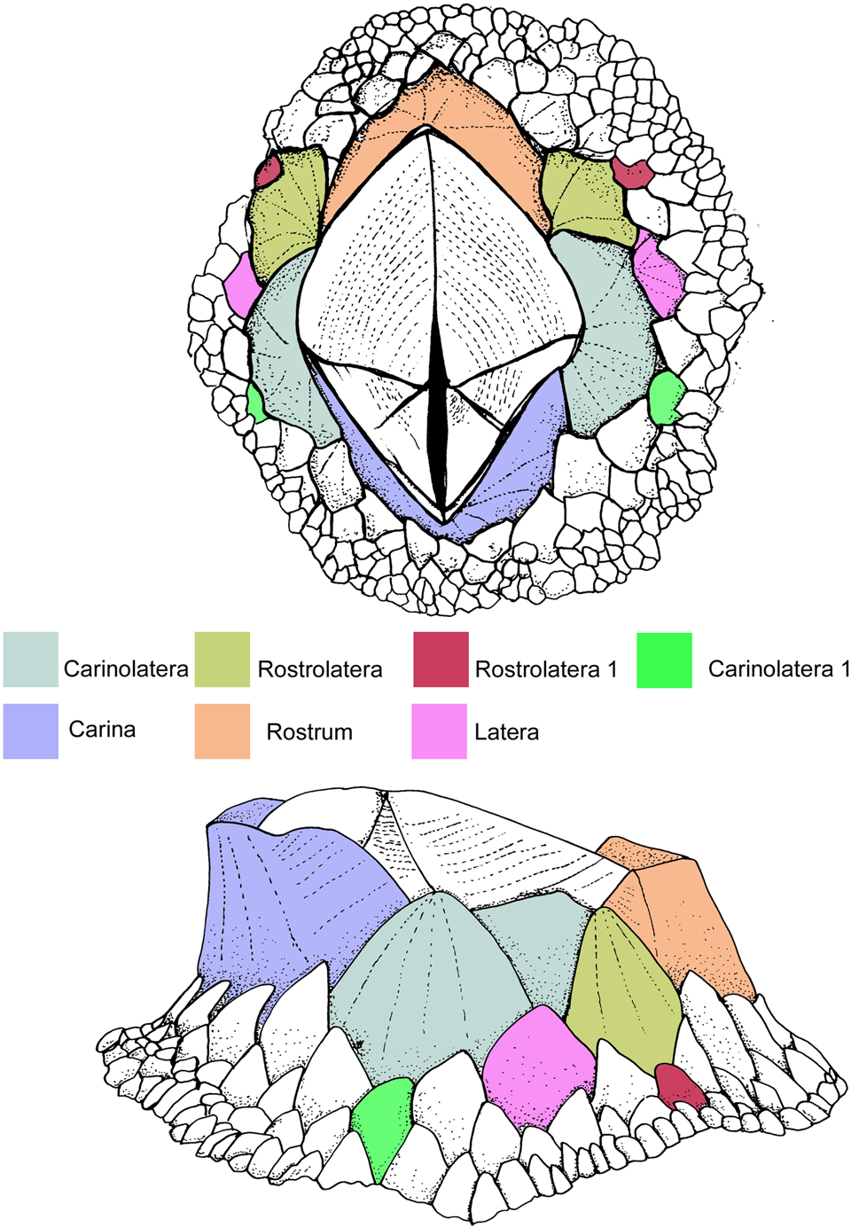
Drawing of Eochionelasmus coreana sp. nov. (A) Plan view and (B) Lateral view, showing the arrangement of shell plates and imbricating plates. Note latera appears on the third whorl of imbricating plates (also see Fig. 3).

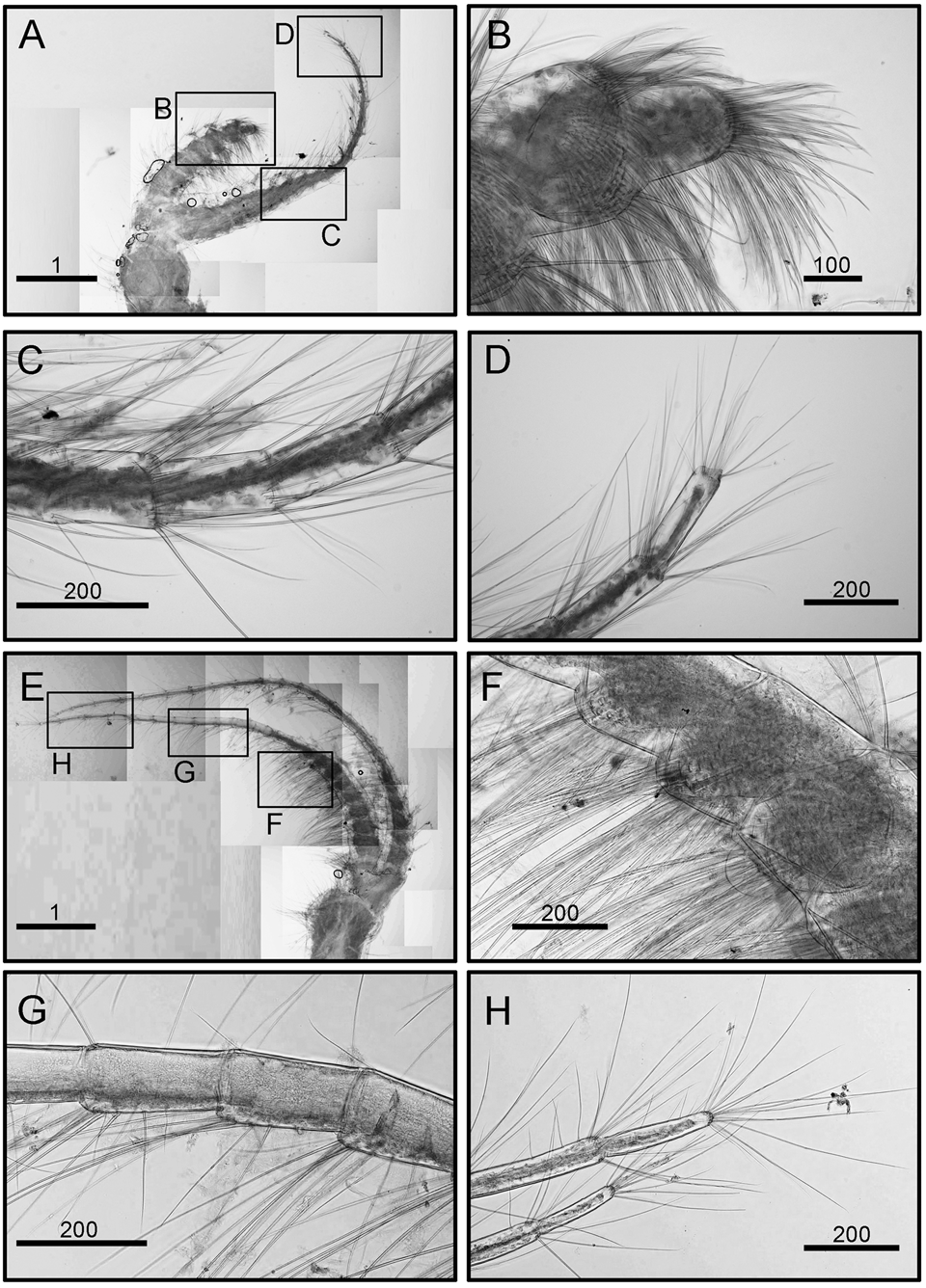
Eochionelasmus coreana sp. nov. (holotype). (A) Cirrus I, showing anterior and posterior ramus. (B) Simple setae on posterior ramus. (C) Simple setae on anterior ramus. (D) Setae on distal end of anterior ramus. (E) Cirrus II. (F) Dense simple setae on the robust segment of anterior ramus. (G) Intermediate segment on posterior ramus. (H) Distal segment of posterior ramus. Scale bars in A, E in mm; others in μm.

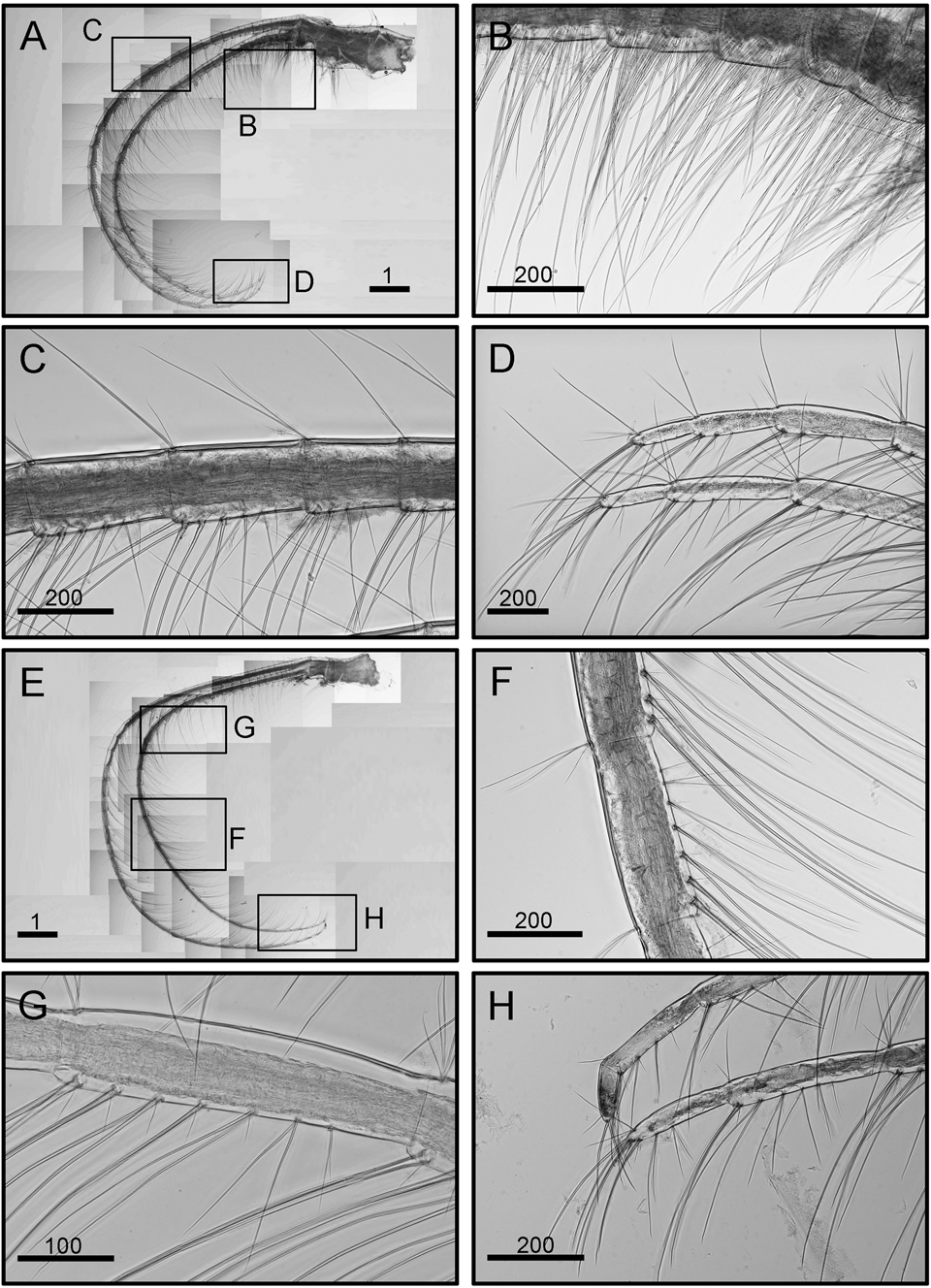
Eochionelasmus coreana sp. nov. (holotype). (A) Cirrus III. (B) Dense setae on proximal region of rami. (C) Intermediate segments. (D) Distal segments. (E) Cirrus IV. (F) Intermediate segment. (G) Proximal segments. (H) Distal segments. Scale bars in A, E in mm; others in μm.

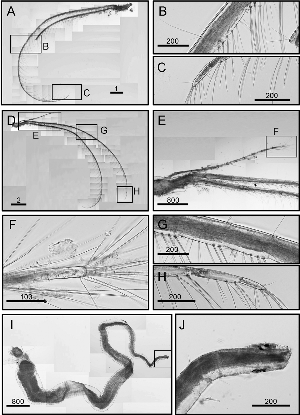
Eochionelasmus coreana sp. nov. (holotype). (A) Cirrus V. (B) Intermediate segments. (C) Distal segments. (D) Cirrus VI and caudal appendages. (E) Caudal appendages. (F) Distal segments. (G) Intermediate segments. (I) Penis. (J) Tip of penis. Scale bars in A, D in mm; others in μm.

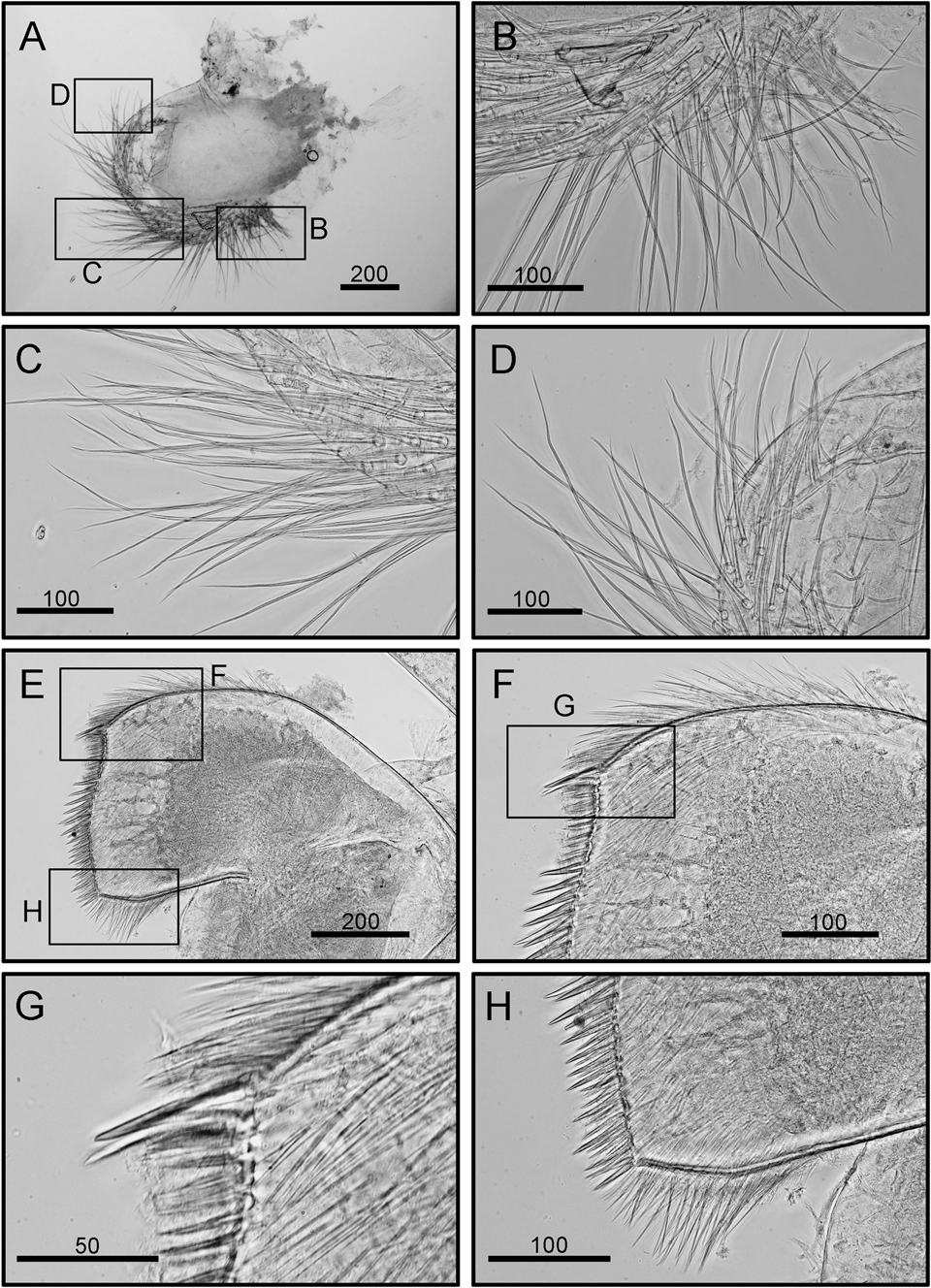
Eochionelasmus coreana sp. nov. (holotype). (A) Maxilla. (B) Setae on exterior margin. (C) Setae on outer margin. (D) Setae on inner margin. (E) Maxillule. (F) Cutting edge of maxillule. (G) Upper portion of cutting edge. (H) Lower portion of cutting edge. Scale bars in μm.

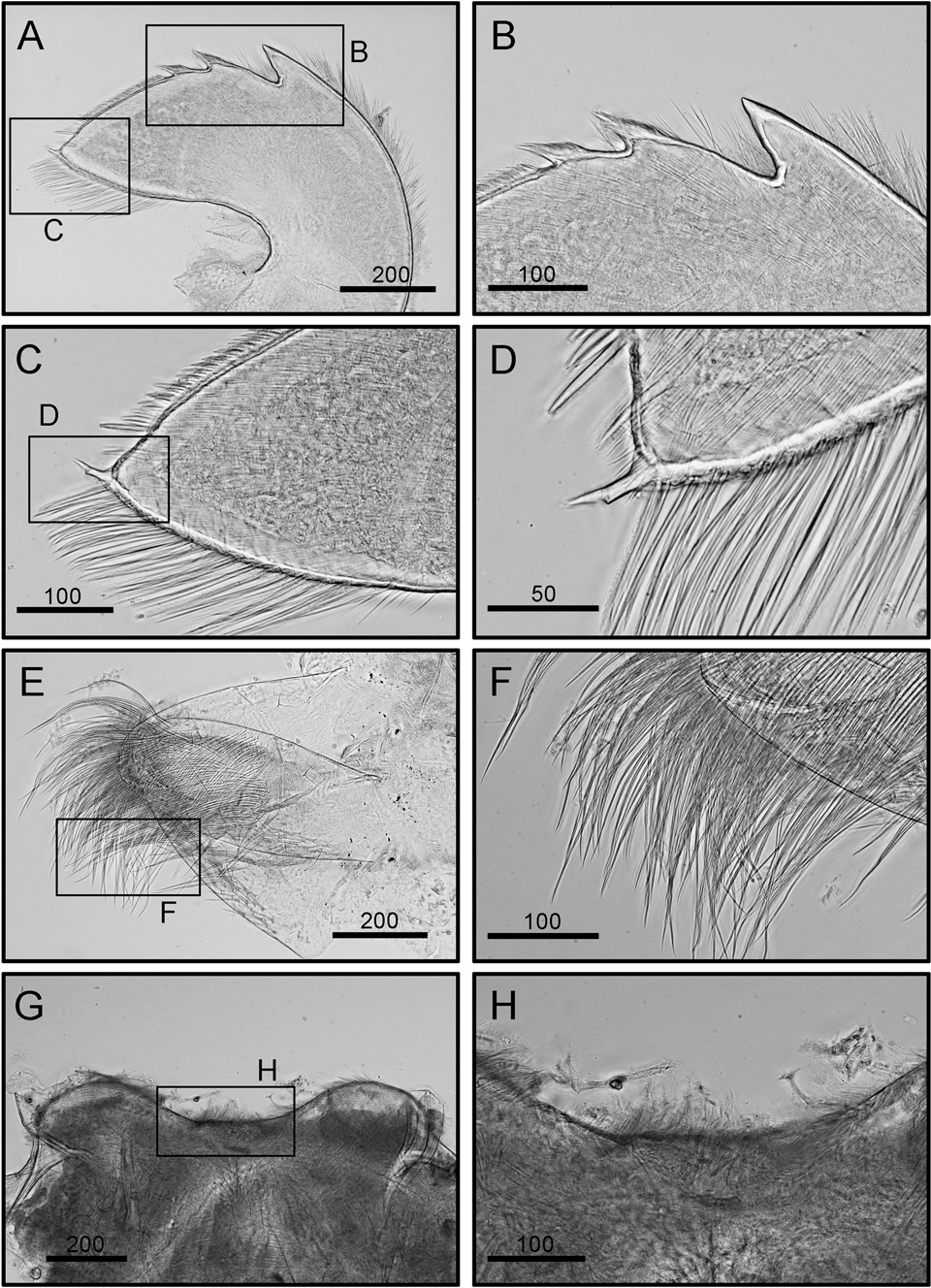
Eochionelasmus coreana sp. nov. (holotype). (A) Mandible. (B) The three teeth of the mandibles. (C) Inferior angle. (D) Outer margin. (E) Mandibulatory palp. (F) Setae on mandibulatory palp. (G) Labrum. (H) Cutting edge of labrum. Scale bars in μm.

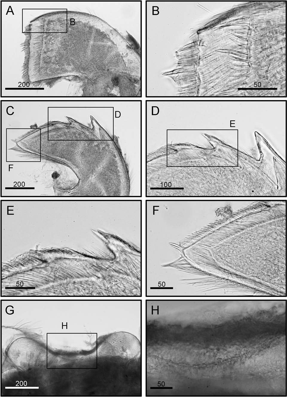
Eochionelasmus coreana sp. nov. (paratype). (A) Maxillule. (B) Upper portion of cutting edge. (C) Mandible. (D) The three teeth of the mandibles. (E) Third tooth of mandible. (F) Inferior angle. (G) Labrum. (H) Cutting edge of labrum. Scale bars in μm.

Type material
Holotype: NIBR-IV0000865932. Solitaire vent fields, Indian Ocean Ridge (19° 33′ 39″ S 65° 50′ 89″ E; water depth: 2625 m); collected using a TV-guided grab sampler, coll. D.-S. Kim, 19 June 2018.
Paratype: ASIZCR-000433, same data as holotype.
Diagnosis
Eochionelasmus with 6–7 whorls of imbricating plates, carinolatera with wide alae, maxillule without notch on upper portion, third tooth of mandibles with numerous denticles on cutting edge. Location of carinolatera 1 and rostrolatera 1 on the third whorl of imbricating plates.
Description
Primary shell wall six-plated; composed of carina, paired carinolatera, paired rostrolatera, and rostrum (c-cl-rl-r-rl-cl); conical and depressed (height ~1/2 of rostro-carino diameter) (Figure 2). Orifice large and rhomboidal (Figure 2). Sheath formed by carina, rostrum, paired carinolatera, but rostrolatera not entered (Figure 2B).
Shells were surrounded by 6–7 whorls of triangular imbricating plates (Figures 2C–F & 3A). Size of imbricating plates increased from the shell rim to the upper whorls (Figure 2C–F). The first whorl of imbricating plates started on the lower portion of primary shell wall (Figures 3A, 4A, B). First whorl with three imbricating plates, carina 1 (c1), latus 1 (l1) and rostrum 1 (r1); second whorl with seven imbricating plates; third whorl with 14 imbricating plates, carinolatera 1 (cl1) and rostrolatera 1 (rl1) occurred; fourth whorl with 27 imbricating plates; fifth whorl with 40 imbricating plates (Figures 3A, 4A, B).
Scutum and tergum strongly articulated (Figure 3B, C). Scutum triangular, dorsal surface with horizontal growth lines. Occludent and basal margins straight. Articular ridge high and articular furrow deep. Tergal margin with V-shaped articular ridge. Adductor muscle and depressor muscle scars in scutum absent (Figure 3D, E). Tergum quadrangular, dorsal surface with horizontal growth lines, scutal margin strongly concaved. Carinal margin straight, basal margin long and straight, spur sharp and triangular. Pits for depressor muscle in tergum absent (Figure 3D, E).
Cirrus I, anterior ramus short, seven segmented, posterior rami long, with antenniform segments starting from half length of rami, 20 segmented (Figure 5A). All segments in both rami bear simple setae (Figure 5B–D). Cirrus II, anterior and posterior rami subsequal, anterior rami 22 segmented, distal half of ramus bear antenniform, proximal half with robust segments (Figure 5E, F). Antenniform segment bears three pairs of long simple setae and one pair of short simple setae (Figure 5G, H). Proximal region of segments bears dense long simple setae (Figure 5F). Posterior ramus 25 segmented, with antenniform segments starting from 1/4 of the proximal region. All segments bear simple setae. Cirri III, IV, V and VI are similar in morphology, all with long and slender rami (segment counts, see Table 1) (Figures 6 & 7). Proximal segments bear dense simple setae (Figure 6B). Intermediate segments of cirrus III bear four pairs of long setae and two pairs of short setae (Figure 6C, D). Intermediate segments of cirrus V and VI bear five pairs of long setae and two pairs of short setae (Figures 6E–H & 7A–G). Caudal appendage long, 16 segmented (Figure 7D, E). Penis long, without basal point (Figure 7I, J).
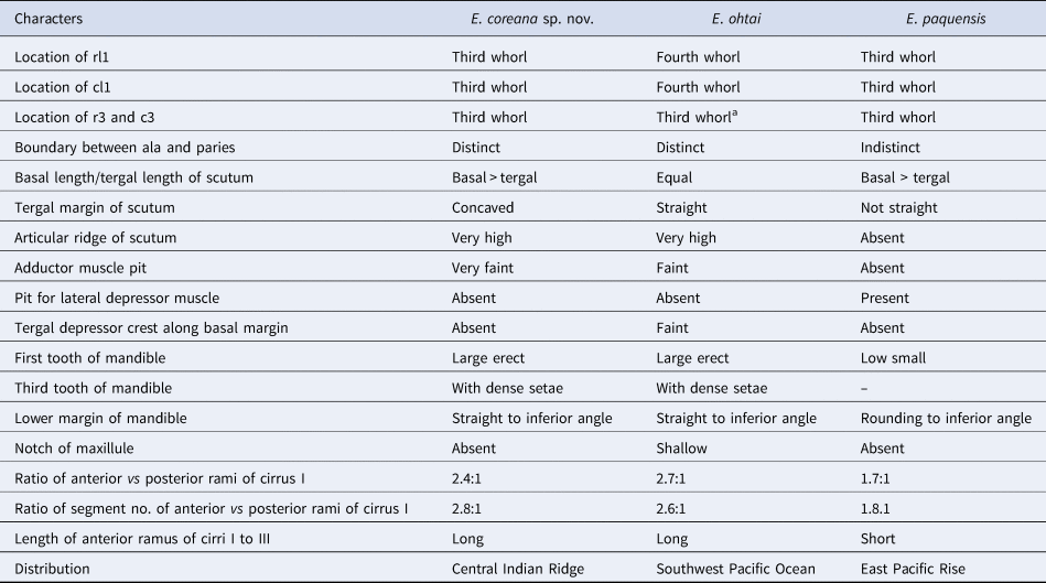
Comparison of morphological characters of Eochionelasmus coreana sp. nov., the generalized E. ohtai from North Fiji Basin, and the holotype of E. paquensis. The contents of the table have been revised from Yamaguchi & Newman (Reference Yamaguchi and Newman1997a, Reference Yamaguchi and Newman1997b)

a In Table 1 of Yamaguchi & Newman (Reference Yamaguchi and Newman1997a, Reference Yamaguchi and Newman1997b), the location of r3 and c3 of E. ohtai was listed as the fourth whorl. In Figure 4 of Yamaguchi & Newman (Reference Yamaguchi and Newman1997a, Reference Yamaguchi and Newman1997b), which showed the generalized pattern of E. ohtai, the r3 and c3 were located on the third whorl. We adopted the arrangement of the generalized pattern of E. ohtai from Figure 4 of Yamaguchi & Newman (Reference Yamaguchi and Newman1997a, Reference Yamaguchi and Newman1997b) and concluded the location of r3 and c3 should be the third whorl.
Maxilla oval, single-lobed, simple setae distally (Figure 8A) and along inferior margin (Figure 8B–D). Maxillule cutting edge slightly convex, without notch, the lower 2/3 portion of cutting edge slightly protruded. Two distinct large setae on top of cutting edge, followed by a row of nine smaller setae on the 1/3 upper portion of cutting edge, protruded 2/3 lower portion of cutting edge with 28–30 large setae (Figure 8E–H) (maxilla morphology consistent between holotype and paratype; Figure 10A, B). Mandible with three teeth (Figure 9A–D), first tooth slightly departed from second and third teeth, lower margin long. First teeth largest and sharp (Figure 9B), second and third with dense row of setae on cutting edge. Lower margin long and straight, with dense row of setae. Inferior angle with two large setae (Figures 9C, D & 10C–F). Mandibular palp triangular, bearing dense serrulate setae distally (Figure 9E, F) and along interior margin. Labrum concaved, notch absent, cutting edge with extreme small teeth (Figure 9G, H; consistent in two specimens, Figure 10G, H).
Distribution
At present, only known from the Solitaire hydrothermal vent field on the Central Indian Ridge.
Etymology
The name coreana represents the Latin name of Korea, ‘corea’. This name acknowledges the Korean deep-sea research team (Korean Institute of Oceanography, Science and Technology, KIOST) that collected the new species in the Solitaire hydrothermal vent field on the Central Indian Ridge.
Remarks
The third Eochionelasmus species, E. coreana, is not only the first record of a sessile vent barnacle from outside the Pacific Ocean, but is also the first discovery of Eochionelasmus from the Indian Ocean; the distributions of E. ohtai and E. paquensis are restricted to the South-west Pacific Ocean and the East Pacific Rise, respectively.
Eochionelasmus coreana and E. ohtai are morphologically similar in their external shell structure, and the shapes of the scutum and tergum. However, Eochionelasmus coreana differs from E. ohtai in the cutting edge on the maxillule (notch absent in E. coreana vs shallow notch in E. ohtai), and the location of rl1 and cl1 on whorls of the imbricating plates (third in E. coreana vs fourth in E. ohtai). Meanwhile, E. coreana is different from E. paquensis in the characteristics of the mandible and the length ratio between the rami parts on cirrus I (Table 1).
GenBank accession numbers of CO1 DNA barcode
MT008257 for the holotype; MT008258 for the paratype.
Phylogenetic position of Eochionelasmus within the balanomorph lineage
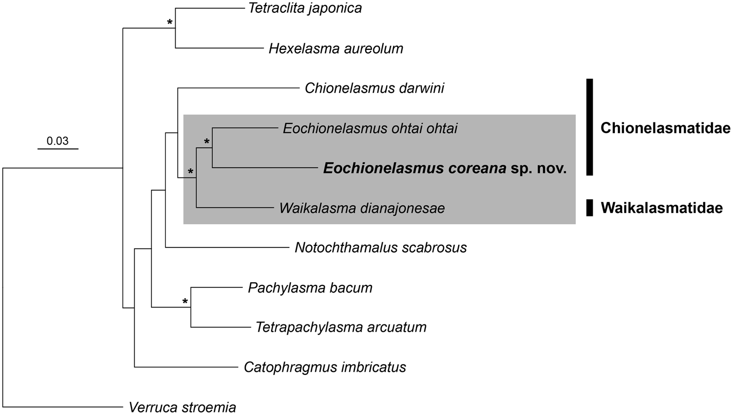
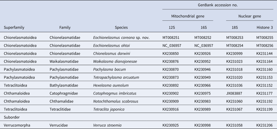
Four newly obtained sequences (12S, 16S, 18S and histone 3) of E. coreana were registered in GenBank (accession nos. MT008251–MT008253, MT008255). Additionally, in this study, the nuclear gene sequences (accession nos. MT008254 for 18S and MT008256 for H3) of E. ohtai were obtained from the same specimen using the complete mitochondrial genome (NC_036957) determined by Kim et al. (Reference Kim, Lee, Kim and Ju2018). We could not include sequences of E. paquensis in all analyses because they were not available from open-access sequence databases. The phylogenetic trees were constructed using the concatenated sequence alignment of four genes from 10 balanomorph genera and one verrucomorph species, Verruca stroemia, as an outgroup (Figure 11; Table 2). The tree strongly supported two Eochionelasmus species, E. coreana and E. ohtai, as monophyletic taxa, with high support values of 97% BP and 1.00 BPP. The chionelasmatid Eochionelasmus species and the waikalasmatid Waikalasma dianajonesae also had strong support for monophyly (97% BP and 1.00 BPP). However, three chionelasmatid species, E. coreana, E. ohtai and Chionelasmus darwini, were not monophyletic.
Phylogenetic tree of Eochionelasmus coreana sp. nov. and the other balanomorph barnacles based on two mitochondrial (12S and 16S rDNA) and two nuclear (18S rDNA and histone 3) genes. Asterisks indicate the maximum likelihood bootstrap proportions >97% and Bayesian posterior probabilities >0.99.

Information of the sequences used for constructing the phylogenetic tree in this study

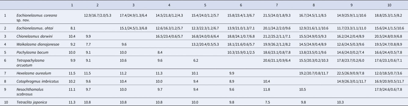
In terms of nucleotide sequence divergence, the four genes showed different variations, in decreasing order: 16S, 12S, H3 and 18S (Table 3). The sequence divergence between E. ohtai and E. coreana was 16.7% for 16S, 12.9% for 12S, 5.3% for H3 and 2.0% for 18S; all values were consistent with the inter-generic variations among balanomorph genera. Based on the concatenated sequences of four genes, the minimum and maximum variations were 6.1% between Pachylasma bacum and Tetrapachylasma arcuatum, and 11.8% between Hexelasma aureolum and Notochthamalus scabrosus, respectively. The variation (8.1%) between two Eochionelasmus species from the concatenated sequences had an intermediate value compared with the range of inter-generic variations among balanomorphs.
Sequence divergence among the partial sequences of two mitochondrial (12S and 16S rDNA) and two nuclear (18S rDNA and histone 3) genes from balanomorph genera

Nucleotide sequence variations (%) were calculated using the p-distance method in Mega 10.0.5. Upper right is for the 12S/16S/18S/H3 genes and lower left the concatenated sequences of the four genes.
Discussion
Geotectonic events, ecological/hydrographic barriers, hydrothermal fluid characteristics, and length of larval development are considered important factors for distribution, migration, settlement and speciation of hydrothermal vent organisms (Watling et al., Reference Watling, Guinotte, Clark and Smith2013). However, the origin, migration and distribution of hydrothermal vent fauna among disconnected vent habitats with repeating cycles of creation and extinction are still not fully understood. In this study, we discovered the first sessile Eochionelasmus barnacle, E. coreana, in the Indian Ocean. This raises an interesting question of how three Eochionelasmus species in three different oceans originated (Figure 1). Thus, if we can understand their distributions and environmental adaptation strategies, we may gain new insights into the association between adaptive radiation of hydrothermal vents organisms and geotectonic events on Earth.
Despite morphological and phylogenetic affinities of the genus Eochionelasmus with Balanomorpha, the phylogenetic position of Eochionelasmus within the balanomorphs is uncertain because of lineage-specific evolutionary rates and/or taxonomic under-sampling (Pérez-Losada et al., Reference Pérez-Losada, Høeg, Simon-Blecher, Achituv, Jones and Crandall2014; Herrera et al., Reference Herrera, Watanabe and Shank2015). In this study, we identified a new species in Eochionelasmus from the Indian Ocean, and then re-constructed the phylogenetic tree with related genera, Waikalasma Buckeridge, Reference Buckeridge1983 and Chionelasmus Pilsbry, 1907 (Figure 10). Based on the tree, the chionelasmatid Eochionelasmus was closely related to the waikalasmatid Waikalasma with high support rather than the other chionelasmatid Chionelasmus. Although chionelasmatids and waikalasmatids were clustered in a single clade in the present study, the bootstrap value was not high enough to support the common ancestry of the Eochionelasmus–Waikalasma clade and the genus Chionelasmus. Thus, the phylogenetic relationship between chionelasmatid and waikalasmatid members remains uncertain and will require additional molecular markers for analysis.
Conversely, based on traditional taxonomy, these three genera have been considered the most basal taxa of balanomorph barnacles, with Eochionelasmus considered more closely related to Chionelasmus than Waikalasma (Jones, Reference Jones and Crosnier2000; Chan et al., Reference Chan, Corbari, Rodriguez and Tsang2017). However, in terms of habitat, Eochionelasmus, which lives in hydrothermal vents, differs from Chionelasmus and Waikalasma, which live in general deep-sea environments. Considering that principal food sources change under different habitat conditions, it has been assumed that Eochionelasmus developed fine setae and weaker spines on its mouth parts to obtain chemosynthetic bacteria as a food source through filter-feeding. Such morphological specialization in mouth parts were also observed in neolepadid barnacles which live in hydrothermal vents (Newman, Reference Newman1979; Buckeridge, Reference Buckeridge2000). Conversely, Chionelasmus and Waikalasma may have developed stronger spines on their mouth parts for capturing and feeding on zooplankton (Jones, Reference Jones and Crosnier2000; Chan et al., Reference Chan, Corbari, Rodriguez and Tsang2017). To improve our understanding of the position and the relationship of these basal groups in the balanomorph phylogeny, further research is needed, including discovery of additional taxa, and mitogenome-based phylogenetic and biogeographic studies.
Financial support
This work was supported by the R & D projects (KIMST #20170411(PM61740) & 19992001) funded by the Korean Ministry of Ocean and Fisheries and a Korea Research Institute of Bioscience and Biotechnology (KRIBB) Research Initiative Program grant.