INTRODUCTION
Clinical malaria caused by Plasmodium falciparum arises during the asexual phase of its life cycle in which it rapidly multiplies in host erythrocytes. P. falciparum differs from the other major human Plasmodium species, P. vivax, in its capacity to efficiently invade both reticulocytes and erythrocytes, which allows it to achieve high levels of parasitaemia. Both enhanced replicative potential and lack of erythrocyte selectivity by P. falciparum isolates have been associated with clinical severity (Chotivanich et al. Reference Chotivanich, Udomsangpetch, Simpson, Newton, Pukrittayakamee, Looareesuwan and White2000). The parasite and erythrocyte molecules underlying these phenotypes remain unknown.
P. falciparum expresses multiple adhesive proteins that localize to the apical invasion organelles of the parasite, most notably members of 2 multigene families: the Erythrocyte Binding Antigen (EBA) family (Sim et al. Reference Sim, Orlandi, Haynes, Klotz, Carter, Camus, Zegans and Chulay1990; Peterson and Wellems, Reference Peterson and Wellems2000; Thompson et al. Reference Thompson, Triglia, Reed and Cowman2001; Gilberger et al. Reference Gilberger, Thompson, Triglia, Good, Duraisingh and Cowman2003), and the P. falciparum Reticulocyte Binding Protein homologue (PfRh) family (Rayner et al. Reference Rayner, Galinski, Ingravallo and Barnwell2000, Reference Rayner, Vargas-Serrato, Huber, Galinski and Barnwell2001; Triglia et al. Reference Triglia, Thompson, Caruana, Delorenzi, Speed and Cowman2001; Kaneko et al. Reference Kaneko, Mu, Tsuboi, Su and Torii2002; Cowman and Crabb, Reference Cowman and Crabb2006). Differential expression of these proteins, particularly of members of the PfRh family, allows P. falciparum to engage alternative receptors for erythrocyte invasion, termed invasion pathways (Taylor et al. Reference Taylor, Grainger and Holder2002; Duraisingh et al. Reference Duraisingh, Maier, Triglia and Cowman2003a, Reference Duraisingh, Triglia, Ralph, Rayner, Barnwell, McFadden and Cowmanb; Stubbs et al. Reference Stubbs, Simpson, Triglia, Plouffe, Tonkin, Duraisingh, Maier, Winzeler and Cowman2005; Triglia et al. Reference Triglia, Duraisingh, Good and Cowman2005). In vitro invasion experiments using antibody inhibition, enzymatically modified erythrocytes, and erythrocytes from rare blood groups have revealed several discrete invasion pathways (Hadley et al. Reference Hadley, Klotz, Pasvol, Haynes and McGinniss1987; Mitchell et al. Reference Mitchell, Hadley, McGinniss, Klotz and Miller1986; Dolan et al. Reference Dolan, Miller and Wellems1990; Rayner et al. Reference Rayner, Vargas-Serrato, Huber, Galinski and Barnwell2001). Several invasion pathways require sialic acid on the erythrocyte surface. The sialic acid-dependent parasite ligands are PfRh1, whose erythrocyte receptor is unknown, and EBA-175 and EBA-140, which bind to the erythrocyte receptors glycophorin A and glycophorin C respectively (Sim et al. Reference Sim, Chitnis, Wasniowska, Hadley and Miller1994; Rayner et al. Reference Rayner, Vargas-Serrato, Huber, Galinski and Barnwell2001; Lobo et al. Reference Lobo, Rodriguez, Reid and Lustigman2003; Maier et al. Reference Maier, Duraisingh, Reeder, Patel, Kazura, Zimmerman and Cowman2003). Both laboratory-adapted and field isolates of P. falciparum can invade erythrocytes that have been depleted of sialic acid. Deans and colleagues recently reported that invasion pathways in Kenyan isolates of P. falciparum were not associated with the severity of malaria (Deans et al. Reference Deans, Nery, Conway, Kai, Marsh and Rowe2007). Interestingly, parasites drawn from patients with type B blood were found to preferentially use sialic acid-independent invasion pathways (Deans et al. Reference Deans, Nery, Conway, Kai, Marsh and Rowe2007).
PfRh2b is a major P. falciparum sialic acid-independent invasion ligand (Duraisingh et al. Reference Duraisingh, Triglia, Ralph, Rayner, Barnwell, McFadden and Cowman2003b). Genetic deletion of PfRh2b results in parasites that invade less efficiently by sialic acid-independent pathways, but with enhanced invasion by protease-resistant receptors (Duraisingh et al. Reference Duraisingh, Triglia, Ralph, Rayner, Barnwell, McFadden and Cowman2003b; Baum et al. Reference Baum, Maier, Good, Simpson and Cowman2005). A similar knockout in the closely related molecule PfRh2a has no effect on invasion pathway utilization. As PfRh2a and PfRh2b differ only in their C-terminal regions, it appears that this ‘unique’ region defines the differential invasion phenotypes of PfRh2b and PfRh2a (Duraisingh et al. Reference Duraisingh, Triglia, Ralph, Rayner, Barnwell, McFadden and Cowman2003b). Specific amino-acid sequence polymorphisms in PfRh2b have previously been associated with specific invasion pathways used by Brazilian P. falciparum isolates (Lobo et al. Reference Lobo, Rodriguez, Struchiner, Zalis and Lustigman2006), but these are rare in Senegalese isolates (Jennings et al. Reference Jennings, Ahouidi, Zilversmit, Bei, Rayner, Sarr, Ndir, Wirth, Mboup and Duraisingh2007). We have recently reported a ~194 amino acid deletion in the unique region of PfRh2b (PfRh2bDEL) that was common among Senegalese isolates of P. falciparum (Jennings et al. Reference Jennings, Ahouidi, Zilversmit, Bei, Rayner, Sarr, Ndir, Wirth, Mboup and Duraisingh2007).
In this paper we describe the analysis of a new series of Senegalese P. falciparum isolates in which the PfRh2bDEL polymorphism is highly prevalent collected over one transmission season in Senegal. We tested the hypothesis that PfRh2bDEL has a role in defining alternative invasion pathways. Our studies used uncultured isolates that removes potential bias resulting from culture adaptation, for instance due to in vitro selection for fast-growing parasites, and at a molecular level in changes in expression or genetic mutation of invasion ligands. First round invasion assays demonstrated that specific invasion pathways are significantly associated with parasite multiplication rate and the presence of PfRh2bDEL, and identify a potential modulatory role for blood group O.
MATERIALS AND METHODS
Sample collection
This study was approved by the Institutional Review Board of the Harvard School of Public Health and by the Ethics Committee of the Ministry of Health in Senegal. Blood samples (5 ml of venous blood in heparanized tubes, stored at 4°C for between 2 and 6 h until processing) were collected between October and December 2005, following informed consent from patients or their legal guardians. Study enrollment was offered to patients with microscopically confirmed P. falciparum malaria presenting to outpatient clinical sites in Thies (Service de Lutte Anti-Parasitaire) and Pikine (Poste de Sante Touba Diacksao), Senegal.
Erythrocyte invasion assay
Invasion efficiency into enzyme-treated erythrocytes was carried out as previously described (Duraisingh et al. Reference Duraisingh, Triglia, Ralph, Rayner, Barnwell, McFadden and Cowman2003b; Jennings et al. Reference Jennings, Ahouidi, Zilversmit, Bei, Rayner, Sarr, Ndir, Wirth, Mboup and Duraisingh2007). Briefly, to prepare acceptor cells, aliquots of banked blood from malaria-uninfected donors (type O+), were washed in RPMI. Cells were spun down (700 g for 10 min), the supernatant was removed, and 200 μl of packed cells (estimated at 2×10⁹ cells) were treated with Vibrio cholerae neuraminidase (Nm)- 66·7 mU (Calbiochem), trypsin (Tr)- 1·0 mg/ml (TPCK-treated, Sigma), low trypsin (lowTr)- 0·04 mg/ml, chymotrypsin (Ch)- 1·0 mg/ml (TLCK-treated, Worthington), and combinations of low trypsin-neuraminidase (lowTr/Nm) and low trypsin-chymotrypsin (lowTr/Ch). Trypsin-neuraminidase-treated (Tr/Nm) cells were used as negative controls and untreated erythrocytes as positive controls for erythrocyte invasion. Enzyme treatments were performed for 1 h at 37°C with gentle shaking. Infected donor cells with greater than 1·5% parasitaemia were diluted to 1% using uninfected RBCs and treated with Tr/Nm to prevent re-invasion. Following enzyme treatment, acceptor cells and parasitized donor cells were diluted in complete RPMI to a final haematocrit of 2%.
Parasitized donor cells were mixed 1:1 with acceptor cells for a final volume of 100 μl per well in 96-well plates. All invasion assays were performed in triplicate for each enzyme treatment and with 6 positive controls. Assay plates were incubated in a modular incubator chamber (Billups-Rothenberg) with 5% CO2/1% O2 at 37°C. Parasite growth was monitored microscopically and assays were terminated upon complete re-invasion. Thin films were made from each well and parasitaemia was determined by counting at least 750 red blood cells per smear using a Miller reticle. Parasitaemia was counted by determining the proportion of erythrocytes containing at least 1 parasite.
PCR typing of PfRh2bDEL, clonality and protein expression
Genomic DNA was isolated from P. falciparum parasite pellets using a QIAmp Blood Kit mini (Qiagen). The PfRh2b gene was amplified by polymerase chain reaction using primers flanking the C-terminal region (forward primer: 5′ TGA TTA TAG TGA AAG TAG CAACA 3′, reverse primer: 5′ GTT ATG GTT TGA ATA CCT TTCA 3′). The PCR products were separated by gel electrophoresis on 1% agarose, with the PfRh2bDEL polymorphism distinguishable from the full length, PfRh2bFULL, by a ~0·58 kbp reduction in fragment size. Clonality was determined by using a nested Msp-2 typing method (Viriyakosol et al. Reference Viriyakosol, Siripoon, Zhu, Jarra, Seugorn, Brown and Snounou1994).
PfRh2b protein expression was determined by Western blot analysis of parasite protein, which was obtained from culture supernatants, as previously described (Bei et al. Reference Bei, Membi, Rayner, Mubi, Ngasala, Sultan, Premji and Duraisingh2007).
Parasite multiplication rate and erythrocyte selectivity index
Previously published methods were used to calculate parasite multiplication rate (PMR), and Selectivity Index (SI) (Simpson et al. Reference Simpson, Silamut, Chotivanich, Pukrittayakamee and White1999; Deans et al. Reference Deans, Lyke, Thera, Plowe, Kone, Doumbo, Kai, Marsh, Mackinnon, Raza and Rowe2006). Parasitaemia was used to calculate the Parasite Multiplication Rate (PMR), as the ratio of the parasitaemia before and after invasion. Selectivity Index is a measure of the deviation of the observed number of multiply-infected red cells from the number expected with random invasion (Simpson et al. Reference Simpson, Silamut, Chotivanich, Pukrittayakamee and White1999). For Selectivity Index calculations, the numbers of parasites in each of 300 parasite-infected erythrocytes were counted. In our invasion assays, we did not use purified schizonts, but infected ‘donor’ erythrocytes that were treated with Tr/Nm to prevent re-invasion and diluted 50:50 with the ‘acceptor’ erythrocytes. This results in an ‘observed’ parasitaemia in our wells. These values are half the ‘effective’ parasitaemia, which results from counting the number of parasites in ‘acceptor’ erythrocytes alone, which make up half the total number of erythrocytes read from a Giemsa-stained slide. For calculating PMR and invasion efficiency, we divided the final observed parasitaemia by the starting observed parasitaemia (0·5%). For SI measurements of in vitro parasites, we used the ‘effective’ parasitaemia in acceptor erythrocytes (×2 ‘observed’ parasitaemia) for calculations, as it results from including only those erythrocytes available for invasion. SI measurements for clinical parasites were carried out as previously described (Simpson et al. Reference Simpson, Silamut, Chotivanich, Pukrittayakamee and White1999).
Blood type determination
The ABO blood type was determined by reverse-typing using serum from source patients and type A1 and type B reagent blood cells (Immucor Gamma reagent blood cells for ABO serum grouping).
RESULTS
Uncultured Senegalese isolates of P. falciparum commonly use multiple invasion pathways
Invasion assays were performed to determine invasion pathway utilization with different enzymes and combinations. Different enzyme treatments leave behind varying erythrocyte receptor repertoires, defining alternative invasion pathways. In addition to Tr, Nm and Ch, as previously reported (Okoyeh et al. Reference Okoyeh, Pillai and Chitnis1999; Baum et al. Reference Baum, Pinder and Conway2003; Bei et al. Reference Bei, Membi, Rayner, Mubi, Ngasala, Sultan, Premji and Duraisingh2007; Lobo et al. Reference Lobo, De Frazao, Rodriguez, Reid, Zalis and Lustigman2004; Jennings et al. Reference Jennings, Ahouidi, Zilversmit, Bei, Rayner, Sarr, Ndir, Wirth, Mboup and Duraisingh2007), we have included treatments that define the PfRh2b-mediated invasion pathways in studies with laboratory isolates, lowTr, and combinations of treatment with lowTr/Ch, and lowTr/Nm (Duraisingh et al. Reference Duraisingh, Triglia, Ralph, Rayner, Barnwell, McFadden and Cowman2003b). We found that 34 of 66 invasion assays (52%) were regarded as successful (achieved at least 0·5% parasitaemia in the positive control cells), comparable to that observed in other studies (Baum et al. Reference Baum, Pinder and Conway2003; Jennings et al. Reference Jennings, Ahouidi, Zilversmit, Bei, Rayner, Sarr, Ndir, Wirth, Mboup and Duraisingh2007). Of the isolates tested 88% were single clone infections (23/26), similar to that previously reported (Jennings et al. Reference Jennings, Ahouidi, Zilversmit, Bei, Rayner, Sarr, Ndir, Wirth, Mboup and Duraisingh2007).
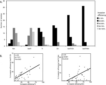
There was considerable heterogeneity in invasion phenotypes among the parasite isolates (Fig. 1A). Variation in use of the different invasion pathways was measured by calculating coefficients of variation – Nm (0·4), lowTr (0·33), and Ch (0·44) were less variable than Tr (0·74), lowTr/Ch (0·76) and lowTr/Nm (1·1 The mean percentage re-invasion into Tr-treated erythrocytes is higher in this study (28%) compared to other studies in West Africa (15% (Baum et al. Reference Baum, Pinder and Conway2003) and 2% (Jennings et al. Reference Jennings, Ahouidi, Zilversmit, Bei, Rayner, Sarr, Ndir, Wirth, Mboup and Duraisingh2007)) may represent natural variations between populations of parasites or alternatively be due to the use of different batches of Tr with variable specific activities. Spearman's rank correlation was used to determine associations between the use of different invasion pathways (Fig. 1B). Significant correlations were identified between Tr and lowTr/Ch (rho=0·63, P<0·001) and Nm and lowTr/Nm (rho=0·54, P=0·001), with weaker correlations between Tr and lowTr (rho=0·4, P=0·02) and Ch and lowTr/Ch (rho=0·39, P=0·02).
(A) Invasion phenotypes of Senegalese Plasmodium falciparum field isolates indicate that parasites routinely use alternative invasion pathways. Efficiency of invasion pathway usage by P. falciparum isolates, defined by treatment of erythrocytes with neuraminidase (Nm), low trypsin (lowTr), trypsin (Tr), chymotrypsin (Ch), lowTr/Ch or lowTr/Nm, to reveal alternative receptor repertoires. (B) Positive Spearmans Rank Correlations between Tr and lowTr/Ch (rho=0·63, P<0·001) and between Nm and lowTr/Nm (rho=0·54, P=0·001).

Parasite multiplication rate, selectivity and age
The parasite muliplication rate (PMR) and red blood cell selectivity have been proposed as determinants of the severity of malaria (Chotivanich et al. Reference Chotivanich, Udomsangpetch, Simpson, Newton, Pukrittayakamee, Looareesuwan and White2000). We hypothesized that this was due to altered repertoires of available erythrocyte receptors, and thus invasion pathways.
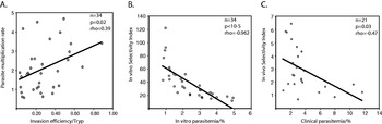
In vitro PMRs measured varied dramatically in the Senegalese isolates (0·08–4·92), with a median value of 1·5. Overall these values are comparable to those seen in uncultured subSaharan isolates, but low compared to those measured with Southeast Asian isolates (Chotivanich et al. Reference Chotivanich, Udomsangpetch, Simpson, Newton, Pukrittayakamee, Looareesuwan and White2000). Invasion assay data were obtained for those patients with PMR >1, due to inaccuracies in counting very low parasitaemias in enzyme-treated samples. We determined correlations between PMR and invasion pathways. The only correlation found was between PMR and invasion into Tr-treated cells (rho −0·350, P=0·02) (Fig. 2A). No invasion pathway was correlated with clinical parasitaemia.
Selectivity index (SI) was calculated for all 34 specimens according to the methods previously published (Chotivanich et al. Reference Chotivanich, Udomsangpetch, Simpson, Newton, Pukrittayakamee, Looareesuwan and White2000), using the control slides which measure invasion into untreated cells in vitro. The median SI for these 34 samples was 10·2 (range 2·5–175). SI was very strongly correlated with PMR (rho −0·962, P<0·01) (Fig. 2B). No significant correlations were found between the use of specific invasion pathways and in vitro SI values.
A positive correlation between in vivo SI, calculated using slides made using the clinical samples, and in vitro PMR has previously been found (Deans et al. Reference Deans, Lyke, Thera, Plowe, Kone, Doumbo, Kai, Marsh, Mackinnon, Raza and Rowe2006). Evaluable clinical slides were available for 23 of the 34 isolates, and these were also counted for selectivity index. The median clinical SI was 3·22 (range 0·98 to 11·7) (Fig. 2C). These values were very comparable to previously reported values from parasites of uncomplicated African isolates (Deans et al. Reference Deans, Lyke, Thera, Plowe, Kone, Doumbo, Kai, Marsh, Mackinnon, Raza and Rowe2006). In vivo SI values were much lower than in vitro SI values, presumably due to our use of static assays. There was no correlation between the clinical SI and the SI after one round of invasion in vitro or in vitro PMR.
Individuals may acquire immune responses against invasion ligands with age, and one could hypothesize the differential usage of invasion pathways in parasites obtained from patients with different age. No correlation was found between age and efficiency of invasion pathway usage, PMR, selectivity index or clinical parasitaemia.
(A) Parasite multiplication rate is significantly associated with the use of the trypsin-resistant invasion pathway (n=34, rho=0·39, P=0·02). (B) In vitro selectivity index is strongly correlated with in vitro parasitaemia (n=34, rho=−0·96, P<10–5). (C) In vivo selectivity index is strongly correlated with in vivo parasitaemia (n=21, rho=0·47 P=0·03).

The majority of Senegalese isolates express PfRh2b with a major sequence deletion
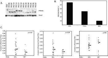
Expression of PfRh2b protein was evaluated by Western blot and confirmed PfRh2b protein expression in all samples (Fig. 3A). PCR typing of DNA from parasites revealed that 10 (29%) had full length PfRh2b, 22 (65%) had the PfRh2b deletion, and 2 (6%) were mixed. There was no correlation between the presence of the deletion and patient age, collection site, or clinical parasitaemia.
(A) PfRh2b is expressed is all Senegalese isolates. Western blot of PfRh2b protein and SERA-5 control in culture supernatants normalized for total protein content. Size differences result from a combination of the presence of repeat region size polymorphisms and PfRh2bDEL(Jennings et al. Reference Jennings, Ahouidi, Zilversmit, Bei, Rayner, Sarr, Ndir, Wirth, Mboup and Duraisingh2007). (B) Prevalence of the PfRh2bDEL polymorphism in the Senegalese isolates. (C) Distribution of invasion efficiencies in Tr and lowTr/Ch-treated erythrocytes in isolates with and without the PfRh2bDEL polymorphism. PfRh2bDEL is associated with enhanced invasion by erythrocyte receptor repertoires revealed by treatment with Tr (P=0·02, Mann-Whitney) and lowTr/Ch (P=0·08, Mann-Whitney), but not neuraminidase. Median values are indicated by the thick bar.

PfRh2bDEL is associated with altered invasion phenotype
We compared the invasion characteristics of our isolates based on the presence or absence of the PfRh2bDEL polymorphism (Fig. 3). Isolates with PfRh2bFULL had a median invasion efficiency of 14% in Tr-treated erythrocytes as compared with 27% among isolates with PfRh2bDEL (P=0·02, Mann-Whitney U). In lowTr/Ch-treated cells, median invasion was 12% for PfRh2bFULL and 18% for PfRh2bDEL (P=0·08, Mann-Whitney U). Invasion into Nm, lowTr/Nm, Ch, and lowTr-treated cells was not significantly associated with PfRh2b polymorphism.
Blood type of patient and associations between PfRh2bDEL and invasion pathways
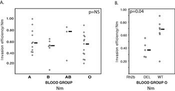
Among our samples, major blood group type, A, B or O, was not associated with any particular invasion pathway (Fig. 4A). Sialic acid-independent invasion was not associated with blood group B as previously reported (Deans et al. Reference Deans, Nery, Conway, Kai, Marsh and Rowe2007). Because our invasion assays were performed in type O cells, we re-examined the invasion assays looking only at isolates from type O patients. Among these isolates, those with PfRh2bDEL grew less efficiently in neuraminidase-treated cells when compared to PfRh2bFULL (P=0·04, Mann-Whitney U) (Fig. 4B), as well as more efficiently into lowTr/Ch-treated (P=0·03 Mann-Whitney U) and Tr-treated erythrocytes (P=0·08 Mann-Whitney U).
Invasion pathways and the ABO blood group system. (A) Sialic acid-independent invasion is not linked to blood group B in the Senegalese isolates. Median values are indicated by the thick bar. (B) Invasion by sialic acid-independent pathways (erythrocytes treated with neuraminidase) is significantly reduced in isolates with the PfRh2bDEL in patients from blood group O (P=0·04, Mann-Whitney U-test). Median values are indicated by the thick bar.

DISCUSSION
In this study we describe the erythrocyte invasion phenotypes in a large cohort of uncultured Plasmodium falciparum field isolates from a region of low transmission in Senegal. The use of alternative invasion pathways has been well described in laboratory isolates of P. falciparum (Hadley et al. Reference Hadley, Klotz, Pasvol, Haynes and McGinniss1987; Dolan et al. Reference Dolan, Miller and Wellems1990, Reference Dolan, Proctor, Alling, Okubo, Wellems and Miller1994). In recent years, several field studies have reported that the variation observed in laboratory isolates also exists in field isolates (Okoyeh et al. Reference Okoyeh, Pillai and Chitnis1999; Baum et al. Reference Baum, Pinder and Conway2003; Lobo et al. Reference Lobo, De Frazao, Rodriguez, Reid, Zalis and Lustigman2004; Bei et al. Reference Bei, Membi, Rayner, Mubi, Ngasala, Sultan, Premji and Duraisingh2007; Jennings et al. Reference Jennings, Ahouidi, Zilversmit, Bei, Rayner, Sarr, Ndir, Wirth, Mboup and Duraisingh2007). The isolates in this study had a nearly identical sialic acid-dependent invasion profile to cohorts previously reported from West Africa (Baum et al. Reference Baum, Pinder and Conway2003; Jennings et al. Reference Jennings, Ahouidi, Zilversmit, Bei, Rayner, Sarr, Ndir, Wirth, Mboup and Duraisingh2007), in contrast to isolates from Brazil and India, that are largely sialic acid-independent (Okoyeh et al. Reference Okoyeh, Pillai and Chitnis1999; Lobo et al. Reference Lobo, De Frazao, Rodriguez, Reid, Zalis and Lustigman2004). Most isolates in our study were strongly dependent on protease-sensitive receptors, which is consistent with other field studies (Baum et al. Reference Baum, Pinder and Conway2003; Lobo et al. Reference Lobo, De Frazao, Rodriguez, Reid, Zalis and Lustigman2004; Jennings et al. Reference Jennings, Ahouidi, Zilversmit, Bei, Rayner, Sarr, Ndir, Wirth, Mboup and Duraisingh2007), and in contrast to some laboratory isolates that exhibit invasion >50% into Tr-treated erythrocytes (Gaur et al. Reference Gaur, Storry, Reid, Barnwell and Miller2003). Laboratory studies indicate that the lowTr/Ch and Tr invasion pathways are distinct, with the former being defined by the PfRh2b ligand (Duraisingh et al. Reference Duraisingh, Triglia, Ralph, Rayner, Barnwell, McFadden and Cowman2003b). In contrast, in the Senegalese field isolates we find an association between these pathways, suggesting that both enzyme treatments reveal common erythrocyte receptors used by Senegalese parasites. The association between the use of Nm and Nm/lowTr invasion pathways is consistent with both treatments defining similar invasion pathways to those described for PfRh2b in laboratory lines (Duraisingh et al. Reference Duraisingh, Triglia, Ralph, Rayner, Barnwell, McFadden and Cowman2003b).
Parasite multiplication rate and selectivity index have been identified as possible determinants of virulence in one study in Thailand (Chotivanich et al. Reference Chotivanich, Udomsangpetch, Simpson, Newton, Pukrittayakamee, Looareesuwan and White2000), but not in another from Kenya (Deans et al. Reference Deans, Nery, Conway, Kai, Marsh and Rowe2007). In our study from Senegal we have used parasites from uncomplicated malaria patients, and find that they have much lower parasite multiplication rates as reported before for Kenyan isolates when compared to Southeast Asian isolates. We find an association, albeit weak, between enhanced invasion by a Tr-resistant invasion pathway and parasite multiplication rate in our Senegalese isolates, suggesting that the ligand-receptor interactions for this pathway may be a virulence determinant. No association was found between any invasion pathway and selectivity index.
We found that although all parasite lines express PfRh2b, approximately 2/3 of P. falciparum isolates collected in Senegal have a previously reported ~0·58 kb deletion in the PfRh2b gene, PfRh2bDEL. This polymorphism occurs in the only region of the PfRh2b gene that differs from the PfRh2a gene. Isolates with PfRh2bDEL achieved invasion parasitaemia nearly twice that of PfRh2bFULL when grown on Tr-treated and lowTr/Ch-treated erythrocytes. PfRh2bDEL parasites utilize an invasion pathway that resembles that of PfRh2b knockouts in laboratory lines, i.e. less reliance on the protease-sensitive PfRh2b receptor. Interestingly, PfRh2bDEL also appears to mirror the phenotype observed in Tanzanian isolates with reduced expression of PfRh2b, that of enhanced invasion into Tr- and Ch-treated erythrocytes (Bei et al. Reference Bei, Membi, Rayner, Mubi, Ngasala, Sultan, Premji and Duraisingh2007). PfRh2bDEL may either directly influence invasion pathway usage by abrogating PfRh2b function, or alternatively may be a marker of reduced PfRh2b function, for its presence may reflect parasites in hosts with prior immune responses against PfRh2b, and hence rely on other invasion ligands for invasion.
We were unable to reproduce in our Senegalese isolates a previous finding that invasion by sialic acid-independent receptors was significantly higher when isolates were drawn from patients with type B blood in Kenyan isolates, although we only had 7 isolates in this group (Deans et al. Reference Deans, Nery, Conway, Kai, Marsh and Rowe2007). Instead, when we examined isolates drawn from patients with type O blood, we found that those with PfRh2bDEL had significantly reduced invasion by sialic acid-independent invasion when compared with PfRh2bFULL-containing isolates, as well as enhanced invasion by protease-resistant receptors.
Why is the association of PfRh2b polymorphism with sialic acid-dependence observed in isolates from type O patients? It is possible that each major blood group selects for different sets of invasion ligands in vivo. This certainly merits follow-up in larger studies due to the association between malaria disease severity and ABO blood group (Cserti and Dzik, Reference Cserti and Dzik2007). Blood group O is the most prevalent blood group in Senegal (~52%) and may therefore provide the strongest selective force on Senegalese isolates. The receptor for PfRh2b is unknown, but two possible candidates are band 3 and glycophorin B (Duraisingh et al. Reference Duraisingh, Triglia, Ralph, Rayner, Barnwell, McFadden and Cowman2003b). Interestingly, the vast majority of ABO blood group determinants are bound to band 3, an abundant erythrocyte-membrane protein that is cleaved by both trypsin and chymotrypsin (Jay, Reference Jay1986).
Numerous erythrocyte mutations have been identified that are thought to confer protection against clinical malaria, and it will be of interest to study their effects on parasite invasion in further studies. These include deficiency of glycophorin C and polymorphisms in band 3 (Southeast Asian Ovalocytosis) (Kwiatkowski and Luoni, Reference Kwiatkowski and Luoni2006). Much remains to be learned about the receptor-ligand interactions involved in erythrocyte invasion. It is clear, however, that in Africa P. falciparum has evolved to become an extremely effective human pathogen, and malaria in turn has been a powerful influence on human evolution. A greater understanding of both parasite and host polymorphisms in endemic settings will shed further light on the biologically important interactions between parasite and host.
We thank D. Ndiaye, Y. Diedhiou, L. Ndiaye O. Ly and A. Mbaye for collecting samples, and the patients of Thies and Pikine for their support of this project. This work was supported by an American Society of Tropical Medicine and Hygiene/Burroughs-Wellcome Fund Fellowship to P.L., National Institute of Allergy and Infectious Diseases (R01 AI057919) supports M. T. D. and a Fogarty International Training Grant (5D43TW001503) to D. F. W.