Introduction
Melioidosis is a life-threatening infectious disease caused by the bacterium Burkholderia pseudomallei. Infection may be asymptomatic, or various clinical manifestations may be apparent including multiple localised abscesses, chronic infection and septic shock. The infectious agent is found in the soil and water of endemic areas.
The highest prevalence of this disease is in tropical areas, including Southeast Asia (especially Thailand, Singapore and Malaysia) and northern Australia. In Thailand, melioidosis is most frequently reported from the north-eastern region, where B pseudomallei is responsible for 20 per cent of all community-acquired bacteraemia and causes death in 40 per cent of treated patients.Reference Chaowagul, White, Dance, Wattanagoon, Naigowit and Davis1 Other epidemiological studies have reported an annual incidence of 4.4 cases per 100 000, in Ubon Ratchathani Province,Reference Suputtamongkol, Hall, Dance, Chaowagul, Rajchanuvong and Smith2 and an average annual prevalence of 9.97 per 100 000, in Nakhon Phanom Province.Reference Tanwisaid and Surin3 These two provinces are located in north-eastern Thailand, bordering Laos.
The antibiotic regimen for melioidosis differs from that for other common bacterial infections in the head and neck. Therefore, awareness of the possibility of melioidosis and of its clinical manifestations is necessary for early diagnosis in order to avoid failure of medication therapy which may lead to high morbidity and mortality.
Because melioidosis in the head and neck is uncommon, there is little information on the clinical manifestations. A literature review identified few reports. Lim et al. reported four cases of melioidosis presenting with parotid abscess, acute sinusitis, acute suppurative lymphadenitis and chronic suppurative otitis media.Reference Lim, Gurdeep and Norain4 Another study reported a case with strange and unusual melioidosis in the head and neck.Reference Loh, Latis, Crossland and Patel5 Therefore, we developed this study to present the clinical manifestations of melioidosis of the head and neck for improved diagnosis and management.
Materials and methods
Patients admitted to Mukdahan Hospital, Thailand, with B pseudomallei infections in the head and neck between 1 January 2013 and 31 October 2016 were reviewed. Patients’ medical records provided data on demographics, clinical presentation, underlying disease, screening laboratory investigations, treatment and clinical outcomes.
Acute melioidosis was defined as an infection of 14 days' duration or less, whereas chronic melioidosis was a long-standing infection of 60 days or more. Subacute melioidosis was an infection of 14–60 days' duration. Complete remission was defined as no melioidosis episode occurring within five years after completing therapy.
The study was approved by the Mukdahan Ethics Committee for Human Research (MEC007/59). In addition, this study was registered with the ClinicalTrial.gov (NCT03048513).
Results
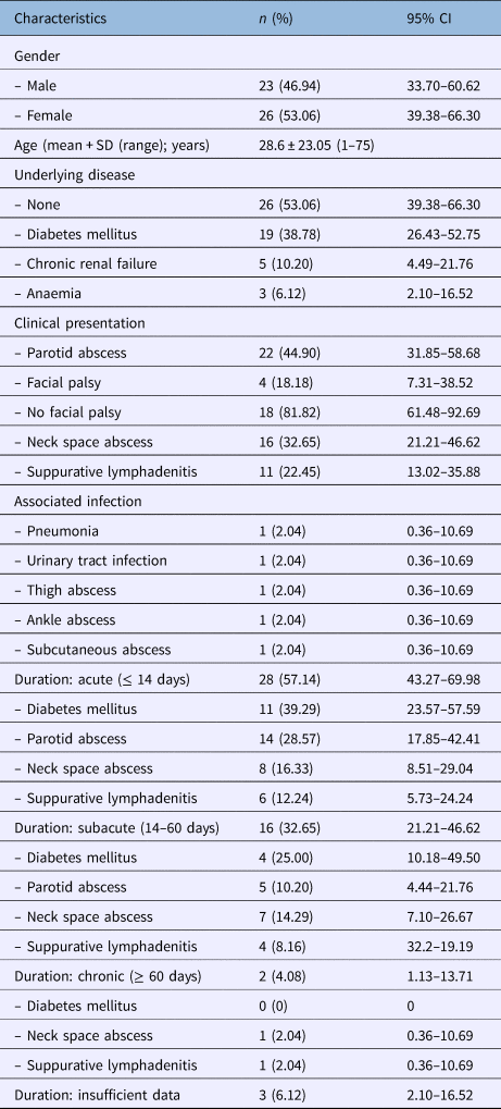
The medical records of 49 patients, 23 males and 26 females, were analysed. The mean age was 28.6 years (range, 1–75 years). There was no obviously different infection rate between immunocompetent and immunocompromised patients. Clinical presentations included 22 parotid abscesses, 16 neck abscesses and 11 cases of suppurative lymphadenitis. Systemic melioidosis infection was also observed that involved multiple organs including lung, urinary tract, thigh, ankle and subcutaneous tissue.
Forty-three cases with sufficient data were classified by infection duration. Most presentations (57.14 per cent) had symptoms with acute infection duration. Parotid abscess (28.57 per cent) was the most common clinical presentation in acute melioidosis cases, whereas neck space abscess (14.29 per cent) was the most common in subacute melioidosis cases. There were only two cases (neck space abscess and lymphadenitis) with chronic duration (Table 1).
Demographic data

CI = confidence interval; SD = standard deviation
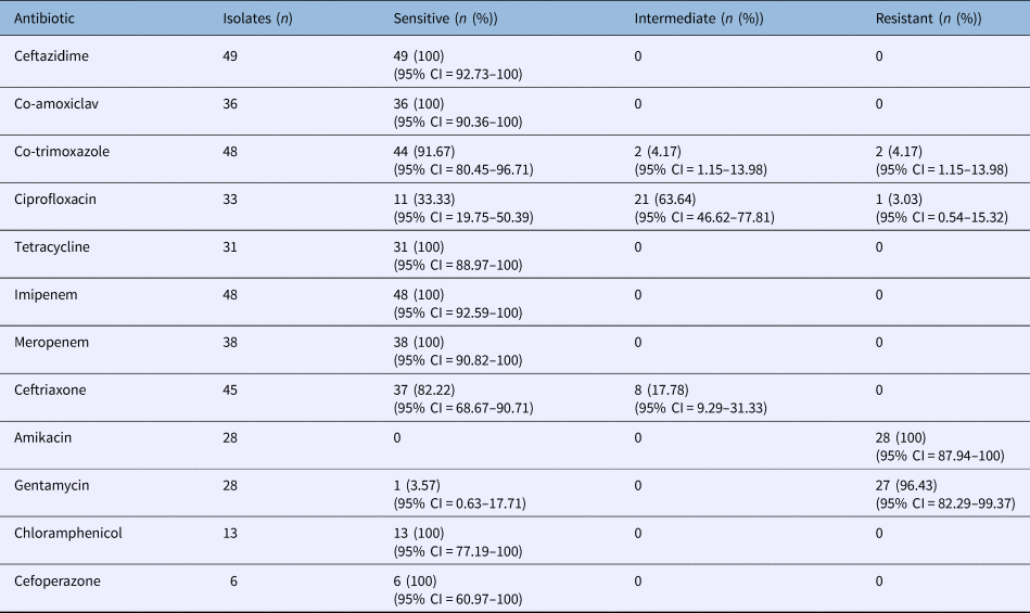
All cases required surgical drainage. B pseudomallei was isolated from pus cultures in all specimens, but only 10 of 14 patients (71.43 per cent; 95 per cent confidence interval (CI) = 45.35–88.28) had high indirect haemagglutination assay results of ≥ 1:160. Antimicrobial susceptibility testing results are described in Table 2.
Antimicrobial susceptibility of Burkholderia pseudomallei isolates*

*Tested at Mukdahan Hospital, Thailand. CI = confidence interval
Various antimicrobial regimens were prescribed. However, almost half of our patients received intravenous ceftazidime, with a subsequent switch to oral co-trimoxazole. Mean treatment duration was 11.11 ± 6.89 days in the initial phase and 122.58 ± 46.78 days in the eradication phase. Forty-four of 46 patients remained in remission without relapse (95.65 per cent; 95 per cent CI = 85.47–98.80) (Table 3).
Antibiotic regimens during initial acute and eradication phases

SD = standard deviation; CI = confidence interval
Discussion
Melioidosis can manifest in any part of the body, but there have not been many reports concerning the head and neck region. A literature review yields only ‘strange’ clinical manifestations in this region of the body. Loh et al. reported a patient who presented with right-sided facial soft tissue infection, mastoid effusion and temporal lobe cerebritis.Reference Loh, Latis, Crossland and Patel5 Another study reported chronic rhinosinusitis secondary to melioidosis.Reference Phillips, Cervin, Earnshaw and Sidjabat6 The usual clinical manifestations of melioidosis can vary, but fall roughly into four classes: asymptomatic carrier in the latent period, prolonged fever without any apparent site of infection, localised infection, and fulminant septicaemia.Reference Lim, Gurdeep and Norain4
In our study, almost all patients had localised infection in the head and neck region. Only two cases had bacteraemia involving other organs. The parotid was the most common site of melioidosis in the head and neck. Four cases with parotid abscesses developed facial nerve palsy. The average infection duration was 17 days. In the parotid abscess cases, two case had multiple-site involvement, with conditions including pneumonia, subcutaneous abscess, liver abscess and urinary tract infection. After treatment, three facial nerve palsy cases patients recovered well, but did not gain normal function of the facial nerve. Unfortunately, another patient with underlying poorly controlled diabetes mellitus developed fatal septic shock due to multiple organ involvement. Other common melioidosis manifestations included 16 neck space abscesses (cheek, submandibular, carotid and retropharynx) and 11 cases of cervical suppurative lymphadenitis.
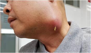
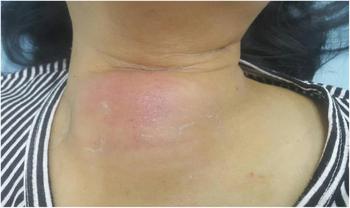
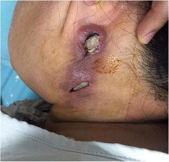
Manifestations in the head and neck can be diagnostically challenging because of variations in infection duration, ranging from acute to chronic infection (range, 3–90 days). Regarding acute infection, patients often present with rapidly progressive inflammation and abscess formation. In our series, acute abscesses were observed in 14 parotid glands (28.57 per cent) and 8 neck spaces (16.33 per cent), with 6 cases of suppurative cervical lymphadenitis (12.24 per cent) (Figure 1). However, abscesses can present with slowly progressive symptoms that may confound the diagnosis, or mimic another subacute or chronic disease. We found subacute abscesses in seven neck spaces (14.29 per cent) (Figure 2) and five parotid glands (10.20 per cent), with four cases of suppurative cervical lymphadenitis (8.16 per cent). Only two cases presented with a chronic neck abscess and chronic suppurative cervical lymphadenitis (Figure 3). Infection duration may depend on host immunity. Eleven of 28 of acute melioidosis patients presented with diabetes mellitus, whereas 4 of 16 of subacute melioidosis patients had diabetes mellitus (Table 1). Thus, host immunity appears to be an important factor for disease progression.
Acute suppurative submandibular lymphadenitis.

Subacute melioidosis abscess in anterior cervical space.

Chronic melioidosis abscess after drainage with tissue necrosis in left post-auricular lymph node and upper jugular lymph node.

Microbiological culture is accepted as the ‘gold standard’ for a melioidosis diagnosis, but it might take several days for the results.Reference Limmathurotsakul, Jamsen, Arayawichanont, Simpson, White and Lee7 Thus, serology tests have been developed to complement direct pathogen detection. The indirect haemagglutination assay is a well-known serodiagnostic test for melioidosis.Reference Wiersinga, Currie and Peacock8–Reference Suttisunhakul, Chantratita, Wikraiphat, Wuthiekanun, Douglas and Day10 In endemic areas, a suitable cut-off titre for the indirect haemagglutination assay is ≥ 1:160. This titre provided the best diagnostic values, with sensitivity of 70 per cent, specificity of 67 per cent, a positive predictive value of 80 per cent and a negative predictive value of 55 per cent.Reference Teparrugkul11 In our study, 10 of 14 melioidosis patients presented with an indirect haemagglutination assay titre of ≥ 1:160. A low titre was observed in four patients with a positive melioidosis culture. Therefore, interpretation of indirect haemagglutination assay titre requires caution and awareness of the possibility of false negatives.
Recently, protein microarrays have been introduced to investigate melioidosis. Kohler et al. reported that this approach provides a high specificity, of 97 per cent, when distinguishing between sera from melioidosis patients and normal controls.Reference Kohler, Dunachie, Müller, Kohler, Jenjaroen and Teparrukkul12 In addition, the array allowed a higher sensitivity than the indirect haemagglutination assay in melioidosis patients (cut-off indirect haemagglutination assay titre ≥ 1:160: indirect haemagglutination assay 57.3 per cent, protein array: 86.7 per cent; p = 0.0001). However, further multicentre studies are needed to determine the true sensitivity and specificity of the protein array.
Melioidosis treatment can be divided into two phases: the initial acute phase and the eradication phase.Reference Dance13 During the initial phase, an antibiotic is prescribed to prevent complications and moribundity. Surgical drainage is reserved for patients who present with macro abscesses. Ceftazidime is still recommended because of its high efficacy against B pseudomallei. Dutta et al. reported that all of 20 isolates of B pseudomallei were uniformly sensitive (100 per cent) to ceftazidime.Reference Dutta, Haq, Hasan and Haq14 This is in line with our findings. However, Ahmad et al. found that one isolate of B pseudomallei (0.6 per cent) was resistant to ceftazidime.Reference Ahmad, Hashim and Mohd Noor15 Alternative antibiotics are reserved for severe infection or resistance to first-line antibiotics. Imipenem or meropenem are recommended as second-line drugs, which were 100 per cent effective in our study.
The eradication phase aims to reduce the melioidosis relapse rate. Co-trimoxazole monotherapy is a treatment of choice in this phase.Reference Fisher and Harris16 However, B pseudomallei showed intermediate resistance to co-trimoxazole in two cases and showed complete resistance in two cases. In such cases, co-amoxiclav is the preferred second-line choice.Reference Cheng, Chierakul, Chaowagul, Chetchotisakd, Limmathurotsakul and Dance17 Dutta et al. found that B pseudomallei isolates were 100 per cent sensitive to co-amoxiclav,Reference Dutta, Haq, Hasan and Haq14 as we also found. Other antimicrobial-resistant isolates were observed in our study (3.03 per cent were resistant to ciprofloxacin, 100 per cent to amikacin and 96.43 per cent to gentamycin). Thus, the antibiotic susceptibility pattern should be recognised, for proper treatment.
Treatment duration depends on clinical severity. Initial-phase treatment usually continues for more than 10 days.Reference Dance13 Eradication treatment should be given for 12–20 weeks, depending on clinical progression.Reference Limmathurotsakul, Chaowagul, Chierakul, Stepniewska, Maharjan and Wuthiekanun18 Our patients were administered ceftazidime in the initial phase for about 8–11 days. Eradication therapy varied in duration according to the antibiotic regimen employed. For co-trimoxazole alone, eradication therapy continued for a mean of 123 days, for co-trimoxazole combined with doxycycline the mean duration was 54 days, and for co-amoxiclav it was 98 days.
Oral therapy alone has been reported,Reference Limmathurotsakul, Chaowagul, Chierakul, Stepniewska, Maharjan and Wuthiekanun18 but it is unclear whether this provides adequate treatment.Reference Dance13 In our study, 13 patients with mild localised melioidosis were prescribed only oral regimens; 5 received co-trimoxazole, and 8 received co-trimoxazole combined with doxycycline. Treatment duration was around 144 days for co-trimoxazole, and 75 days for co-trimoxazole combined with doxycycline.
The outcome of the regimens employed seems excellent. All patients were in remission. However, there are limitations to our study. The retrospective nature of the study and the small sample size may have influenced analysis and conclusions. Further studies are required for clarity.
• Clinical manifestations of melioidosis may vary
• The parotid gland was a common site of melioidosis infection in the head and neck
• In endemic areas, a suitable cut-off titre of indirect haemagglutination assay is ≥ 1:160 for melioidosis diagnosis
• Indirect haemagglutination assay titre interpretation requires caution and awareness of possibility of false negatives
• Four patients with positive B pseudomallei culture had a low titre
• Intravenous ceftazidime and subsequent oral co-trimoxazole is the mainstay of treatment, but mild localised melioidosis may be treated with oral regimens only
Conclusion
The clinical symptoms of melioidosis can vary; thus, careful clinical correlation and proper investigation are required to establish early diagnosis. Furthermore, antimicrobial susceptibility should be considered when deciding on the treatment regimen.
Acknowledgement
The authors thank Prof David Blair (under the aegis of the Publication Clinic Research Affairs, Faculty of Medicine, Khon Kaen University), for assistance with the English-language presentation.
Competing interests
None declared