There has been significant disagreement recently in the pages of the journal concerning the definition and meaning of aortic overriding.Reference Anderson 1 , Reference Bozok, Kestelli and Akyuz 2 In the work that engendered the recent exchange, the authors wrote “dextroposition of the aorta occurs when the aorta overrides the interventricular septum to the right ventricular side”.Reference Bozok, Kestelli and Ilhan 3 In their discussion, however, they then stated “overriding of the aorta over the right ventricle, which occurs in the normal heart, should not be confused with dextroposition”. This begs the question as to whether the aorta does override the right ventricle in the normal heart? Part of the right coronary aortic valvar sinus is certainly positioned cranial to the crest of the muscular ventricular septum in the normal heart. However, the essence of normality is that the entirety of the circumference of the aortic valvar leaflets is hinged within the left ventricle. If aortic overriding is defined on the basis of support of the aortic valvar leaflets within the right ventricle, in other words, a biventricular connection for the aortic valve, then it is obvious that such overriding cannot exist when the ventricular septum is intact. Defining overriding on the basis of aortic biventricular connection removes the need to seek to distinguish between “true” and “false” dextroposition. This is more important, as the rightward location of the aorta in the setting of transposition would not be considered as “dextroposition” if we followed the suggestion of our Turkish colleagues.Reference Bozok, Kestelli and Ilhan 3 Furthermore, in response to the letter questioning the value of “dextroposition”, the Turkish authors opined that, as cardiac surgeons, they required “the necessity to clearly define dextroposition of aorta”.Reference Bozok, Kestelli and Akyuz 2 Unfortunately, the required clear definition was not provided in their response.Reference Bozok, Kestelli and Akyuz 2 An added value of using the concept of overriding according to the biventricular connection of the aortic valve is that, as we will show, it now provides a reproducible means of distinguishing between double outlet and concordant ventriculo-arterial connections, another topic of potential contention.Reference Restivo, Unolt, Putotto and Marino 4 , Reference Anderson 5 When discussing the potential distinction of “true” as opposed to “false” dextroposition, the Turkish authors had also suggested that “new diagnostic modalities” are required for “proper planning of treatment”.Reference Bozok, Kestelli and Ilhan 3 In this review, we will show that such modalities already exist. We submit that they serve to validate the use of the degree of aortic overriding, rather than “dextroposition”, to inform the required decision making.
Does the normal aortic valve override the ventricular septum?
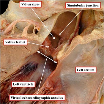
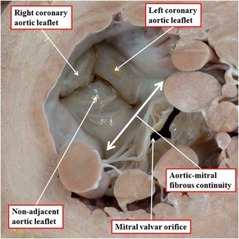
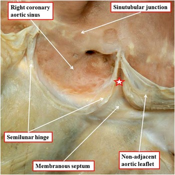
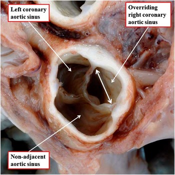
The aortic valve, in the normal heart, guards the outflow tract from the left ventricle. Part of the essence of such normality is the integrity of the ventricular septum. Understanding why some might consider overriding to exist in the normal heartReference Bozok, Kestelli and Akyuz 2 requires an appreciation of the components of the aortic valve, specifically those parts considered to be positioned above the ventricular septum. The normal aortic valve is made up of leaflets, valvar sinuses, and supporting left ventricular structures (Fig 1). Much has been written concerning the “annulus” of the aortic valve, this being another area of potential confusion.Reference Anderson 6 There is no ring-like structure that supports the leaflets of the valve. The so-called “annulus”, as measured by clinicians, is no more than the diameter of the entrance to the aortic root (Fig 1). It is usually measured from the opposite basal hinges of the valvar leaflets.Reference Anderson 6 In the normal situation, these hinges are exclusively left ventricular (Fig 2). Thus, there is no overriding of the aortic valvar orifice in the normal heart. Some surgeons, in contrast, take the overall attachment of the semilunar hinges of the valvar leaflets to represent the “annulus”.Reference Sievers, Hemmer and Beyersdorf 7 These attachments extend distally to the level of the sinutubular junction, with each leaflet supported within its own valvar sinus of Valsalva (Fig 3). It is part of the right coronary aortic sinus that, in the normal heart, is positioned cranial to the crest of the muscular ventricular septum (Fig 4). This relationship can be exaggerated in the setting of the “sigmoid septum”. Even in this setting, however, it is the cavity of the sinus that overrides, rather than the aortic valvar orifice (Fig 4). The valvar orifice remains committed to the left ventricle even in the setting of the sigmoid septum – otherwise it would not be possible for the ventricular septum to be intact. It follows that the essence of normality is univentricular connection of the aortic valve. To emphasise again, when defined on the basis of its connections within the cardiac base, there is no overriding of the aortic valve in the normal heart.
The image shows the aortic root as seen in the parasternal echocardiographic long axis section. The root extends from the plane produced by joining together the proximal attachments of the valvar leaflets, the so-called echocardiographic annulus, to the sinutubular junction.

The image shows the ventricular aspect of the aortic root as viewed in short axis from the apex of the left ventricle. The leaflets of the aortic valve are supported exclusively within the cavity of the left ventricle. Therefore, there is no overriding of the aortic valvar orifice relative to the ventricular septum.

The right aortic leaflet of the aortic valve has been cut back to its semilunar hinge, which supports it within the right coronary aortic sinus. As can be seen, the semilunar line of attachment extends proximally from an attachment to the musculature of the ventricular septum to a distal attachment at the sinutubular junction. The triangle between the right coronary and non-adjacent sinuses is filled by the membranous septum and the fibrous interleaflet triangle (star). Some surgeons identify these semilunar lines of attachment of the three leaflets as the valvar annulus.

The aortic root has been transected at the level of the sinutubular junction, and is viewed from the arterial aspect. The double-headed arrow shows the plane of attachment of the right coronary aortic leaflet relative to the muscular ventricular septum. The leaflet is attached exclusively within the left ventricle. However, the cavity of the valvar sinus does override the plane of the muscular septum. This does not mean that the valvar orifice itself is overriding (see Fig 2).

How does the aortic valve achieve its position within the left ventricle?
With the development of episcopic microscopy,Reference Mohun and Weninger 8 knowledge of the precise changes occurring during cardiac development now permits significant inferences to be made regarding the biventricular, as opposed to univentricular, connections of the aortic valve. The observed changes are also pertinent to the definition of the channels that permit interventricular shunting.Reference Bailliard, Spicer, Mohun, Henry and Anderson 9 In their presentation, the Turkish authors commented “the spatial position of the aortic root can only be the result of a true anomaly developed during embryogenesis”.Reference Bozok, Kestelli and Ilhan 3 This statement is not true. The spatial location of the aortic root can be altered simply in consequence of an abnormal rotation of the heart itself. This is yet another reason why it is better to consider the abnormal situations in terms of overriding defined on the basis of a biventricular connection of the aortic root. Nonetheless, if we consider the comment of the Turkish authorsReference Bozok, Kestelli and Ilhan 3 relative to the ventricular support provided for the developing aortic root, their statement does remain valid. Using the episcopic data sets prepared from the developing mouse, we are now able to demonstrate precisely how the leaflets of the aortic valve achieve their exclusively left ventricular support.
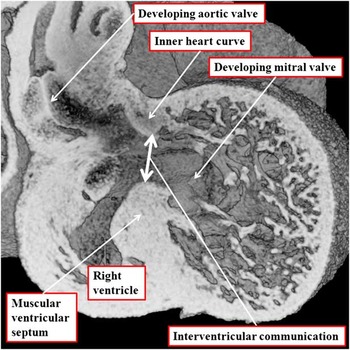
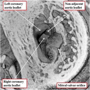
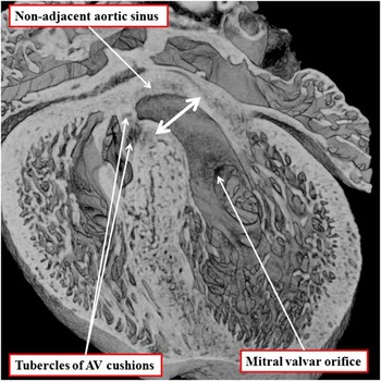
When the proximal part of the developing cardiac outflow tract becomes separated into the components destined to form the outlets of the right and left ventricles, the entire circumference of the outflow tract, which includes the developing aortic root, is supported exclusively above the cavity of the developing right ventricle (Fig 5). This means that the blood ejected by the left ventricle must exit through the embryonic interventricular communication, which is roofed by the inner heart curvature, so as to reach the developing aortic outflow channel (Fig 5). The arch produced by fusion of the proximal outflow cushions, when first formed, is also positioned exclusively above the cavity of the right ventricle. It is during the 12th and 13th days of development in the mouse heart, equivalent to the 7th week of development in the human heart, that the developing aortic root is transferred to take origin from the left ventricle. As the transfer takes place, the arch provided by the fused proximal cushions is brought in line with the apical muscular ventricular septum. This process then realigns the plane of the embryonic interventricular communication such that it ceases to be interventricular. Instead, it becomes the exit from the left ventricle. Then, during the 13th day of development in the mouse, when the developing aortic valvar leaflets are all positioned above the cavity of the left ventricle (Fig 6), the rightward tubercles of the atrioventricular endocardial cushions close the persisting communication between the root and the cavity of the right ventricle, thus forming the primordium of the membranous part of the ventricular septum (Fig 7).
The image is a coronal section through an episcopic data set prepared from the developing heart of the mouse at embryonic day (E) 12.5. The developing aortic root at this stage is supported exclusively by the developing right ventricle. The left ventricular blood has to traverse the interventricular communication so as to enter the subaortic outlet. Note that the interventricular communication extends between the crest of the muscular ventricular septum and the inner heart curvature, the latter representing the tissues between the developing leaflets of the aortic and mitral valves.

The image, from an episcopic data set of the developing murine heart at E13.5, shows that the circumference of the aortic root is now supported exclusively by the walls of the left ventricle (compared with Fig 2).

The coronal section through an episcopic data set from a mouse heart at E14.5 shows how the rightward tubercles of the AV cushions have walled the aortic root into the left ventricle. The plane that initially represented the embryonic interventricular communication (double-headed white arrow – see Fig 5) has become realigned to form the border between the aortic root and the remainder of the left ventricle. The tubercles of the cushions will eventually become the membranous part of the ventricular septum. AV=atrioventricular.

During the intermediate stages of this transfer, it is possible to distinguish several planes of space subtended beneath the developing root. These images show how the plane first seen as the embryonic interventricular communication (Fig 5) is in the process of reorientation to mark the boundary between the initial cavity of the left ventricle and the aortic root (Fig 8). The tubercles of the atrioventricular cushions, which serve to complete the process of ventricular septation, close the communication between the root and the cavity of the right ventricle. However, during the intermediate stage of this process of transfer, the geometric interventricular communication is represented by the cranial continuation of the long axis of the muscular ventricular septum. This plane bisects the overriding root (Fig 8). Recognition of these planes as seen in the developing heart provides important inferences when considering the naming of the channels that permit interventricular shunting in the setting of deficient ventricular septation, particularly when associated with overriding of the aortic root.Reference Bailliard, Spicer, Mohun, Henry and Anderson 9
The image is a coronal section from an episcopic data set prepared from a mouse heart at E13.5. The aortic root is in the process of being transferred from the right to the LV. At this stage, its rightward border remains supported by right ventricular structures, but its leftward border is now supported in the roof of the LV. The geometric interventricular communication is now the cranial continuation of the long axis of the muscular ventricular septum, which transects the roof of the root. The plane representing the initial embryonic interventricular communication (see Fig 5) is becoming transformed into the outlet for the LV, whereas the remaining hole in the rightward margin of the root will be eventually closed by the rightward tubercles of the atrioventricular cushions (see Fig 7). LV=left ventricle.

When does aortic overriding exist in the congenitally malformed heart?
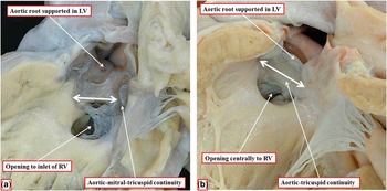
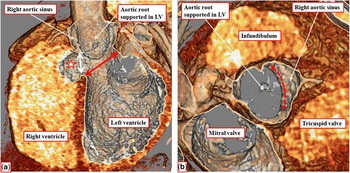
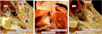
As we have shown, it is only the right aortic valvar sinus that overrides the crest of the muscular ventricular septum when the heart is normally septated at ventricular level (Fig 4). This is because the leaflets of the valve are attached exclusively within the left ventricle. Therefore, it follows that overriding of the aortic valvar orifice can only be found when ventricular septation itself is deficient. However, it is only when the root is positioned directly above a hole between the ventricles that the aortic valve has biventricular connections. Small defects within the apical muscular part of the septum are the most common examples of holes between the ventricles. These defects do not disturb the exclusive left ventricular support of the aortic valvar leaflets at the ventricular base. It is in the setting of the so-called membranous defects, or when the defect is doubly committed, that the potential is provided for aortic valvar overriding. As long ago as the early part of the 19th century, Peacock pointed out that the so-called membranous defect was the consequence of deficiency of the muscular septum that formed the borders of the hole between the ventricles.Reference Peacock 10 This concept was subsequently endorsed by Becu et al,Reference Becu, Fontana and Dushane 11 and by Sherman.Reference Sherman 12 The concept, in turn, also formed the basis for describing the defects as being perimembranous, rather than membranous.Reference Soto, Becker, Moulaert, Lie and Anderson 13 Depending on which part of the muscular septum is deficient, the defects that exist adjacent to the membranous part of the septum can open to different parts of the right ventricle. When the defects open to the right ventricular inlet, or open centrally into the right ventricle, the orifice of the aortic root remains committed exclusively to the cavity of the left ventricle. This is because the plane of the muscular outlet septum, supporting the free-standing subpulmonary infundibular sleeve, remains aligned with the plane of the apical muscular ventricular septum (Figs 9 and 10).
The images show the LV aspect of perimembranous defects opening into the inlet ( a ) and the central parts ( b ) of the RV. Both defects are present because of deficient musculature of the apical septum, with fibrous continuity postero-inferiorly between the leaflets of the aortic, mitral, and tricuspid valve in the defect opening to the inlet, and between the leaflets of the aortic and tricuspid valves in the defect opening centrally. Note that, although part of the aortic root is now supported by the borders of the RV in the roof of the defect, the plane of the muscular outlet septum (double-headed white arrow) remains aligned with the apical ventricular septum. It is an interventricular structure, and therefore there is no overriding of the aortic root. LV=left ventricle; RV=right ventricle.

The images are taken from a CT angiogram prepared from a patient with a perimembranous ventricular septal defect (double-headed white arrow) opening centrally to the right ventricle that has been closed spontaneously by a tricuspid valve pouch (white star). ( a ) showing the long axis of the ventricular septum, reveals how the aortic root remains committed exclusively to the LV, with the initial embryonic interventricular communication (double-headed red arrow) reorientated to form the path between the LV and the root. Although the aortic root is supported exclusively by the LV, the short-axis section, shown in ( b ), shows how the right coronary aortic sinus overrode the crest of the muscular ventricular septum (dashed double-headed red arrow). This should be compared with the situation in the normal heart (Fig 4). LV=left ventricle.

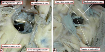
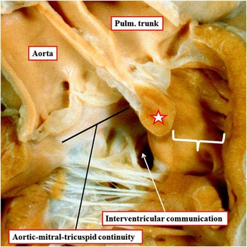
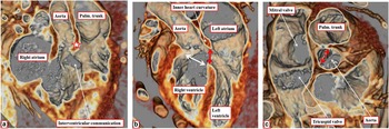
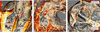
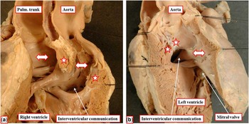
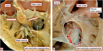
In contrast, when defects open to the outlet of the right ventricle, there is usually malalignment of the muscular outlet septum. When malaligned in cranial direction, it becomes an exclusively right ventricular structure (Fig 11a). In this setting, a proportion of the circumference of the aortic root, along with the valvar leaflets guarding that part of the aortic root, is then supported by right ventricular, rather than left ventricular, structures (Fig 11b). The defect associated with such malalignment was emphasised initially by Eisenmenger,Reference Eisenmenger 14 and is now widely known as the Eisenmenger defect.Reference Oppenheimer-Dekker, Gittenberger-de Groot, Bartelings, Moene and van der Harten 15 In the past, before widespread therapeutic closure, this type of defect promoted the early development of pulmonary hypertension. This is because, despite the malalignment of the muscular outlet septum, there is no obstruction of flow into the subpulmonary outflow tract. Nonetheless, the phenotypic feature of the defect is the presence of fibrous continuity postero-inferiorly between the leaflets of the aortic and tricuspid valves. Owing of this feature, the atrioventricular conduction axis is at risk during surgical correction of such defects, as it is in all other perimembranous defects.Reference Soto, Becker, Moulaert, Lie and Anderson 13 However, the precise degree of override of the aortic root can vary markedly in the setting of such defects. In most instances, the greater part of the aortic root is usually supported by the left ventricle (Fig 11). In other examples, the root can be supported in more or less equal parts by the two ventricles (Fig 12). This arrangement would still be diagnosed in terms of concordant ventriculo-arterial connections. If the override, and hence the commitment of the aortic root, is such that more than half of the circumference is supported within the right ventricle, however, then the ventriculo-arterial connection becomes one of double outlet (Fig 13). In the heart illustrated, there is not only fibrous continuity between the leaflets of the aortic and tricuspid valve, and also between the leaflets of the aortic and mitral valves in the roof of the defect. The ventriculo-arterial connection, nonetheless, is double outlet from the right ventricle (Fig 13). Similar hearts, of course, can also be found with completely muscular bilateral infundibulums (Fig 14). In the presence of aortic-mitral continuity, nonetheless, and with the interventricular communication in subaortic location, the hearts represent the endpoint of the spectrum of commitment of the overriding aortic root to the right ventricle in the setting of a perimembranous defect, but in the absence of subpulmonary obstruction, in other words the Eisenmenger defect. In all these settings, as in the developing heart, when the aortic root is supported predominantly or exclusively by the right ventricle, the hole between the ventricles is better described as an interventricular communication. This is because the plane of putative ventricular septation, roofed by the malaligned outlet septum, is within the cavity of the right ventricle (Fig 14). Indeed, whenever the aortic root is overriding, this plane is within the cavity of the right ventricle (Figs 12–14). Moreover, it is this plane of deficient septation, rather than the geometric interventricular communication, which will be closed by the surgeon so as to reconnect the aortic root with the left ventricle.
The images show a perimembranous defect that opens to the outlet of the RV. As shown in ( a ), which shows the left ventricular aspect, there is now malalignment between the muscular outlet septum and the apical ventricular septum, and the aortic root overrides the crest of the muscular septum, being supported partly by the RV structures. This is well seen in ( b ), which shows the RV aspect. The muscular outlet septum (double-headed white arrow) is located exclusively within the RV. The parts of the aortic valve supported above the cavity of the RV are evident. However, less than half of the root is supported within the RV, and thus the ventriculo-arterial connections remain concordant. RV=right ventricle.

The images are from a CT data set prepared from a patient with an Eisenmenger ventricular septal defect. ( a ) shows the “en-face” view of the ventricular septum, revealing the plane of the interventricular communication, and showing the antero-cephalad deviation of the muscular outlet septum. ( b ) shows a “four-chamber” section, revealing the overriding of the aortic root, and showing the planes of space subtended beneath the aortic root. The short-axis cut ( c ) shows that the aortic valve is equally supported by the right and left ventricles, and therefore that the ventriculo-arterial connections remain concordant. Pulm.=pulmonary.

The image shows a close-up of the outflow tracts of a heart with double outlet from the right ventricle. The aortic root arises almost exclusively from the right ventricle, but with fibrous continuity between the leaflets of the aortic, mitral, and tricuspid valves. The hole between the ventricles is now an interventricular communication, rather than a “ventricular septal defect”. As can be seen, the outlet septum (star) is exclusively a right ventricular structure. Hence, the plane of putative ventricular septation, in other words the ventricular septal defect, is within the cavity of the right ventricle. Note the unobstructed subpulmonary outlet (white bracket). Plum.=pulmonary.

The images, from another CT angiogram, are this time from a patient with double outlet right ventricular and subaortic interventricular communication. ( a ) shows the origin of both arterial trunks from the right ventricle, with the attachment of the muscular outlet septum (white star) committing the hole between the ventricles to the aortic root. As shown in ( b ), this hole (double-headed red arrow) is the interventricular communication, as its roof is the inner heart curvature. The plane to the underside of the outlet septum (double-headed white arrow) is exclusively within the cavity of the right ventricle. ( c ), taken in the short axis, shows that, although the aortic root overrides the crest of the muscular ventricular septum (dashed double-headed red arrow), its larger part is supported above the right ventricle. Plum.=pulmonary.

What about tetralogy of Fallot?
Biventricular connection of the aortic valve is an integral part of the Eisenmenger defect. It is much more frequently encountered as part of the tetralogy of Fallot. Indeed, it was the setting of tetralogy that promoted the Turkish surgeons to discuss the potential “true” and “false” settings for aortic dextroposition.Reference Bozok, Kestelli and Ilhan 3 When considered in terms of aortic overriding, or biventricular connection of the aortic root, there is no need to indulge in such linguistic sophistry. As was recognised by Fallot himself,Reference Fallot 16 when describing the rightward location of the aorta, the anatomic feature of this component of the lesion is biventricular connection of the aortic valve. As was also emphasised by Fallot, when discussing his first three cases, the aortic valve can be supported predominantly by the left ventricle, in the greater part by the right ventricle, or exclusively from the right ventricle. However, it is not the aortic overriding that is the phenotypic feature of the tetralogy. This feature is also an integral part of the Eisenmenger defect (Figs 11 and 12). The phenotypic feature of tetralogy is malalignment of the outlet septum, but in combination with abnormal septoparietal trabeculations such that there is subpulmonary muscular obstruction (Fig 15a).Reference Anderson, Spicer, Giroud and Mohun 17 In some examples of tetralogy, nonetheless, again with biventricular connection of the aortic valve, and hence aortic overriding, the outlet septum can be fibrous. In this latter setting, the pulmonary obstruction is directly juxta-arterial. It is this arrangement that tests the rule for diagnosis of tetralogy, as the cases with doubly committed defects, while having hypertrophied septoparietal trabeculations (Fig 15b), lack the obvious squeeze at the mouth of the narrowed subpulmonary outflow tract (Fig 15a).
The images show the outflow tracts in three variants of tetralogy of Fallot. ( a ) shows the most common variant, in which there is aortic-tricuspid continuity in the postero-inferior margin of the interventricular communication, with malalignment of the muscular outlet septum (white star) together with anomalous septoparietal trabeculations (red stars) combining to produce a muscular squeeze (bracket) at the mouth of the subpulmonary infundibulum. In ( b ), the outlet septum (double-headed black arrow) is fibrous rather than muscular. There are still hypertrophied septoparietal trabeculations present, even though the subpulmonary obstruction is juxta-arterial. Note also the aortic-tricuspid continuity in the postero-inferior margin of the interventricular communication. ( c ) shows the typical muscular squeeze at the mouth of the subpulmonary infundibulum, but in this example the postero-inferior margin of the interventricular communication is muscular. This does not affect the biventricular communication of the aortic valve, which continues to override the crest of the muscular ventricular septum. Plum.=pulmonary.

The examples of tetralogy with fibrous rather than muscular outlet septums are rare in Caucasians, but much commoner in Occidental populations.Reference Anderson, Spicer, Giroud and Mohun 17 There is then another variant in patients having the phenotypic feature of tetralogy. This is when the postero-inferior margin of the interventricular communication is muscular, rather than being made up of fibrous continuity between the leaflets of the aortic and tricuspid valves. In this variant, found in around one-fifth of Caucasian populations with tetralogy, the margins of the plane of deficient ventricular septation, when viewed from the right ventricle, are exclusively muscular (Fig 15c). The presence of the muscular rim does not detract from the hearts having unequivocal biventricular connection of the aortic root, and hence obvious overriding of the aortic valve. It was this variant that accounted for one of the examples said by the Turkish surgeons to have “false” dextroposition.Reference Bozok, Kestelli and Ilhan 3 The overriding of the aorta is just as obvious in these examples as when the interventricular communication is perimembranous (compare Figs 15a and c). The Turkish surgeons also suggested that, in some patients, “false” dextroposition could exist should the aorta and the ventricular septal defect be “adjacent”.Reference Bozok, Kestelli and Ilhan 3 This reflects their failure to distinguish between the planes subtended beneath the overriding aortic root. The situation is exemplified by images now obtained using CT angiography (Fig 16).
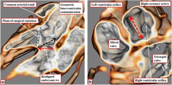
The images show different cuts through a CT angiographic data set prepared from a patient with tetralogy of Fallot. ( a ) shows the planes subtended between the overriding aortic root and the crest of the apical muscular ventricular septum (compared with Fig 8). The long black arrow shows the plane of the apical septum. The geometric interventricular communication is the cranial continuation of this plane. The double-headed white arrow shows the plane of deficient ventricular septation, or the ventricular septal defect, as shown in ( b ). The double-headed red arrow shows the outflow tract from the left ventricle. This plane is equivalent to the embryonic interventricular communication (see Fig 8). ( b ), from the same data set, shows the plane of deficient ventricular septation, between the crest of the muscular apical septum and the undersurface of the malaligned muscular outlet septum (white star). This plane shows also the squeeze (black bracket) between the malaligned outlet septum and the hypertrophied septoparietal trabeculations (red star). ( c ) then shows how the data set can be sectioned to show the precise degree of overriding of the aortic root. This is determined by showing the cord of the apical muscular septum (dashed double-headed arrow) relative to the circumference of the overriding aortic root. In this case, the commitment is virtually equal between the right and left ventricles, and hence in this instance the ventriculo-arterial connections would still be considered concordant. Pulm.=pulmonary.

The CT sections show how the planes subtended beneath the overriding aortic root can be compared with those seen during transfer of the aorta to the left ventricle during cardiac development, and to those seen in the Eisenmenger defect (compare Figs 8, 12, and 16). It is the cranial continuation of the long axis of the plane of the apical muscular ventricular septum that is the geometric interventricular communication. The plane from the crest of the muscular septum to the malaligned muscular outlet septum is the plane of deficient ventricular septation, and hence the ventricular septal defect. This is the plane that will be closed by the surgeon so as to reconnect the overriding aortic root with the left ventricle. It will not be changed by differences in the angulation of the apical muscular septum, as implied in the cartoons produced by the Turkish surgeons.Reference Bozok, Kestelli and Ilhan 3 The third plane represents the interventricular communication seen during cardiac development at the stage when both the arterial trunks arise exclusively from the right ventricle (Fig 5). This plane, subsequent to its realignment in patients with tetralogy of Fallot and the Eisenmenger defect, becomes the outlet for the morphologically left ventricle. It remains the outlet for the left ventricle whenever there is double outlet right ventricle (Fig 14b). Interrogation of the CT data set shows that the extent of aortic overriding is determined by the cord subtended by the muscular ventricular septum relative to the circumference of the overriding aortic root. This feature is independent of the angulation of the apical muscular septum (Figs 10, 14, and 15). Assessing the extent of the root committed to the right as opposed to the left ventricle in this manner now makes it possible to determine with precision the ventriculo-arterial connection. In the illustrated example in the patient with tetralogy of Fallot (Fig 16c), the proportions of the overriding root are supported virtually equally by the two ventricles. Therefore, in this instance, with half of the aortic root supported by the left ventricle, as in our illustrated example of the Eisenmenger defect (Fig 12), the connection will be deemed to be concordant. In the patient shown with the perimembranous ventricular septal defect (Fig 10), the ventriculo-arterial connections are obviously concordant, whereas in other patients illustrated (Fig 15) the connections are equally obviously double outlet from the right ventricle, despite the minor degree of support for the aortic root provided by the left ventricle.
Overriding of a left-sided aorta
“Dextroposition”, when qualifying aortic position, and if translated literally, simply means a right-sided aorta. Such right-sided aortas are a feature of the most common examples of transposition. Indeed, the right-sided aorta in this setting is sufficiently common for the lesion itself frequently to be described as “d-transposition”. Therefore, another potential problem with the use of “dextroposition” is that the rightwardly located aorta does not always override the crest of the deficient ventricular septum. Furthermore, the aorta is not always right-sided when the arterial trunks are transposed. It can be left-sided in a small proportion of patients with usual atrial arrangement, concordant atrioventricular, and discordant ventriculo-arterial connections. The aorta can similarly be left-sided when it, along with the pulmonary trunk, is supported exclusively by the right ventricle. The latter combination is typically associated with a subaortic interventricular communication, which can then be overridden by an aorta that is not right-sided.Reference Lincoln, Anderson, Shinebourne, English and Wilkinson 18 It is this finding that makes it feasible to patch the subaortic outlet into the left ventricle so as to achieve anatomic repair.Reference Lincoln, Anderson, Shinebourne, English and Wilkinson 18 Should the aorta have been transferred to the left ventricle during development of this pattern, the ventriculo-arterial connections would have been concordant, but with parallel arterial trunks (Fig 17).Reference Cavalle-Garrido, Bernasconi, Perrin and Anderson 19 This combination is often described as “anatomically corrected malposition”. Its variant with the usual atrial arrangement, concordant atrioventricular connections, and left-sided aorta is readily interpreted on the basis of the spectrums that are to be found whenever there is overriding of the aortic root, irrespective of its position.
The images show a heart with usual atrial arrangement, concordant atrioventricular connections, and concordant ventriculo-arterial connections, but with parallel rather than spiralling arterial trunks. ( a ) shows the view from the right ventricle, with a cut having been made across the muscular outlet septum (stars) to show the subaortic location of the interventricular communication. Note the presence of bilateral completely muscular infundibulums (double-headed white arrows). ( b ) shows the view from the left ventricle, having reconstituted the muscular outlet septum (stars). The aorta is originating from the cavity of the left ventricle, despite being supported by an extensive muscular infundibulum (double-headed arrow). The heart was photographed, and is reproduced, by kind permission of Mr Donald Perrin. It is housed in the Robert M. Freedom archive of the Hospital for Sick Children, Toronto, Canada. Pulm.=pulmonary.

Overriding of other arterial trunks
Once it is recognised that the major feature of overriding is biventricular connection of an arterial root, it follows that the definition also can be applied to the pulmonary trunk and to a common arterial trunk. Overriding of the pulmonary root is the essence of the Taussig–Bing malformation.Reference Taussig and Bing 20 Uncertainty has existed as to whether the index lesion had transposition or double outlet right ventricle.Reference Van Praagh 21 This problem was solved by an elegant radiographic study. This showed that, in the index case, the circumference of the pulmonary trunk was shared more or less equally between the right and left ventricles.Reference Hinkes, Rosenquist and White 22 It was also suggested that bilateral infundibulums were necessary so as to diagnose double outlet right ventricle in this setting.Reference Van Praagh 21 Examples certainly exist in which the pulmonary valve is supported by an exclusively muscular infundibulum (Fig 18). As with the Eisenmenger defect, however, hearts also exist when the pulmonary trunk, directly related to an interventricular communication, is supported exclusively above the right ventricle, but with fibrous continuity between the leaflets of the pulmonary and mitral valves.Reference Stellin, Zuberbuhler, Anderson and Siewers 23
The images show overriding of the pulmonary (Pulm. – a ) and both ( b ) arterial trunks. In both the hearts, the larger parts of the overriding trunks are supported by the right ventricle, and therefore the ventriculo-arterial connection is one of double outlet. Both the hearts are photographed from the right ventricular aspect. In ( a ), the muscular outlet septum (star) is attached to the inner heart curve, so that the interventricular communication is subpulmonary. This is the double outlet end of the spectrum representing the Taussig–Bing heart. Note the muscular postero-inferior rim to the interventricular defect, and the presence of bilateral infundibulums (double-headed white arrows with red borders). In ( b ), both arterial trunks are overriding, with fibrous continuity between both arterial valves and the mitral valve, and a fibrous outlet septum. There is also fibrous continuity between the leaflets of the tricuspid and mitral valves (double-headed dashed red arrow). The red dashed line shows the extent of the interventricular communication. The spectrum of overriding seen with the doubly committed defect extends to double outlet from the left ventricle. In this heart, however, there is double outlet right ventricle, with the plane of deficient ventricular septation (white dashed line) within the cavity of the right ventricle.

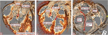
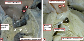
The existence of these hearts (Figs 18 and 19) is further evidence that double outlet right ventricle is not a phenotype, but rather one form of ventriculo-arterial connection.Reference Anderson 5 Furthermore, they show that bilateral infundibulums are not an essential part of the diagnosis, although they certainly can, and do, exist in this setting (Fig 18). Recognition of these facts facilitates the understanding of the spectrums seen in the setting of overriding arterial trunks. Indeed, a further spectrum is found when both the arterial trunks are overriding the ventricular septum (Fig 18b).Reference Lev, Bharati, Meng, Liberthson, Paul and Idriss 24 This spectrum was described by Brandt et al as “double outlet both ventricles”.Reference Brandt, Calder, Barratt-Boyes and Neutze 25 Its essential feature is the doubly committed nature of the interventricular communication.Reference Ueda and Becker 26 In this spectrum, when the arterial trunks are committed predominantly to the left ventricle, the ventriculo-arterial connection is obviously double outlet from the left ventricle.Reference Brandt, Calder, Barratt-Boyes and Neutze 25 A still further spectrum in this setting, with a defect that is directly juxta-arterial, is found when there is a common arterial trunk (Fig 20). In most examples, common truncal valves override the crest of the muscular ventricular septum (Fig 20). In some instances, the trunk and its valve can be supported predominantly (Fig 21) or exclusively by the right ventricle (Fig 20b). In rarer instances, the common trunk can have an exclusive left ventricular origin.
The images are taken from a CT angiographic data set prepared from a patient with the Taussig–Bing malformation. As can be seen in ( a ), the attachment of the muscular outlet septum (white star) to the inner heart curve commits the interventricular communication to the pulmonary root. In this patient, there are bilateral infundibulums. ( b ) shows that the plane of deficient ventricular septation (white double-headed arrow) is exclusively within the cavity of the right ventricle, with the interventricular communication (double-headed red arrow) providing the outlet for the left ventricle. The short-axis section ( c ) shows that the pulmonary (pulm.) trunk arises exclusively to the right side of the muscular ventricular septum (dashed double-headed red arrow).

The images show the spectrum of overriding when there is a common arterial trunk. Both the hearts have aortic dominance, with the pulmonary arteries (stars) arising from the leftward and posterior aspect of the common trunk. In ( a ), the truncal valve is committed equally to both the ventricles, whereas in ( b ) the trunk is supported exclusively by the RV, albeit with fibrous continuity in the roof of the interventricular communication between the leaflets of the mitral and truncal valves. Note that, in ( a ), there is a muscle bar interposed between the leaflets of the tricuspid and truncal valves, whereas in ( b ) the valvar leaflets are in continuity, making the latter defect perimembranous (double-headed white arrows). RV=right ventricle.

The images are from a CT angiographic data set prepared from a patient with common arterial trunk. ( a ) shows the long-axis cut, indicating that the truncal valve is supported predominantly by the right ventricle. This fact is conformed by the short-axis cut looking from the head directly down the barrel of the trunk ( b ). As can be seen, the cord of the ventricular septum taken relative to the orifice of the trunk (double-headed red dashed arrow) shows that the greater part of the overriding valvar circumference is supported by the right ventricle. It is the short-axis cut that shows best the ventriculio-arterial connection, as it is independent of the angulation of the apical muscular ventricular septum.

Discussion
Defining overriding of the arterial trunks based on their biventricular connections has many advantages when compared with describing the same feature on the basis of “aortic dextroposition”. In the first instance, it is not only the aorta that can override the crest of the muscular ventricular septum. In the second place, aortas that are right-sided do not always override the ventricular septal crest. Most importantly, defining overriding on the basis of the biventricular connections of the arterial roots emphasises the spectrums that exist between double outlet and one-to-one ventriculo-arterial connections. All of these aspects have been contentious in the past, and some continue to be so.Reference Anderson 1 , Reference Bozok, Kestelli and Akyuz 2 , Reference Restivo, Unolt, Putotto and Marino 4 , Reference Anderson 5 Use of the definitions as proposed in our review provides simple resolutions to these various disagreements. Perhaps most importantly, concentration on the degree of connection of the overriding root as revealed by CT analysis in the short axis provides a simple and rigorous means of defining double outlet right ventricle (Fig 15), and distinguishing this entity from the hearts with concordant or discordant ventriculo-arterial connections (Figs 10, 12, and 16). As was emphasised by Lev et al,Reference Lev, Bharati, Meng, Liberthson, Paul and Idriss 24 the interventricular communication in this setting is usually directly related to one, or both, of the arterial roots. Thus, if during surgical correction, the surgeon considers that it has been necessary to tunnel the defect to one or other of the arterial roots, then the patient must initially have had double outlet ventriculo-arterial connection. In contrast, if the surgeon considers that it has been feasible to close the septal defect, then the ventriculo-arterial connection must have initially been concordant or discordant. If discordant, the surgical repair will also require incorporation of an arterial or atrial switch into the operative procedure. Although the hole between the ventricles is usually described as the ventricular septal defect in both these instances, the plane identified as the defect is fundamentally different. When there is double outlet, it is a disaster to close the channel permitting interventricular shunting. The patch placed to tunnel the defect to the arterial root occupies the plane of deficient ventricular septation, and is within the cavity of the right ventricle. It is this plane that is also closed when the ventriculo-arterial connections are deemed to be concordant or discordant. In the latter instances, the hole that represented the interventricular communication in the setting of double outlet has become the plane between the overriding part of the trunk supported by the left ventricle and the remainder of the left ventricular cavity. It is likely, nonetheless, that many investigators will continue to describe this plane as the “ventricular septal defect” when describing patients with double outlet right ventricle, presumably because they are aware intuitively that the hole cannot be closed. It would be more logical to describe the plane as the interventricular communication. It is equally logical to consider the plane of placement of the patch used for correction as the “septal defect”, not least because the patch is secured to the underside of the outlet septum. Only time will tell as to whether logic will eventually prevail.
The other great advantage of describing aortic overriding as opposed to dextroposition is that it removes the need to seek to distinguish true as opposed to false attributes of the term.Reference Bozok, Kestelli and Ilhan 3 , Reference Restivo, Unolt, Putotto and Marino 4 Aortic overriding is always present when the aortic root is supported by both ventricles, rather than having an exclusively left or right ventricular origin. It is incorrect to suggest that the aortic root overrides in the normally septated heart.Reference Bozok, Kestelli and Ilhan 3 , Reference Restivo, Unolt, Putotto and Marino 4 An aortic valvar sinus can override the septal crest when the ventricular septum is intact, but of necessity the valvar leaflets have an exclusively left ventricular origin in this setting. The same is true for patients having ventricular septal defects in the absence of overriding of the aortic root. In offering their tips for assessing the significance of “dextroposition”, the Turkish authors argued that new diagnostic modalities would be required to demonstrate the salient features. As we have shown, such modalities already exist, notably CT angiography. This technique can now be used with minimal exposure to radiation, and produces exquisite images. These show all the necessary features, not only of tetralogy of Fallot, but all lesions with overriding arterial roots. Similar images can be produced using MRI and three-dimensional echocardiography, although perhaps not yet with the precision of CT. Therefore, the paediatric cardiologist already possesses all the techniques required to demonstrate the features rightly emphasised by the Turkish authors.Reference Bozok, Kestelli and Akyuz 2 , Reference Bozok, Kestelli and Ilhan 3 The surgeons argued that the techniques would “change the timing and technique of the surgery”. In the case of tetralogy, these will be determined by the length and morphology of the subpulmonary obstruction, and the precise degree of commitment of the aortic valve to the right ventricle. It remains to be seen whether these features are interrelated, or whether all patients can be successfully corrected without recourse to placement of a patch across the ventriculo-pulmonary junction.Reference Vida, Guariento and Castaldi 27