Introduction
Primary arterial switch operation is the gold standard management for transposition of the great arteries with an intact interventricular septum. In patients with transposition of the great arteries with an intact interventricular septum, the ability of the left ventricle to function against systemic pressure declines after 2–3 weeks of age. Therefore, the assessment of left ventricular preparedness is particularly important in patients with late-presenting transposition of the great arteries with an intact interventricular septum. Reference Sologashvili, Wannaz, Beghetti, Aggoun, Prêtre and Myers1
Left ventricular preparedness before arterial switch operation is affected by several factors, including age at presentation, pulmonary vascular resistance, patency of the ductus arteriosus, size of the atrial septal defect, and presence of left ventricular outflow tract obstruction. Reference Lacour-Gayet, Piot and Zoghbi2 However, a single preoperative factor that can reliably predict postoperative outcomes is still challenging to identify. This difficulty is particularly relevant in developing countries where late diagnosis, delayed referral, and limited availability of advanced diagnostic modalities are common.
Conventional echocardiographic parameters, such as left ventricular mass index, interventricular septal thickness and configuration, and left ventricular cavity shape, are frequently used to assess left ventricular preparedness before arterial switch operation. Although useful, tissue Doppler imaging is limited by operator dependence, frame rate, and angle of insonation, which affect the accuracy of velocity measurement. Speckle-tracking echocardiography is angle-independent, and it allows quantitative assessment of myocardial deformation. Therefore, it may provide additional information regarding left ventricular function. Reference Foran, Sullivan, Elliott and de Leval3
There are limited data regarding the usefulness of preoperative left ventricular strain in predicting early postoperative outcomes after arterial switch operation in patients with late-presenting transposition of the great arteries with intact interventricular septum. Therefore, the current study aimed to evaluate whether preoperative echocardiographic parameters, including age at surgery, left ventricular mass index, interventricular septal thickness, direction of interventricular septum movement, and left ventricular strain pattern, can predict early postoperative outcomes after arterial switch operation.
Material and methods
Study design
This prospective observational study was performed at the Department of Cardiothoracic and Vascular Surgery of our institution between August 2020 and September 2021.
The current study was approved by the Institutional Ethics Committee (reference no. IECPG-224/ 24.06.2020, RT-21/ 22.07.2020). A written informed consent was obtained from the parents of all patients.
Inclusion criteria
The study population included 35 patients (23 men and 12 women) with d-transposition of great arteries with intact ventricular septum who did not present with other cardiac or extracardiac anomalies. After preoperative optimisation, these patients underwent primary arterial switch operation with standardised pre- and postoperative management protocols.
Exclusion criteria
Patients with associated intra- and/or extracardiac anomalies, those with preoperative sepsis, and those receiving mechanical ventricular support were excluded from the study.
Preoperative factors
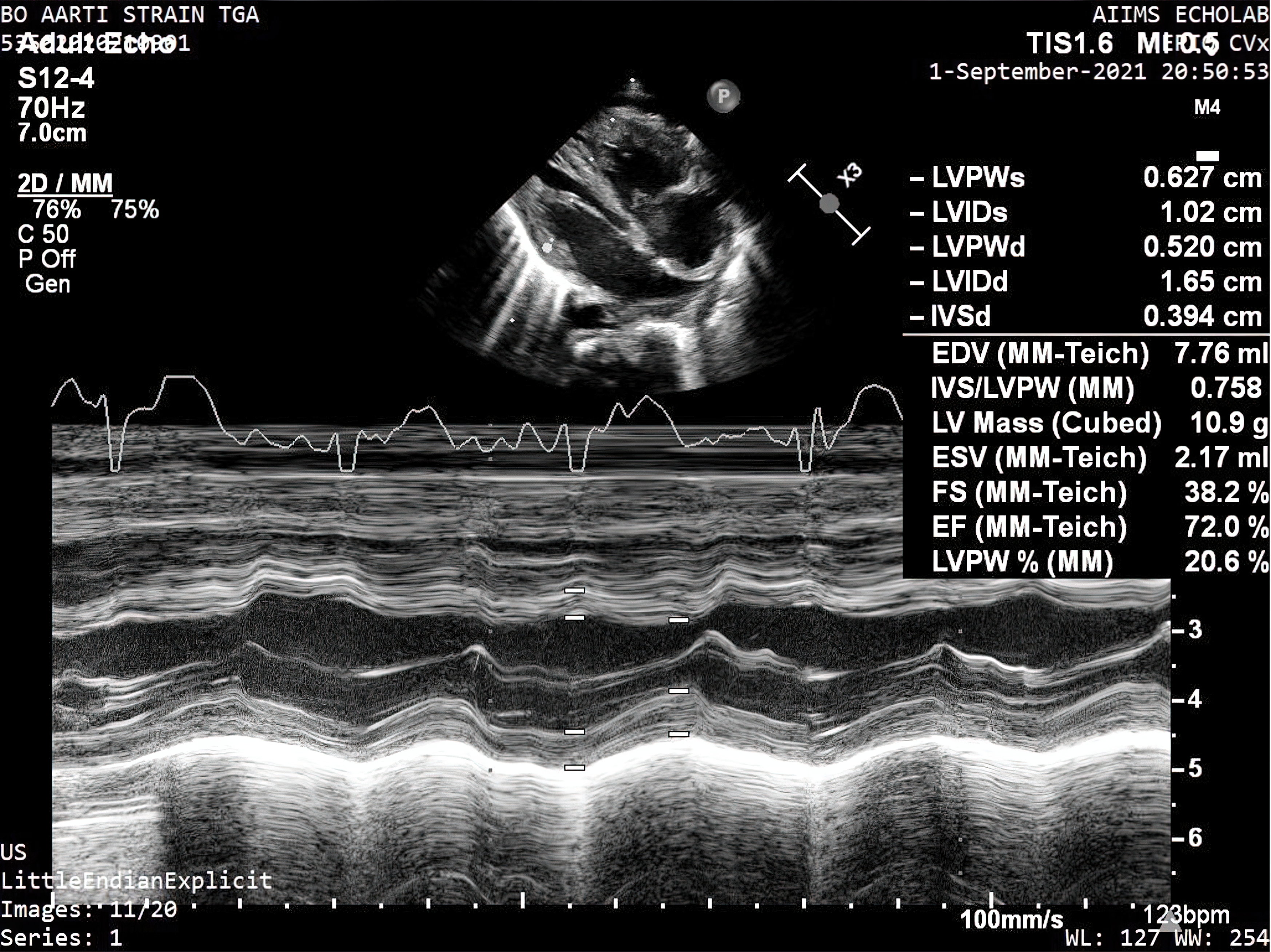
Baseline demographic factors, such as age at the time of surgery, weight, and height, were recorded. As part of the standard preoperative assessment protocol, preoperative echocardiography was performed within 24–48 h prior to surgery. Using the Philips Epic Vx echocardiography machine (Koninklijke, Philips NV 2004–2021, Amsterdam, Netherlands) with a 12 to 4 MHz multi-frequency phased array transducer (S12-4), the diagnosis was confirmed (Figure 1).
Transthoracic echocardiography in M-mode for left ventricular dimensions.

Figure 1. Long description
Panel A: An echocardiogram image displaying a cross-sectional view of the heart. The image includes measurements of various left ventricular dimensions, such as LVPWs, LVIDs, LVPWd, LVIDd, and IVSd, with values provided in centimeters. Panel B: A graph showing the echocardiogram data over time, with the x-axis representing time and the y-axis representing the measurement values. Key measurements include EDV, IVS/LVPW, LV Mass, ESV, FS, EF, and LVPW percent, with specific values provided.
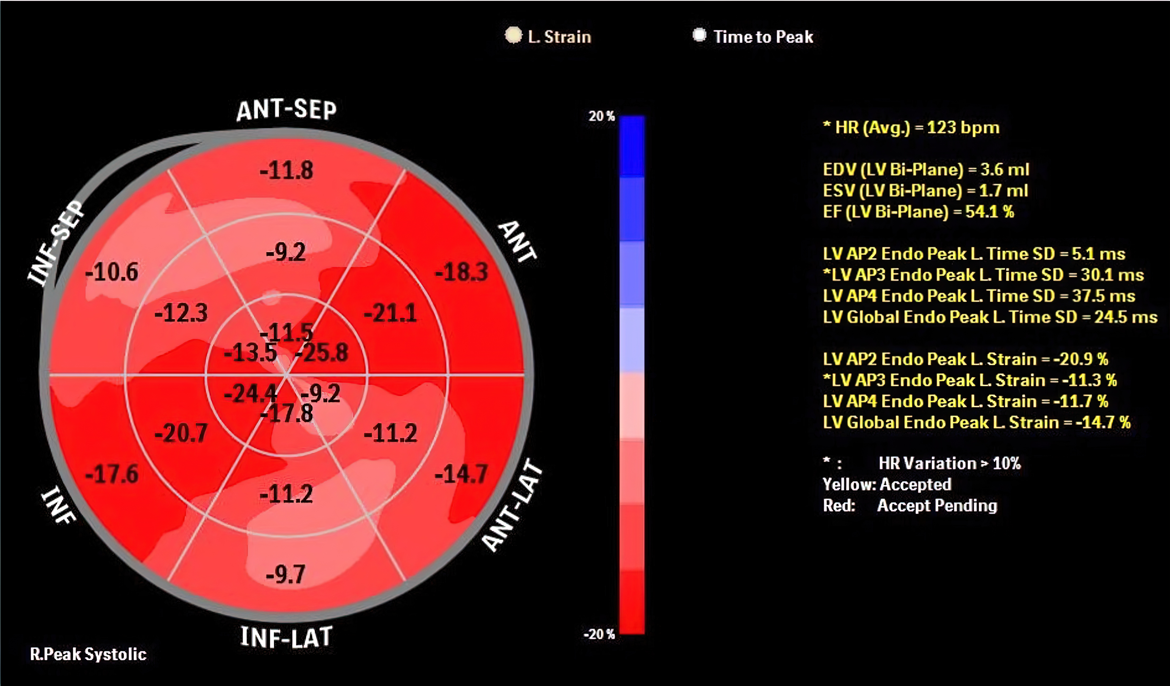
Preoperative factors such as left ventricular end systolic and end diastolic volumes were examined in the subcostal, parasternal short- and long-axis views and apical 2-, 3-, and 4-chamber views (Figure 2).
Measurement of the left ventricular strain pattern—strain measured in all segments of the heart.

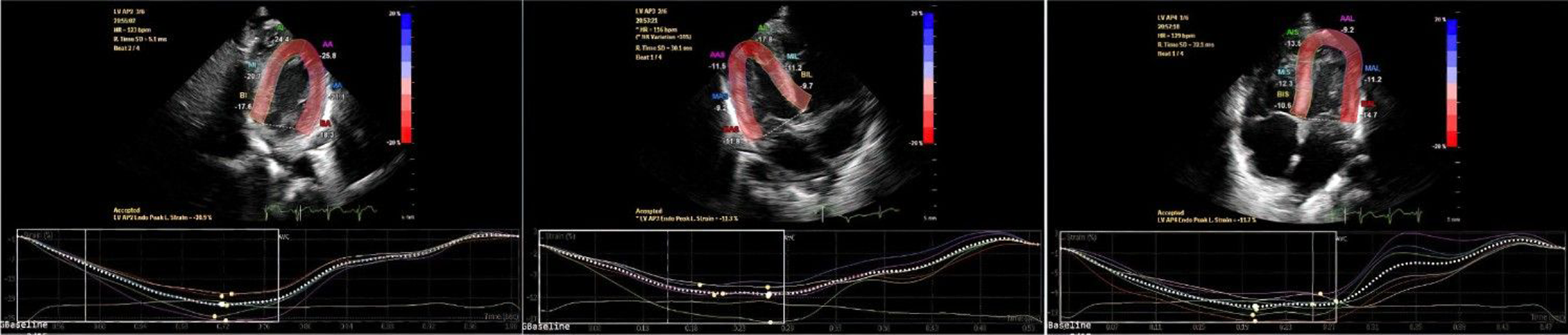
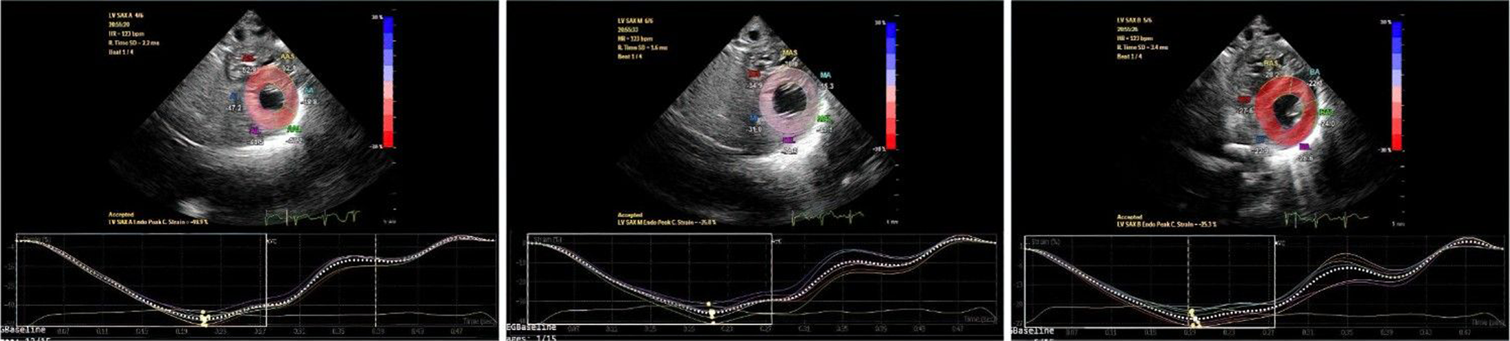
The endocardial border of the left ventricle was automatically traced using the in-built Philips anatomical intelligence ultrasound platform software. However, manual correction by the operator was feasible whenever necessary. They were indexed to body surface area. Preoperative thickness of the interventricular septum, direction of movement of the interventricular septum, and left ventricular cavity shape were assessed. Thickness of the interventricular septum and direction of movement of the interventricular septum were examined in end diastole at the mid-cavity level. The left ventricular cavity shape was assessed at end systole in the subcostal short-axis view. In each view, all walls were automatically subdivided into six segments. In each view, opposite walls were comparatively examined. The global peak systolic strain for each wall was measured by obtaining the average values of the segments of that wall and was presented as a mean of +/− 2 standard deviation. The global strain of each individual patient was calculated by acquiring the average strain values obtained from apical 2-, 3-, and 4-chamber views (Figures 3 and 4).
Longitudinal strain measured in the apical 2-, 3-, and 4-chamber views.

Circumferential strain measured in the short-axis view at the level of the apex, mid-zone, and base of the heart.

Figure 4. Long description
Panel A: Echocardiogram image with a color-coded region highlighting the left ventricle. The corresponding graph below shows a line plot with the x-axis labeled ‘Time (s)’ and the y-axis labeled ‘Strain (%)’. The line represents the strain measurements over time. Panel B: Echocardiogram image with a color-coded region highlighting the left ventricle. The corresponding graph below shows a line plot with the x-axis labeled ‘Time (s)’ and the y-axis labeled ‘Strain (%)’. The line represents the strain measurements over time. Panel C: Echocardiogram image with a color-coded region highlighting the left ventricle. The corresponding graph below shows a line plot with the x-axis labeled ‘Time (s)’ and the y-axis labeled ‘Strain (%)’. The line represents the strain measurements over time.
Left ventricular strain was included as an exploratory parameter to assess myocardial deformation and to determine whether strain-derived indices can be an additional marker of left ventricular preparedness before arterial switch operation. Normative strain data in unoperated transposition of great arteries with intact ventricular septum are limited. However, strain analysis was conducted to evaluate its potential role as a predictive parameter of early postoperative outcomes.
The adequacy of mixing through a patent foramen ovale or ductus arteriosus, coronary artery anatomy, and additional defects were also assessed.
I ntraoperative details
Arterial switch operation was performed using a uniform standard surgical technique on all patients. Intraoperative factors such as cardiopulmonary bypass time, aortic cross-clamp time, and inotropic score were recorded. The inotropic score was quantified using the vasoactive inotropic score described by Wernovsky et al. Reference Wernovsky, Mayer and Jonas4
The inotropic score was calculated within the first 48 h after surgery using the following formula: inotropic score = dopamine dose (µg/kg/min) + dobutamine dose (µg/kg/min) + [10 × milrinone dose (µg/kg/min)] + [100 × adrenaline dose (µg/kg/min)] + [100 × noradrenaline (µg/kg/min)] + [10,000 × vasopressin dose (U/kg/min)]. Reference Wernovsky, Mayer and Jonas4
Postoperative data
Patients were managed postoperatively using a uniform management protocol. The type, amount, and duration of inotropic/chronotropic/after-load-reducing agents used within the first 48 h, length of ICU stay and postoperative hospital stay, and use of extracorporeal membrane oxygenation were recorded from the day of surgery up to hospital discharge. Two-dimensional transthoracic echocardiography for left ventricular function and speckle-tracking echocardiography for left ventricular strain pattern were reassessed before discharge from the hospital.
The primary end point was early survival, and the secondary end points were length of hospital stay, ICU stay, extracorporeal membrane oxygenation support, and inotropic score.
Preoperative variables, including age at the time of surgery, thickness of the interventricular septum, direction of movement of the interventricular septum, left ventricular mass index, and left ventricular strain pattern, were correlated with postoperative parameters, such as length of ICU stay and overall postoperative hospital stay, use and duration of extracorporeal membrane oxygenation support, and inotropic score.
Statistical analysis
Statistical analysis was performed using Stata 16.0 (Stata Corp. LLC 1996–2021, 4905 Lake Way Drive, College Station, Texas, USA). Continuous variables were presented as the mean ± standard deviation or the median with interquartile range, as appropriate. Discrete variables were expressed as percentages. Continuous monotonic variables, such as preoperative age, thickness of the interventricular septum, left ventricular mass index, and left ventricular strain parameters, were analysed using the Spearman rank-order correlation test. The Spearman correlation coefficient (ρ) ranged from −1 (perfect negative correlation) to +1 (perfect positive correlation), and a value of 0 indicated no correlation.
Categorical variables, such as the direction of movement of the interventricular septum, were analysed using the Wilcoxon rank-sum (Mann–Whitney U) test. All statistical tests were two-tailed, and a p-value of <0.05 indicated statistically significant differences.
The correlation between preoperative variables (age at surgery, interventricular septal thickness, left ventricular mass index, and left ventricular strain parameters) and postoperative outcome variables (length of ICU stay, hospital stay, and extracorporeal membrane oxygenation support and inotropic score) was assessed via Spearman correlation analysis.
Results
Demographic characteristics of the patients and preoperative variables
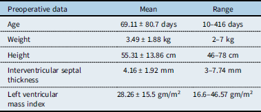
Table 1 shows the demographic characteristics of the patients and preoperative variables. The current study included 35 patients with transposition of great arteries with intact ventricular septum who underwent primary arterial switch operation. Of them, 23 were men, and 12 were women. The median age at surgery was 45 (interquartile range: 22.5–72; range: 10–416) days, and the median weight was 3.4 (interquartile range: 2.9–3.79) kg. The median interventricular septal thickness was 4.1 (interquartile range: 3.75–5.85) mm, and the median left ventricular mass index was 26.4 (interquartile range: 22.75–34.29) g/m2.
Demographic characteristics of the patients

Table 1. Long description
A table with two columns and five rows, detailing preoperative data of patients. The columns are labeled ‘Preoperative data’ and ‘Mean’ and ‘Range’. The rows are labeled ‘Age’, ‘Weight’, ‘Height’, ‘Interventricular septal thickness’, and ‘Left ventricular mass index’. Row 1: Age, Mean: 69.11 ± 80.7 days, Range: 10–416 days. Row 2: Weight, Mean: 3.49 ± 1.88 kg, Range: 2–7 kg. Row 3: Height, Mean: 55.31 ± 13.86 centimeters, Range: 46–78 centimeters. Row 4: Interventricular septal thickness, Mean: 4.16 ± 1.92 millimeters, Range: 3–7.74 millimeters. Row 5: Left ventricular mass index, Mean: 28.26 ± 15.5 grams per square meter, Range: 16.6–46.57 grams per square meter.
Left ventricular strain and operative variables
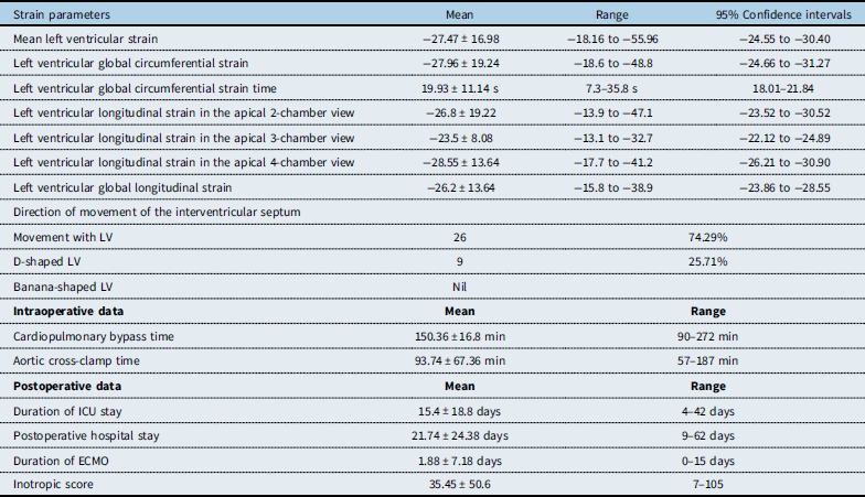
Table 2 shows the left ventricular strain parameters and operative variables. The median average left ventricular strain was −24.19 (interquartile range: −31.15 to −21.90). The mean cardiopulmonary bypass time was 150.36 ± 16.8 min, and the mean aortic cross-clamp time was 93.74 ± 67.36 min.
Left ventricular parameters

Table 2. Long description
The table presents data on left ventricular strain parameters and operative variables. It has 15 rows and 5 columns. The columns are labeled Strain parameters, Mean, Range, and 95 percent Confidence intervals. The rows are labeled with different strain parameters and operative variables. Each row provides the mean, range, and 95 percent confidence intervals for each parameter. Notable parameters include mean left ventricular strain, left ventricular global circumferential strain, left ventricular global circumferential strain time, left ventricular longitudinal strain in various views, direction of movement of the interventricular septum, cardiopulmonary bypass time, aortic cross-clamp time, duration of ICU stay, postoperative hospital stay, duration of ECMO, and inotropic score.
ICU = intensive care unit; ECMO = extracorporeal membrane oxygenation; LV = left ventricle.
Early postoperative outcomes
There were no cases of mortality in the study population. Thirteen (37.14%) patients required extracorporeal membrane oxygenation support. Of them, five were transitioned directly from cardiopulmonary bypass to ECMO (integrated extracorporeal membrane oxygenation). Meanwhile, the remaining patients required extracorporeal membrane oxygenation in the ICU during the early postoperative period. In patients requiring extracorporeal membrane oxygenation, the duration of support ranged from 1 to 15 days, with a median duration of 4 days.
In the whole cohort, the median ICU stay was 12 (interquartile range: 9–17.5) days, and the median postoperative hospital stay was 17 (interquartile range: 14–27.5) days. The median duration of extracorporeal membrane oxygenation support in the whole cohort was 0 (interquartile range: 0–2) days. Among the patients requiring extracorporeal membrane oxygenation, the median duration was 4 (interquartile range: 2–6) days. The median ICU stay was 12 (interquartile range: 9–17.5) days, and the median postoperative hospital stay was 17 (interquartile range: 14–27.5) days. The median IS was 28 (interquartile range: 15.4–50.5). There was no case of early mortality (Tables 6 and 7).
Correlation of age and interventricular septal thickness with postoperative outcomes
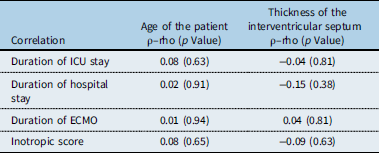
Table 3 presents the correlation of age and interventricular septal thickness with postoperative outcomes. Age at surgery showed a weak positive correlation with length of ICU stay, hospital stay, and extracorporeal membrane oxygenation support and inotropic score. Thickness of the interventricular septum showed a weak negative correlation with length of ICU stay and hospital stay and inotropic score and a weak positive correlation with length of extracorporeal membrane oxygenation support. However, none of these correlations were statistically significant (p > 0.05).
Correlation of age and interventricular septal thickness with postoperative outcomes

Table 3. Long description
The table presents the correlation of age of the patient and thickness of the interventricular septum with various postoperative outcomes. It has four rows and three columns. The columns are labeled Correlation, Age of the patient, and Thickness of the interventricular septum. The rows are labeled Duration of ICU stay, Duration of hospital stay, Duration of ECMO, and Inotropic score. Each cell contains a rho value and a p value. Row 1: Duration of ICU stay, Age of the patient rho 0.08 p 0.63, Thickness of the interventricular septum rho -0.04 p 0.81. Row 2: Duration of hospital stay, Age of the patient rho 0.02 p 0.91, Thickness of the interventricular septum rho -0.15 p 0.38. Row 3: Duration of ECMO, Age of the patient rho 0.01 p 0.94, Thickness of the interventricular septum rho 0.04 p 0.81. Row 4: Inotropic score, Age of the patient rho 0.08 p 0.65, Thickness of the interventricular septum rho -0.09 p 0.63.
ICU = intensive care unit; ECMO = extracorporeal membrane oxygenation.
Association of septal configuration with postoperative outcomes
The association between the direction of movement of the interventricular septum and postoperative outcomes was analysed using the Wilcoxon rank-sum test. Table 4 shows this association. Patients with a D-shaped left ventricle had a longer length of ICU stay and extracorporeal membrane oxygenation support and a higher inotropic score than those with a septum moving with the left ventricle. However, these differences were not statistically significant.
Association between septal configuration and postoperative outcomes

Table 4. Long description
A table comparing postoperative outcomes and their variances. The table has four rows and five columns. The columns are labeled Postoperative outcomes, Unadjusted variance, Adjustment for ties, Adjusted variance, z-score, and Prob [z]. The rows are labeled Duration of ICU stay, Duration of hospital stay, Inotropic score, and Duration of ECMO support. Each row provides values for each column. For example, the Duration of ICU stay has an Unadjusted variance of 702.00, Adjustment for ties of -3.05, Adjusted variance of 698.95, z-score of -0.79, and Prob [z] of 0.43. The table presents data on the association between septal configuration and various postoperative outcomes.
ICU = intensive care unit; ECMO = extracorporeal membrane oxygenation; IVS = interventricular septum.
Correlation of left ventricular mass index and strain with postoperative outcomes
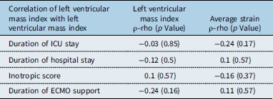
Table 5 shows the correlation of left ventricular mass index and left ventricular strain with postoperative outcomes. Left ventricular mass index showed a weak negative correlation with length of ICU stay, hospital stay, and extracorporeal membrane oxygenation support, and a weak positive correlation with IS. Left ventricular strain showed a variable correlation with length of ICU stay, hospital stay, and extracorporeal membrane oxygenation support and inotropic score. However, none of these correlations were statistically significant.
Correlation of left ventricular mass index and left ventricle strain with postoperative outcomes

Table 5. Long description
A table with four rows and three columns. The columns are labeled ‘Correlation of left ventricular mass index with left ventricular mass index’, ‘Left ventricular mass index’, and ‘Average strain’. The rows are labeled ‘Duration of ICU stay’, ‘Duration of hospital stay’, ‘Inotropic score’, and ‘Duration of ECMO support’. The table presents p-rho values and p-values for each correlation. Row 1: Duration of ICU stay, -0.03 (0.85), -0.24 (0.17). Row 2: Duration of hospital stay, -0.12 (0.5), 0.1 (0.57). Row 3: Inotropic score, 0.1 (0.57), -0.16 (0.37). Row 4: Duration of ECMO support, -0.24 (0.16), 0.11 (0.57).
ICU = intensive care unit; ECMO = extracorporeal membrane oxygenation.
Age at surgery, thickness of the interventricular septum, direction of septal movement, left ventricular mass index, and left ventricular strain parameters showed weak correlations with postoperative outcome variables such as length of ICU stay, hospital stay, and extracorporeal membrane oxygenation support and inotropic score. None of the preoperative echocardiographic parameters, including left ventricular strain, showed a statistically significant correlation with early postoperative outcomes after arterial switch operation in this cohort.
Coronary artery anatomy
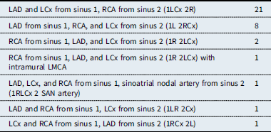
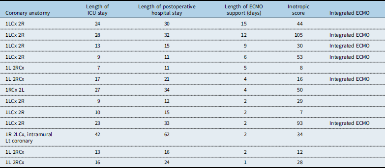
Coronary artery anatomy was classified according to the Leiden Convention. Reference Gittenberger-de Groot, Koenraadt and Bartelings5 The distribution of coronary artery patterns is presented in Table 6. No direct association was observed between coronary pattern and duration of extracorporeal membrane oxygenation support, ICU stay, postoperative hospital stay, or inotropic score in this cohort (Table 7).
Coronary anatomy–Leiden classification

Table 6. Long description
The table categorizes coronary artery origins based on the sinus of Valsalva, detailing different configurations and their frequencies. It has six rows and three columns. The columns are labeled ‘Coronary Configuration’, ‘Leiden Classification’, and ‘Frequency’. The rows list specific configurations of the left anterior descending (LAD), left circumflex (LCx), and right coronary arteries (RCA) originating from different sinuses, along with their corresponding Leiden classifications and frequencies. For example, the first row indicates that LAD and LCx originate from sinus 1, and RCA from sinus 2, with a frequency of 21. The table provides a detailed breakdown of various coronary artery configurations and their occurrences.
RCA = right coronary artery; LAD = left anterior descending artery; LCx = left circumflex artery; SAN = sinoatrial nodal artery; LMCA = left main coronary artery.
Association of coronary anatomy and utilisation of extracorporeal membrane oxygenation

Table 7. Long description
A table with 13 rows and 6 columns comparing coronary anatomy and utilization of extracorporeal membrane oxygenation. The columns are labeled: Coronary anatomy, Length of ICU stay, Length of postoperative hospital stay, Length of ECMO support (days), Inotropic score, and Integrated ECMO. Row 1: 1LCx 2R, 24, 30, 15, 44. Row 2: 1LCx 2R, 28, 32, 12, 105, Integrated ECMO. Row 3: 1LCx 2R, 13, 15, 9, 30, Integrated ECMO. Row 4: 1LCx 2R, 9, 11, 6, 53, Integrated ECMO. Row 5: 1L 2RCx, 7, 11, 5, 8. Row 6: 1L 2RCx, 17, 21, 4, 16, Integrated ECMO. Row 7: 1RCx 2L, 27, 34, 4, 50. Row 8: 1LCx 2R, 9, 12, 2, 29. Row 9: 1LCx 2R, 10, 15, 2, 7. Row 10: 1LCx 2R, 23, 33, 2, 93, Integrated ECMO. Row 11: 1R 2LCx, intramural Lt coronary, 42, 62, 2, 34. Row 12: 1L 2RCx, 13, 16, 2, 12. Row 13: 1L 2RCx, 16, 24, 1, 28.
ICU = intensive care unit; ECMO = extracorporeal membrane oxygenation; LAD = left anterior descending coronary artery; LCx = left circumflex coronary artery; RCA = right coronary artery; LMCA = left main coronary artery.
Coronary anatomy may increase the technical complexity of coronary button transfer during arterial switch operation. However, no direct association was observed between coronary pattern and length of extracorporeal membrane oxygenation support, ICU stay, and postoperative hospital stay and inotropic score in this cohort.
Discussion
The current study evaluated whether preoperative echocardiographic parameters such as interventricular septal thickness, interventricular septal configuration, left ventricular mass index, and left ventricular strain can predict early postoperative outcomes after arterial switch operation in patients with transposition of the great arteries with an intact ventricular septum. The main finding of this study is that none of the preoperative variables, including left ventricular strain, showed a statistically significant correlation with early postoperative outcome variables, such as length of ICU stay, hospital stay, and extracorporeal membrane oxygenation support and inotropic score.
Age at surgery showed a positive correlation with all postoperative outcome variables. Based on this finding, patients who underwent surgery at a later age were more likely to have a longer ICU stay and hospital stay, a higher inotropic requirement, and an increased need for extracorporeal membrane oxygenation. However, this correlation was not statistically significant. Reference Sologashvili, Wannaz, Beghetti, Aggoun, Prêtre and Myers1,Reference Foran, Sullivan, Elliott and de Leval3,Reference Wernovsky, Mayer and Jonas4 Previous studies have well described the regression of the left ventricle after the first 2–3 weeks of life and its effect on postoperative outcomes. Reference Sologashvili, Wannaz, Beghetti, Aggoun, Prêtre and Myers1–Reference Wernovsky, Mayer and Jonas4,Reference Bisoi, Malankar, Chauhan, Das, Ray and Das6,Reference Hui, Chau, Leung, Chiu and Cheung7 These studies suggest that with increasing age, left ventricle regression occurs, and the left ventricle may take longer to adapt to systemic circulation after arterial switch operation. Nevertheless, acceptable outcomes can still be achieved in patients who received appropriate perioperative management and underwent late arterial switch operation. Reference Hui, Chau, Leung, Chiu and Cheung7–Reference Leong, Ahmed Alhassan, Sivalingam and Alwi9
In the current study, most patients underwent arterial switch operation after the neonatal period, representing a late-presenting and high-risk cohort. In this subgroup, reliable preoperative predictors are particularly important. The finding that none of the preoperative echocardiographic parameters showed a statistically significant correlation with postoperative outcome is, therefore, clinically important.
Interventricular septal thickness and configuration are parameters commonly used for assessing left ventricular preparedness. Reference Malakan Rad, Ghandi, Kocharian and Mirzaaghayan10–Reference Shah, Chryssos and Parker13 In the current study, patients with increased interventricular septal thickness and a septum moving with the left ventricle tended to have better postoperative outcomes, such as a shorter ICU and hospital stay and a lower inotropic requirement. Meanwhile, patients with a D-shaped left ventricle required a longer ICU and hospital stay, a higher inotropic requirement, and more frequent extracorporeal membrane oxygenation support. However, these correlations were not statistically significant, indicating that interventricular septal geometry alone may not be reliable in predicting postoperative outcomes. Previous studies evaluating left ventricular preparedness before arterial switch operation have reported similar observations. Reference Lacour-Gayet, Piot and Zoghbi2,Reference Yacoub, Radley-Smith and Maclaurin14–Reference Sarris, Balmer and Bonou16
Left ventricular mass index showed a weak negative correlation with length of ICU stay, hospital stay, and extracorporeal membrane oxygenation support, suggesting that patients with a higher left ventricular mass index had a relatively better postoperative recovery. However, this correlation was not statistically significant. Some studies have also reported that left ventricular mass index and left ventricular wall thickness are useful indicators of left ventricular preparedness. However, they may not independently predict postoperative outcomes. Reference Yacoub, Radley-Smith and Maclaurin14,Reference Villalba, Lafuente and María17,Reference Kang, de Leval and Elliott18
To evaluate myocardial deformation, left ventricular strain was assessed using speckle-tracking echocardiography. In the current study, left ventricular strain parameters showed a variable correlation with postoperative outcome variables. Some left ventricular strain parameters had a weak positive correlation with extracorporeal membrane oxygenation support and length of hospital stay. Meanwhile, others showed a weak negative correlation with length of ICU stay and inotropic score. However, none of the left ventricular strain parameters showed a statistically significant correlation with postoperative outcomes. This finding suggested that preoperative left ventricular strain may not be a reliable predictor of early postoperative outcomes after arterial switch operation in patients with transposition of great arteries with intact ventricular septum. Previous studies evaluating left ventricular strain and function after arterial switch operation have shown contrasting results. Reference Malakan Rad, Ghandi, Kocharian and Mirzaaghayan10–Reference Bengel, Hauser and Duvernoy12,Reference Yasui, Kado and Yonenaga19,Reference Öner, Özdemir, Güven, Yılmazer, Doksöz and Meşe20 Some studies have reported normalisation of left ventricular function after arterial switch operation, and others have revealed persistent abnormalities in myocardial mechanics despite normal global left ventricular function. Reference Leong, Ahmed Alhassan, Sivalingam and Alwi9,Reference Yasui, Kado and Yonenaga19,Reference Öner, Özdemir, Güven, Yılmazer, Doksöz and Meşe20 These results indicate that myocardial deformation parameters may not be directly correlated with early postoperative clinical outcomes.
Coronary artery anatomy is a known predictor of postoperative outcomes after arterial switch operation. In the current study, the coronary artery anatomy of all patients was documented. The two patients who required prolonged extracorporeal membrane oxygenation support had a complex perioperative course. However, coronary anatomy alone did not appear to be the primary determinant of outcome in these patients. Due to the small sample size, coronary anatomy could not be analysed as an independent predictor in this study.
The current study suggests that postoperative outcomes in patients with late-presenting transposition of great arteries with intact ventricular septum may be affected by multiple perioperative factors rather than a single preoperative echocardiographic parameter. Therefore, preoperative echocardiographic parameters alone, including left ventricular strain, may not be sufficient to predict early postoperative outcomes in this patient group.
Although no evaluated preoperative parameters reached statistical significance in the present cohort, the relatively small sample size may have limited the ability to detect meaningful associations. Some of these factors may demonstrate statistically significant relationships with postoperative outcomes when evaluated in a larger cohort of late-presenting patients.
Clinical implications
Primary arterial switch operation can be performed on patients with late-presenting transposition of great arteries with intact ventricular septum who had acceptable early outcomes. The absence of a strong correlation between preoperative echocardiographic parameters and postoperative outcomes indicates that surgical decision-making should not be based on a single echocardiographic parameter alone. The availability of extracorporeal membrane oxygenation and cautious perioperative management remain important factors in the management of these patients.
In conclusion, age, interventricular septal thickness, interventricular septal configuration, left ventricular mass index, and left ventricular strain showed weak correlations with postoperative outcome variables. However, none of these correlations reached statistical significance. Based on these findings, no single preoperative echocardiographic parameter can be reliable in predicting early postoperative outcomes after arterial switch operation in patients with late-presenting transposition of the great arteries with an intact ventricular septum.
Limitations
This study has several limitations. Its sample size was small, and the age distribution was heterogeneous in a large proportion of patients with late-presenting transposition of great arteries with intact ventricular septum. Only early postoperative outcomes were examined, and long-term left ventricular function was not assessed. In addition, there are limited data on normative left ventricular strain in unoperated transposition of great arteries with an intact ventricular septum. Larger multi-institutional studies should be conducted to further evaluate the role of preoperative echocardiographic parameters in predicting postoperative outcomes.
Conclusions
Preoperative echocardiographic parameters, including left ventricular strain, left ventricular mass index, interventricular septal thickness, and interventricular septal configuration, did not significantly predict early postoperative outcomes after arterial switch operation in late-presenting transposition of great arteries with intact ventricular septum. Hence, postoperative outcome is likely influenced by multiple perioperative factors rather than by a single preoperative echocardiographic parameter. Nevertheless, larger multi-institutional studies must be conducted to identify reliable predictors of outcomes in this patient group.
Acknowledgements
None.
Financial support
None.
Competing interests
None.
Ethical standard
Approved by the institutional ethics committee, All India Institute of Medical Sciences, New Delhi 110029, India.