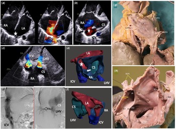
A 41-year-old man with pulmonary arterial hypertension due to an inferior sinus venosus defect was referred for heart–lung transplantation. He complained of congestive heart failure and WHO 3 dyspnoea. Right ventricle ejection fraction on MRI was lowered to 18% with enlarged right cardiac cavities (end diastolic right ventricle volume: 228 ml/m2, right atrium area: 50 mm²). Under advanced pulmonary arterial hypertension therapy (tadalafil and ambrisentan), pulmonary vascular resistance was 6.2 Wood unit.m². Mean atrial pressures were highly elevated at 22 mmHg. Inferior sinus venosus defect consisted of overriding of the inferior caval vein over the interatrial septum, nicely depicted on a 3D model (panels E and F), with a large interatrial defect (panels A, B, E, and F ; star) and continuity between the inferior caval vein and the posterior wall of the left atrium (panel A). Reference Chen, Wang, Hsu, Hsu, Chen and Wu1 A prominent Eustachian valve (panel A; arrow) on the right side of the inferior caval vein directed the flow preferentially to the left atrium. An intriguing left hepatic vein anomalous return to a dilated coronary sinus was diagnosed (panels C and D). Reference Sanders2,Reference Lee and Saremi3 There was an intrahepatic small connection between the inferior caval vein and the left hepatic vein (panel D). The patient died of terminal heart failure while on the waiting list. Autopsy confirmed the diagnosis of inferior sinus venosus defect by visualising partial anomalous connection of the two right lower pulmonary veins to the inferior caval vein at its junction with the right atrium, and the anomalous drainage of the left hepatic vein to coronary sinus (panels G and H).
( a ) Trans-thoracic echocardiography TTE (4 chambers view): vizualisation of the atrial septal defect (star), and the left and right atriums (LA and RA) dilated. The atrial septum overrides the inferior caval vein (ICV). ( b ) Trans-thoracic echocardiography TTE (modified 4 chambers view): vizualisation of the coronary sinus (CS) opening in right atrium (RA). Septal defect is marked by the star, and the septum (arrow) overrides the inferior caval vein (ICV). ( c ) Trans-thoracic echocardiography TTE (sub-costal view): vizualisation of the inferior caval vein (ICV) drainage to the right atrium (RA), overriding the atrial septum. Vizualisation of the left hepatic vein (LHV) drainage to the coronary sinus (CS) opened in the RA. ( d ) Angiographic face view during catheterization. Vizualisation of the inferior caval vein (ICV) drainage to right atrium (RA) and the left hepatic vein (LHV) drainage to the coronary sinus (CS). Note the small vein communication between these two venous returns just above the diaphragm area. ( e and f ) Three-dimensional view of the malformation built from cardiac CT using open-sources modelling softwares (ITK-snap®, Meshmixer®). E: front view. F: posterior view. Coronary sinus (CS) appears in green. Inferior caval vein (ICV) and left hepatic vein (LHV) appear in royal-blue. Right atrium (RA) appears in dark-blue. Left atrium (LA) appears in red. The atrial defect is marked by a star. The arrow shows the prominent Eustachian valve. ( g ) Posterior view of the heart specimen. CS, coronary sinus; ICV, inferior caval vein; LA, left trium; LHV, left hepatic veins; LLPV, left lower pulmonary vein; LUPV, left upper pulmonary vein; RA, right atrium; RLPV, right lower pulmonary veins; RUPV, right upper pulmonary vein. ( h ) Right atrium. The atrial septum overrides the inferior caval vein (inferior sinus venosus defect). AS, atrial septum; CSO, coronary sinus orifice; EV, Eustachian valve; ICV, inferior caval vein; LA, Left atrium; RAA, right atrial appendage; RLPV, right lower pulmonary veins; SCV, superior caval vein; TV, tricuspid valve. Yellow arrow: inferior sinus venosus defect.

Left hepatic vein drainage to the coronary sinus is an extremely rare and benign congenital vascular anomaly often associated with other vascular malformations such as persistent left superior vena cava, anomalous pulmonary venous drainage, interrupted inferior caval vein with azygos continuation. Reference Morshuis, de Lind van Wijngaarden, Kik and Bogers4 In this case, its recognition was important to plan the heart–lung transplantation. Reference Vuran, Ozker, Gumus and Turkoz5,Reference Rao, Varadaraju, Girish and Singh6 Pulmonary arterial hypertension was likely to be related to the sinus venosus defect and the right ventricle volume overload. Genetic study did not identify at-risk anomaly to date. The left hepatic vein drainage to the coronary sinus was not associated with porto-caval fistula.
Supplementary material
To view supplementary material for this article, please visit https://doi.org/10.1017/S1047951122001354
Financial support
This research received no specific grant from any funding agency, commercial or not-for-profit sectors.
Conflicts of interest
None.