




No CrossRef data available.
Article contents
Non-invasive assessment of right ventricular function in the late follow-up of the Senning procedure
Published online by Cambridge University Press: 22 April 2005
Abstract
Deteriorating ventricular function is a major concern after the Senning operation. A Doppler-derived non-geometric measurement, the so-called myocardial performance index, has been described for use in adults and children. We aimed to assess the utility of this index as a method for quantification of right ventricular function in patients in the late follow-up of the Senning procedure, and to correlate the right ventricular ejection fraction and the first derivative of right ventricular pressure as derived using echocardiography with the ejection fraction determined using magnetic resonance imaging.
We studied 44 patients within a mean postoperative period of 15.3 years. We calculated the right ventricular myocardial performance index by pulsed wave Doppler interrogation of tricuspid inflow and aortic outflow, the ejection fraction by Simpson's rule, and the first derivative of right ventricular pressure by continuous wave Doppler from tricuspid regurgitation.
Mean values of right ventricular myocardial performance index, ejection fraction, and the first derivative of right ventricular pressure were 0.50, 39 percent and 1,398 millimetres of mercury per second, respectively. A cut-off value of 0.47 for the right ventricle myocardial performance index was determined, with a sensitivity of 75 percent and a specificity of 62.5 percent. We found no correlation between ejection fraction and the first derivative of right ventricular pressure as estimated by echocardiography and the ejection fraction as shown by magnetic resonance imaging (r2 equal to 0.29 and 0.04 respectively).
We concluded, first, that patients with preserved right ventricular function had values for the right ventricular myocardial performance index lower than 0.47, and second, that ejection fraction and the first derivative of right ventricular pressure as determined echocardiographically did not correlate with values derived using magnetic resonance imaging.
Keywords
Information
- Type
- Original Article
- Information
- Copyright
- © 2005 Cambridge University Press
Right ventricular dysfunction is a major concern, and has been shown to be a risk factor for late death in long-term follow-up after the Senning operation. Echocardiography is the method most widely used in the evaluation of patients with congenital heart disease, but none of the parameters to assess right ventricular function has gained general acceptance,1–3 largely because of the complex geometry of this chamber, and particularly in patients where the ventricle is submitted to a systemic burden.
The calculation of ejection fraction by modified Simpson's rule has several limitations, so that a visual assessment of function is more often applied. In the presence of tricuspid regurgitation the first derivative of right ventricular pressure can be measured by Doppler, and has shown good correlation with the index obtained at cardiac catetherization,4 but its use in the assessment of right ventricles submitted to systemic pressures has yet to be proven.
There are, of course, other methods for calculation of right ventricular ejection fraction, such as angiography or magnetic resonance imaging. One of these methods, however, is invasive and applies ionizing radiation, while the other requires elaborate and time-consuming analysis, precluding thus far repetitive studies.2, 5
More recently, an index derived from Doppler measurements, named the Tei or myocardial performance index, and combining systolic and diastolic time intervals, has been used to assess ventricular function.3, 6–11 Because this Doppler index is relatively independent of geometry, heart rate, preload and afterload, it has been said to be useful in the evaluation of right ventricular function, particularly in ventricles with abnormal shape and pressure overload.3–11 Normal values for the index in the right ventricle submitted to a systemic burden, nonetheless, is unknown, since in normal volunteers the right ventricle is under pulmonary pressure. Thus far, therefore, as far as we know, the clinical utility of the index in assessing right ventricular global function in patients after the Senning procedure has not been demonstrated in a large cohort.
Hence, the purposes of our study were, first, to evaluate the myocardial performance index as a method with which to quantify right ventricular function in patients in the late follow-up of the Senning procedure, and second, to correlate the right ventricular ejection fraction and the first derivative of right ventricular pressure as derived echocardiographically with the ejection fraction determined by magnetic resonance imaging.
Methods
Patients.
From August 1999 to March 2002, 44 patients submitted to Senning procedure were prospectively studied by echocardiography and magnetic resonance imaging. The mean age was 16.7 years, with a range from 12 to 26 years. The mean postoperative period was 15.3 years, ranging from 10 to 23 years. Of the patients, 41 were in the first class of the system devised by the New York Heart Association, with the remaining three in the second class.
Magnetic resonance imaging.
The studies were performed with a LX CV/i system (General Electric Healthcare, United States). End-diastolic and end-systolic images were selected by choosing the image with the largest and the smallest endocardial area, respectively, during the cardiac cycle, using the short axis view. Endocardial contours at end-diastole and end-systole were then traced manually on each slice. Volumes and ejection fraction were displayed automatically, and normal values of right ventricular ejection fraction were considered as being greater than 55 percent. All patients underwent magnetic resonance imaging and echocardiography within an interval of one month.
Echocardiography.
A complete echocardiographic examination was performed in all patients, using commercially available equipment (Philips SONOS 5500 or HDI 5000), with 5.0, 4.0 and 3.5 megahertz transducers. The right ventricular myocardial performance index was calculated using pulsed wave Doppler signals from the tricuspid inflow, or regurgitation, and the right ventricular outflow. The tricuspid inflow was insonated from an apical four-chamber view, with the Doppler sample volume placed at the tips of the leaflets of the tricuspid valve (Fig. 1). We measured the interval from cessation to onset of tricuspid valvar inflow, the so-called “a” interval. If tricuspid regurgitation was present, we used the duration of regurgitant flow (Figs 1 and 2). The “a” interval corresponds to the sum of isovolumetric contraction time, ejection time, and isovolumetric relaxation time. The right ventricular outflow was insonated from a modified parasternal long axis view, or apical five-chamber view, with the Doppler sample volume placed at the proximal attachments of the hinge points of the leaflets of the aortic valve (Fig. 1). The right ventricular ejection time, the so-called “b” interval, was measured from the onset to the end of flow across the right ventricle outflow (Figs 1 and 2). The sum of isovolumetric contraction time and isovolumetric relaxation time was then easily obtained by subtracting “b” from “a”. The index of right ventricular global function was calculated as “a” minus “b” divided by “b”8, 9 (Fig. 2). The measurements of this index were performed by two independent observers, both being unaware of the results obtained using magnetic resonance imaging.

Figure 1. Example of a measurement of the myocardial performance index. Continuous wave Doppler of the tricuspid regurgitation (a) and pulsed wave Doppler of the right ventricular outflow (b). Myocardial performance index = a − b/b = 410 − 260/260 = 0.57.

Figure 2. Scheme showing the Doppler time intervals used to calculate the right ventricular myocardial performance index (RVMPI). ET: ejection time; ICT: isovolumetric contraction time; IRT: isovolumetric relaxation time; RV: right ventricle.
Tricuspid regurgitation was classified as absent, trivial or mild, moderate, or severe. The first derivative of right ventricular pressure was calculated using the signal of tricuspid regurgitation obtained with continuous wave Doppler, where it was measured as the time necessary for the pressure gradient to go from 4 to 36 millimetres of mercury. We selected the same values for velocity as applied to the first derivative of left ventricular pressure, 1 and 3 metres per second, as the right ventricle is under systemic burden after the Senning procedure (Fig. 3). Normal values were considered as being greater than 1,200 millimetres of mercury per second.

Figure 3. The first derivative of right ventricular pressure (dP/dt) calculated by continuous wave Doppler signal from tricuspid regurgitation. dP/dt = (4 × 32 − 4 × 12)/time in seconds. RA: right atrium; RV: right ventricle.
The ejection fraction was calculated by a modified Simpson's rule, planimetering the right ventricle in systole and diastole from a four-chamber view. Normal values were considered as being greater than 55 percent.
Statistical analysis.
The receiver operator characteristic curve was used to select the cut-off value for the right ventricular myocardial performance index to distinguish preserved from deteriorated right ventricular function, considering values of ejection fraction by magnetic resonance imaging above 55 percent as normal.
Comparison of the mean myocardial performance index and the degree of tricuspid regurgitation was made by analysis of variance, and its distribution by Kolmogorov–Smirnov test.
Correlation between right ventricular ejection fraction and the first derivative of right ventricular pressure calculated by echocardiography and the ejection fraction calculated by magnetic resonance imaging was evaluated by Pearson's coefficient.
The correlation between the measurements made by the two observers for the myocardial performance index was also evaluated by Pearson's coefficient.
Results were expressed as values plus and minus standard deviation, and values for p less than 0.05 were considered significant.
Results
Magnetic resonance imaging.
Right ventricular ejection fraction ranged from 16 percent to 85 percent, with a mean of 60 plus or minus 12 percent (Table 1).
Table 1. Results of our parameters as established using echocardiography and magnetic resonance imaging.

Echocardiography.
The calculated right ventricular myocardial performance index ranged from 0.16 to 1.42, with a mean of 0.50 plus or minus 0.24. Only three patients did not have tricuspid regurgitation. In those with regurgitation, it was moderate in 11, but trivial or mild in the other 30 patients. There was no significant difference between the means of myocardial performance index and the different degrees of tricuspid regurgitation (p equal to 0.27), with normal distribution of the myocardial performance index (p equal to 0.24). We determined a cut-off value of 0.47 for the index. Above this value, the right ventricular function was considered to be preserved, with a sensitivity of 75 percent and a specificity of 62.5 percent.
The first derivative of right ventricular pressure was measured in the 28 patients who presented an adequate curve for tricuspid regurgitation from which velocity could be determined using Doppler. It ranged from 457 to 3,213 millimetres of mercury per second, with a mean of 1,398 plus or minus 837. The right ventricular ejection fraction as calculated using the modified Simpson's rule ranged from 6 percent to 63 percent, with a mean of 12 plus or minus 39 percent (Table 1).
Comparison of the right ventricular first derivative of right ventricular pressure and ejection fraction as calculated by echocardiography and right ventricular ejection fraction calculated by magnetic resonance imaging.
There was no correlation between the first derivative of right ventricular pressure and the ejection fraction, both measured by echocardiography, with the ejection fraction measured by magnetic resonance imaging (Figs 4 and 5).

Figure 4. Correlation between the ejection fraction calculated by echocardiography (ECHO) and by magnetic resonance imaging (MRI).

Figure 5. Correlation between the first derivative of right ventricular pressure (dP/dt) and ejection fraction calculated by magnetic resonance imaging (MRI).
Interobserver variability.
There was an excellent correlation between the measurements of the myocardial performance index made by the two observers (r equal to 0.98, p less than 0.001). The mean difference was 0.03 plus or minus 0.15.
Discussion
Disturbances of rhythm and right ventricular dysfunction are known to be major problems in the late postoperative period following the Senning procedure.1 Thus, Kirjavainen et al.1 found right ventricular dysfunction to be a great risk factor for late death, necessitating close follow-up throughout adulthood.
Magnetic resonance imaging has been used to study right ventricular function in normal subjects, adults, and children with congenital cardiac disease, since it provides noninvasive, dimensionally accurate assessment of right ventricular volumes without the need for making geometric assumptions. It is, however, time-consuming,2, 3, 12 and is costly to use in a routine evaluation.
Because of this, it is more usual to use echocardiographic parameters, such as ejection fraction, for measuring right ventricular function. On the other hand, because of its limitations, the accuracy of this method has been questioned.5
Our study, therefore, constituted an attempt to establish a practical, easy to be obtained, reproducible and reliable echocardiographic means of assessing right ventricular function in the postoperative period after the Senning procedure. We found, however, that the right ventricular ejection fraction as calculated by the modified Simpson's rule, and the first derivative of right ventricular pressure, as calculated by Doppler, showed no correlation with the ejection fraction calculated by magnetic resonance imaging. Helbing et al.2 have already explained that right ventricular geometry significantly affects the estimation of the ejection fraction by cross-sectional echocardiography, so the lack of correlation between these methods found in our study is hardly unexpected.
Although the first derivative of right ventricular pressure has been used to assess right ventricular function in adults,4 its value has yet to be established when the right ventricle is working as the systemic ventricle. Since there are no normal values for the right ventricle in this situation, we compared our results with the already established normal values for the left ventricle, but this could be another limitation of using this method. We suggest that the parameter should not be use to define normal as opposed to abnormal right ventricular function, but rather as an index with which to detect changes in ventricular function of a given patient over the course of follow-up.
The same reservation exists with regard to our use of normal values for left ventricular ejection fractions measured by nuclear resonance imaging when comparing values obtained in the systemic right ventricle. At this point, nonetheless, these are the only well established normal values for ventricles under systemic pressure.
Since Tei8 in 1995, first described the myocardial performance index as a parameter to assess combined systolic and diastolic function, some studies have been done in an attempt to determine if this index is dependent or not on preload, afterload, and heart rate.6, 9–11
Moller et al.,10 studying the effect of changes in preload on the index in health volunteers, and in patients with myocardial infarction, found that it was significantly preload-dependent in normal subjects, but relatively preload-independent in patients with depressed left ventricular function. The authors also commented that the magnitude of changes was minor, at less than 10 percent, and that the ability of the index to differentiate individuals in a healthy control group from patients with previous myocardial infarction was maintained during the changes in preload.
Eidem et al.6 studied the index in the pre and postoperative period of patients with large atrial septal defects, representing increased ventricular preload, with isolated pulmonary valvar stenosis, representing increased ventricular afterload, and with congenitally corrected transposition with severe atrioventricular valvar regurgitation, representing the combined effects of increased preload and afterload on the morphologically right ventricle. They compared their various groups of patients with normal children and adults. They found no significant change in the index in any of their groups of patients despite relief of right ventricular volume or pressure overload. Thus, they concluded that this index is a quantitative measurement of right ventricular function that appears to be relatively independent of changes in preload and afterload.
Recently, LaCorte et al.11 studied in a porcine model the correlation between myocardial performance index and invasive measurements of ventricular function, demonstrating a direct correlation between the index and systolic and diastolic invasive measurements of ventricular function.
Although some measurements of ventricular function are dependent on heart rate, such as the first derivative of ventricular pressure and the systolic time intervals, the myocardial performance index is a ratio between isovolumetric time and ejection time. It should therefore be independent of heart rate, and require no correction.9, 10 Poulsen et al.13 studied the effect of heart rate in patients with atrial pacemakers, and observed that for each increase of 10 beats per second, the myocardial performance index increased only 0.02 plus or minus 0.03 seconds, showing no importance in the clinical setting.
Ishii et al.3 studied 35 patients with atrial septal defects, and 8 patients after a Senning operation. In this elegant study, they found values of the myocardial performance index significantly higher in patients after the Senning operation than in normal subjects, as well as in the patients with atrial septal defects. The authors did not compare their results with another method used to quantify right ventricular function, but only with the position within the classification of the New York Heart Association. Since all their patients were in the first class, it is difficult to determine from their study those values for the myocardial performance index that are abnormal.
Hurwitz et al.,14 studying right ventricular systolic function in patients after a Mustard operation, found low ejection fractions in one-sixth of asymptomatic patients. All these results demonstrate the need for more objective parameters.
In our study, the values of the myocardial performance index ranged from 0.16 to 1.42. The patient with the largest value was a 14-year-old boy, in the second class of the system devised by the New York Heart Association, who had an ejection fraction of 42 percent as revealed by magnetic resonance imaging. Even though the patient seemed to be stable, this is a frequent situation in which the patient has few symptoms but deteriorated ventricular function. Tei et al.9 also found values as high as 1.49 for the left ventricle myocardial performance index in adults with dilated cardiomyopathy, and they found good correlation with invasive methods.
Recently, Lissin et al.15 measured the aortic velocity time integral, the aortic ejection time, the excursion of the lateral and septal wall as revealed by M-mode, the first derivative of right ventricular pressure, and the myocardial performance index by Doppler in 18 patients in the late postoperative period of the atrial switch operation, compared their results with the ventricular volumes and ejection fraction calculated by magnetic resonance imaging. They found good correlation of septal wall excursion with right ventricle ejection fraction, and weak correlation of aortic velocity time integral and aortic ejection time with right ventricular ejection fraction. As far as the first derivative of right ventricular pressure was concerned, the authors only reported correlation of impaired systolic function with increased right ventricular volumes. No significant correlation was found between myocardial performance index and the other parameters, except for the first derivative of right ventricular pressure. The main difference between this and our study is that they did not try to determine the value of the myocardial performance index below which a right ventricle submitted to a systemic burden would be considered preserved. The same is valid for the first derivative of right ventricular pressure, as they did not compare this index with the right ventricular ejection fraction by magnetic resonance imaging. On the other hand, they studied the long axis function of the systemic right ventricle, and certainly addition of this method could bring significant further information to our own study.
Vogel et al.16 recently studied the systemic ventricular function in patients with transposition after atrial repair using an index based on tissue Doppler echocardiography, namely isovolemic myocardial acceleration. They found this index to be reduced when compared both to the subpulmonary right ventricle, and to the systemic left ventricle of the controls. We did not use tissue Doppler in our study, and this index certainly would have contributed to the evaluation of our patients.
Our study has demonstrated, nonetheless, that echocardiographic parameters, such as systemic right ventricle ejection fraction and the first derivative of right ventricular pressure, have no correlation with ejection fraction as determined using magnetic resonance imaging. We did not make correlations between the right ventricle myocardial performance index and the ejection fraction as revealed by magnetic resonance imaging. It was used only to establish a cut-off value for the myocardial performance index, permitting us to differentiate preserved from deteriorated systemic right ventricular function. Based on our results, we suggest that values lower than 0.47 for the index show that the patients have preserved systemic right ventricular function. We believe that this index could now become a useful tool in the evaluation of right ventricular function in the postoperative follow-up of patients after the Senning procedure.
References
Kirjavainen M, Happonen JM, Louhimo I. Late results of Senning operation. J Thorac Cardiovasc Surg 1999; 117: 488–495.Google Scholar
Helbing WA, Bosch HG, Maliepaard C, et al. Comparison of echocardiographic methods with magnetic resonance imaging for assessment of right ventricular function in children. Am J Cardiol 1995; 76: 589–594.Google Scholar
Ishii M, Eto G, Tei C, et al. Quantitation of the global right ventricular function in children with normal heart and congenital heart disease: a right ventricular myocardial performance index. Pediatr Cardiol 2000; 21: 416–421.Google Scholar
Imanishi T, Nakatani S, Yamada S, Nakanishi N, Beppu S, Nagata S. Validation of continuous wave Doppler-determined right ventricular peak positive and negative dP/dt: effect of right atrial pressure on measurement. J Am Coll Cardiol 1994; 23: 1638–1643.Google Scholar
Helbing WA, Rebergen AS, Maliepaard C, et al. Quantification of right ventricular function with magnetic resonance imaging in children with normal hearts and with congenital heart disease. Am Heart J 1995; 130: 828–837.Google Scholar
Eidem BW, O'Leary PW, Tei C, Seward JB. Usefulness of the myocardial performance index for assessing right ventricular function in congenital heart disease. Am J Cardiol 2000; 86: 654–658.Google Scholar
Eidem BW, Tei C, O'Leary PW, Cetta F, Seward JB. Nongeometric quantitative assessment of right and left ventricular function: myocardial performance index in normal children and patients with Ebstein anomaly. J Am Soc Echocardiogr 1998; 11: 849–856.Google Scholar
Tei C. New noninvasive index for combined systolic and diastolic ventricular function. J Cardiol 1995; 26: 135–136.Google Scholar
Tei C, Nishimura RA, Seward JB, Tajik AJ. Noninvasive Doppler-derived myocardial performance index: correlation with simultaneous measurements of cardiac catheterization measurements. J Am Soc Echocardiogr 1997; 10: 169–178.Google Scholar
Moller JE, Poulsen SH, Egstrup K. Effect of preload alternations on a new Doppler echocardiographic index of combined systolic and diastolic performance. J Am Soc Echocardiogr 1999; 135: 1065–1072.Google Scholar
LaCorte JC, Cabreriza S, Rabkin DG, et al. Correlation of the Tei Index with invasive measurements of ventricular function in a porcine model. J Am Soc Echocardiogr 2003; 16: 442–447.Google Scholar
Davlouros PA, Kilner PJ, Hornung TS, et al. Right ventricular function in adults with repaired tetralogy of Fallot assessed with cardiovascular magnetic resonance imaging: detrimental role of right ventricular outflow aneurysms or akinesia and adverse right-to-left ventricular interaction. J Am Coll Cardiol 2002; 40: 2044–2052.Google Scholar
Poulsen SH, Nielsen JC, Andersen HR. The influence of heart rate on the Doppler-derived myocardial performance index. J Am Soc Echocardiogr 2000; 13: 379–384.Google Scholar
Hurwitz RA, Caldwell RL, Girod DA, Brown J. Right ventricular systolic function in adolescents and young adults after Mustard operation for transposition of the great arteries. Am J Cardiol 1996; 77: 294–297.Google Scholar
Lissin WL, Wei L, Murphy DJ, et al. Comparison of transthoracic echocardiography versus cardiovascular magnetic resonance imaging for the assessment of ventricular function in adults after atrial switch procedures for complete transposition of the great arteries. Am J Cardiol 2004; 93: 654–657.Google Scholar
Vogel M, Derrick G, White PA, et al. Systemic ventricular function in patients with transposition of great arteries after atrial repair: a tissue Doppler and conductance catheter study. J Am Coll Cardiol 2004; 43: 100–106.Google Scholar

Example of a measurement of the myocardial performance index. Continuous wave Doppler of the tricuspid regurgitation (a) and pulsed wave Doppler of the right ventricular outflow (b). Myocardial performance index = a − b/b = 410 − 260/260 = 0.57.

Scheme showing the Doppler time intervals used to calculate the right ventricular myocardial performance index (RVMPI). ET: ejection time; ICT: isovolumetric contraction time; IRT: isovolumetric relaxation time; RV: right ventricle.

The first derivative of right ventricular pressure (dP/dt) calculated by continuous wave Doppler signal from tricuspid regurgitation. dP/dt = (4 × 32 − 4 × 12)/time in seconds. RA: right atrium; RV: right ventricle.
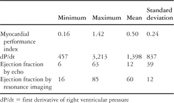
Table 1.

Correlation between the ejection fraction calculated by echocardiography (ECHO) and by magnetic resonance imaging (MRI).

Correlation between the first derivative of right ventricular pressure (dP/dt) and ejection fraction calculated by magnetic resonance imaging (MRI).
 You have
Access
- 4
- Cited by