Introduction
Various treatments can be used to parthenogenetically activate mammalian oocytes by increasing cytoplasmic free calcium concentrations. Among these treatments, while ionomycin (Loi et al., Reference Loi, Ledda, Fulka, Cappai and Moor1998) and strontium (Cuthbertson et al., Reference Cuthbertson, Whittingham and Cobbold1981) promote calcium release from intracellular calcium stores, electrical pulses promote calcium influx from the extracellular medium (Tan et al., 1996; Reference Tan, Liu, Ren, Ni, Sun and He1997). Ethanol treatment is a calcium-dependent method for oocyte activation (Macháty and Prather, Reference Macháty and Prather1998), and has been reported to activate mouse (Cuthbertson et al., Reference Cuthbertson, Whittingham and Cobbold1981; Cuthbertson, Reference Cuthbertson1983; Rickords and White, Reference Rickords and White1992; Winston and Maro, Reference Winston and Maro1995), bovine (Nagai, Reference Nagai1987, Reference Nagai1992; Presicce and Yang, Reference Presicce and Yang1994a, 1994b) and porcine (Didion et al., Reference Didion, Martin and Markert1990) oocytes. However, while Shiina et al. (Reference Shiina, Kaneda, Matsuyama, Tanaka, Hiroi and Doi1993) reported that ethanol exposure promoted both calcium release from intracellular stores and calcium influx from the extracellular medium, Yang (Reference Yang2017) found that treatment with T-type or L-type calcium channel blockers had no effect on ethanol-induced activation (EIA) of mouse oocytes. Furthermore, although Ilyin and Parker (Reference Ilyin and Parker1992) observed that in Xenopus oocytes, extracellular application of ethanol evoked a rapid calcium release from intracellular stores through stimulating the formation of inositol 1,4,5-trisphosphate (IP3) at the plasma membrane, the IP3-mediated calcium release and whether the extracellular calcium is essential remains to be verified during EIA of mammalian oocytes.
It has been shown in somatic cells that elevations in extracellular calcium induce a conformational change in the calcium-sensing receptor (CaSR), which activates phospholipase C (Coburn et al., Reference Coburn, Elangovan, Goodman and Frazaõ1999). The activated phospholipase C promotes the production of IP3 by hydrolyzing the phosphatidylinositol 4,5-bisphosphate, and the resultant IP3 facilitates calcium release from the endoplasmic reticulum (ER) through interactions with IP3 receptors (Hofer and Brown, Reference Hofer and Brown2003). It has been observed that CaSR is expressed in humans (Dell’Aquila et al., Reference Dell’Aquila, De Santis, Cho, Reshkin, Caroli, Maritato, Minoia and Casavola2006), equine (De Santis et al., Reference De Santis, Casavola, Reshkin, Guerra, Ambruosi, Fiandanese, Dalbies-Tran, Goudet and Dell’Aquila2009), porcine (Liu et al., Reference Liu, Wu, Fu, Mo, Zhao, Hu, Zhu and Hou2015) and rat (Yang et al., Reference Yang, Sun, Ji, Zhang, Yuan, Luo, Liu and Tan2018) oocytes. However, we could find reports on neither the expression of CaSR in mouse oocytes nor its role during the artificial oocyte activation of any species. Furthermore, whether the ethanol-stimulated IP3 formation at the plasma membrane observed by Ilyin and Parker (Reference Ilyin and Parker1992) in Xenopus oocytes is through activating CaSR remains an open question.
It is known that the intracellular calcium stores can accumulate, store, release and reaccumulate Ca2+ ions repetitively in response to specific cellular events. Studies have indicated that the intracellular calcium stores were greater in the in vitro matured bovine (Boni et al., Reference Boni, Cuomo and Tosti2002) and ovine oocytes (Boni et al., Reference Boni, Cocchia, Silvestre, Tortora, Lorizio and Tosti2008) that showed a remarkably higher developmental potential. Abnormalities in the molecular machinery such as IP3 receptors within the oocyte are considered likely to affect the pulsatile release of calcium within the ooplasm, leading to activation deficiency in human oocytes (Yeste et al., Reference Yeste, Jones, Amdani, Patel and Coward2016). However, while it is well known that the oocyte susceptibility to activation stimulus increases with extended post-maturation ageing (Kubiak, Reference Kubiak1989; Lan et al., Reference Lan, Ma, Wang, Luo, Chang and Tan2004; Miao et al., Reference Miao, Liu, Qiao, Miao, Luo and Tan2005), Szpila et al. (Reference Szpila, Walewska, Sabat-Pośpiech, Strączyńska, Ishikawa, Milewski, Szczepańska and Ajduk2019) observed that both in vivo and in vitro postovulatory ageing for 9 or 25 h decreased the Ca2+ store in the ER and the expression of Ca2+ pump SERCA2 in mouse oocytes. Therefore, the role of the intracellular calcium stores in the activation of postovulatory ageing oocytes remains to be verified using more convincing models.
Calcium concentration in the oviduct fluid is lower than that in serum and it changes with oestrous stages (Borland et al., Reference Borland, Biggers, Lechene and Taymor1980; Grippo et al., Reference Grippo, Henault, Anderson and Killian1992). Furthermore, although it was found that culture in calcium-free medium inhibited spontaneous activation of hamster oocytes (Sun et al., Reference Sun, Yue, Zhou, Chen and Tan2002), the underlying mechanisms are unknown. Therefore, the role of calcium in oocyte ageing should be studied to elucidate the mechanisms for the postovulatory ageing of oocytes. The objective of this study was to explore the role of calcium in oocyte ageing and the roles of intracellular calcium stores, calcium influx and CaSR in EIA of mouse oocytes to reveal the mechanisms for EIA and postovulatory oocyte ageing.
Materials and methods
The experiments in this study were conducted according to the relevant guidelines and regulations. Animal care and handling were carried out strictly in accordance with the guidelines issued by the Animal Care and Use Committee of the Shandong Agricultural University, China (Permit number: SDAUA-2001–001). All the chemicals and reagents used were purchased from Sigma Chemical Co., unless otherwise specified.
Mice and oocyte recovery
We used Kunming mice, which were originally derived from ICR (CD-1) mice. The mice were raised in a room under a 14 h light:10 h darkness photoperiod, with the dark period starting at 20:00 h. Female mice, 8–12 weeks after birth, were superovulated using 10 IU equine chorionic gonadotropin (eCG, i.p.) and 10 IU human chorionic gonadotropin (hCG, i.p.) at a 48-h interval. Both eCG and hCG were produced by the Ningbo Hormone Product Co., Ltd., China. The superovulated mice were sacrificed 13 h or 19 h after the hCG injection, and cumulus–oocyte complexes (COCs) were recovered by rupturing the oviduct ampullae. After being washed in M2 medium, the COCs were denuded of cumulus cells by incubating for 3 min and pipetting in M2 containing 0.1% hyaluronidase, to obtain cumulus-denuded oocytes (DOs).
Oocyte ageing in vitro
After being washed in M2 and CZB medium, DOs were cultured for ageing in CZB with or without calcium for 6 h or 12 h before further experiments. The ageing culture was performed using culture wells (20–30 DOs per well containing 100 μl medium) at 37.5°C in humidified air containing 5% CO2. The CZB medium consisted of NaCl, 81.62 mM; KCl, 4.83 mM; KH2PO4, 1.18 mM; MgSO4, 1.18 mM; NaHCO3, 25.12 mM; CaCl2·2H2O, 1.7 mM; EDTA-2Na·2H2O, 0.11 mM; l-Gln, 1 mM; sodium lactate, 31.3 mM; sodium pyruvate, 0.27 mM; penicillin, 100 IU/ml, streptomycin, 50 IU/ml; BSA, 5 g/l. CaCl2·2H2O was omitted in the calcium-free CZB.
Measurement for calcium
Intracellular calcium was measured using a Leica live cell imaging system (DMI6000B; Leica) following staining oocytes with calcium-sensitive dye Fluo-2 AM. Briefly, to load the Ca2+ probe, DOs were incubated at room temperature for 30 min in the HEPES-buffered CZB medium containing 1 μM Fluo-2 AM and 0.02% pluronic F-127. After 4-μl drops of HEPES-buffered CZB were made in a Fluoro dish (FD35–100, World Precision Instruments), and were covered with mineral oil, we placed the DOs in the drops (20–25 DOs per drop) and examined them at 37°C under a Leica DMI 6000 inverted microscope. A Fluo-2 fluorescence module was used for excitation, and the fluorescence excitation and emission wavelengths were 488 and 512 nm, respectively. A Leica LAS-AF calcium imaging module was used to calculate the F488/515 ratio to represent the calcium concentrations. The DOs were monitored for, in total, 20 min. During the first 5 min, the F488/515 ratio was recorded every 10 s to indicate the cytoplasmic calcium (cCa) concentration and, at 5 min, 1 μl of ionomycin was injected into the drop and F488/515 ratio was recorded every 10 s to indicate the total calcium (tCa) concentration. The stored calcium (sCa) concentration was calculated by subtracting the cCa value from the tCa value.
Ethanol activation of oocytes
The DOs were first treated for 5 min at room temperature with calcium-free CZB medium containing 10% ethanol. Then, after being washed three times in M2 medium, the DOs were cultured at 37.5°C for 6 h under an humidified atmosphere of 5% CO2 in air, in calcium-containing CZB medium with or without NPS-2143 or nifedipine. At the end of the activation culture, oocytes were examined under a stereomicroscope for activation. In this study, the oocytes that displayed one or two pronuclei, or had two cells each with a nucleus, were judged as activated. Oocytes with ruptured plasma membranes were considered lysed, while oocytes with cleaved cytoplasm but without a pronucleus were considered fragmented. In this study, while the lysed oocytes were not included in the treated oocytes, the fragmented oocytes were considered not activated.
Oocyte SrCl2 activation and embryo culture
To examine oocyte developmental competence, oocytes were activated with SrCl2. The DOs were incubated for 6 h in a calcium-free CZB medium containing 10 mM SrCl2 and 5 mg/ml cytochalasin B. At the end of the activation culture, the Sr2+-activated oocytes were cultured for embryo development at 37.5°C in calcium-containing glucose-free CZB medium (20–25 oocytes/100 μl medium) in humidified air containing 5% CO2. The 4-cell development was examined at 48 h of culture and, at the same time, embryos were transferred to CZB medium containing 5.55 mM glucose for further culture. Blastocyst rates were examined at 96 h of the culture. Percentages of activated oocytes, 4-cell and blastocyst embryos were calculated from SrCl2-treated oocytes, activated oocytes and 4-cell embryos, respectively.
Immunofluorescence microscopy for detection of calcium-sensing receptor (CaSR), cortical granules (CGs) and spindle/chromosomes
All the procedures were performed at room temperature unless otherwise stated. The DOs were always washed three times in the M2 medium between treatments. The DOs were (1) fixed for 30 min in M2 with 3.7% paraformaldehyde; (2) incubated for 10 s in M2 containing 0.5% protease to remove the zona pellucida; (3) permeabilized in M2 with 0.1% Triton X-100 for 10 min at 37.5°C; and (4) blocked in M2 with 3% BSA for 30 min at 37.5°C.
To detect CaSR, the blocked DOs were incubated at 4°C overnight with rabbit polyclonal anti-CaSR (IgG, 1:100, Immunoway) in M2 medium with 3% BSA. Then, the DOs were incubated for 1 h with Cy3-conjugated goat-anti-rabbit IgG (1:800, Jackson ImmunoResearch) in M2 with 3% BSA. Finally, chromatin was stained by incubating the DOs for 10 min in M2 containing 10 μg/ml Hoechst 33342. To stain CGs, the blocked DOs were incubated for 30 min in M2 containing 100 μg/ml of FITC-labelled Lens culinaris agglutinin. To stain spindle/chromosomes, the blocked oocytes were cultured at 37°C for 1 h in M2 containing FITC-conjugated anti-α-tubulin monoclonal antibodies (1:50). Then, the DOs were cultured for 10 min in M2 containing 10 μg/ml Hoechst 33342 to stain chromatin.
The stained DOs were mounted on glass slides for observation under microscopes. While CaSR was observed with a Leica laser scanning confocal microscope (TCS SP2), CGs and spindle/chromosomes were observed with a Leica fluorescence microscope. During CaSR observation, blue diode (405 nm) and helium/neon (543 nm) lasers were used to excite Hoechst and Cy3, respectively. Fluorescence was detected with bandpass emission filters: 420–480 nm for Hoechst and 560–605 nm for Cy3, and the captured signals were recorded as blue and red, respectively.
Data analysis
Each treatment contained at least three replicates and the number of replicates in each treatment was specified in respective figure legends. The software of SPSS 11.5 (SPSS Inc.) was used to carry out all the data analyses in this study. For the percentage data, arc sine transformation was conducted before further analysis. The analysis of variance (ANOVA) was used to analyze data when there were more than two groups in each measure, and an independent sample t-test was conducted when there were only two groups in each measure. The differences were located using the Duncan multiple comparison test. All data were expressed as mean ± standard error of the mean (SEM), and a difference was considered significant for P < 0.05.
Results
Effects of oocyte calcium-free ageing (CFA) on levels of intracellular calcium stores
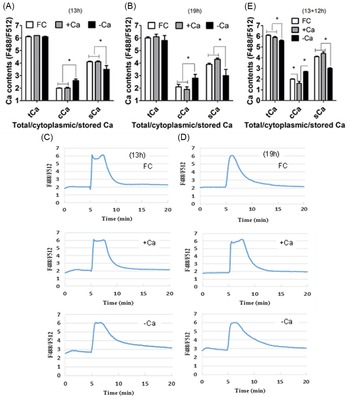
Oocytes recovered at 13 or 19 h post hCG injection were denuded of cumulus cells. The cumulus-denuded oocytes (DOs) were cultured for ageing for 6 or 12 h with (+Ca) or without (−Ca) calcium before calcium measurement. Some DOs were measured for calcium immediately after collection without culture (FC). Following ageing culture for 6 h of both the 13-h (Figure 1A, C) and 19-h recovered oocytes (Figure 1B, D), the level of total calcium (tCa) did not differ among the various treatments. While the level of cytoplasmic calcium (cCa) was higher, the level of stored calcium (sCa) was significantly lower in −Ca than in +Ca aged oocytes, suggesting that some calcium was released into the cytoplasm after CFA. When oocytes recovered 13 h post hCG were aged for 12 h, levels of tCa and sCa were significantly lower in −Ca than in +Ca and FC oocytes (Figure 1E). Levels of sCa did not differ between +Ca and FC 13-h or 19-h oocytes after ageing culture for either 6 or 12 h, and between the 13-h and 19-h FC oocytes (4.09 ± 0.06 vs 3.97 ± 0.15, P = 0.53). The results suggest that CFA significantly decreased sCa in both the 13-h and 19-h oocytes, but sCa levels did not change during culture for 6 or 12 h in the presence of calcium and during in vivo ageing for 6 h from 13 to 19 h post hCG injection.
Effects of oocyte calcium-free ageing (CFA) on levels of intracellular calcium stores. Panels (A/C) and (B/D) show levels (F488/F512) of total (tCa), cytoplasmic (cCa) and stored (sCa) calcium in freshly collected oocytes (FC), or oocytes cultured for 6 h with (+Ca) or without (−Ca) calcium following oocyte recovery at 13 h and 19 h after hCG injection, respectively. Graph (E) shows the concentrations of tCa, cCa and sCa in FC oocytes and oocytes cultured for 12 h +Ca or −Ca after oocyte recovery at 13 h post hCG. Each treatment was repeated three times with each replicate including 20–25 oocytes. *Indicates significant difference (P < 0.05). In each graph of panels (C) and (D), the F488/F512 value from 0 to 5 min represents the cCa, while the peak value that appeared at 5 min when ionomycin was added stands for the tCa. The sCa in calcium stores was calculated by subtracting cCa from tCa.

Effects of CFA on EIA of oocytes
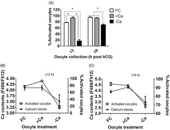
The DOs recovered 13 or 19 h post hCG injection were aged for 6 h with (+Ca) or without (−Ca) calcium before ethanol activation treatment. Some DOs were activated with ethanol immediately after collection without culture (FC). The activation rate decreased significantly from 93% in +Ca oocytes to 17.2% in −Ca oocytes when oocytes were recovered 13 h post hCG, and it decreased from 92.5% in +Ca oocytes to 70.1% in −Ca oocytes when oocytes were recovered 19 h post hCG (Figure 2A). While the activation rate was significantly higher in +Ca than in FC 13-h oocytes, it did not differ between +Ca and FC 19-h oocytes. Furthermore, the activation rate was significantly higher in 19-h than in 13-h FC oocytes (94.28 ± 1.76 vs 77.50 ± 1.44, P = 0.00). The results suggest that CFA significantly impaired EIA in both the 13-h and 19-h oocytes with the impairment more severe in 13-h oocytes, and that the oocyte susceptibility to EIA increased significantly during in vivo ageing from 13 h to 19 h post hCG injection.
Effects of calcium store levels on ethanol-induced activation of oocytes. The DOs recovered 13 or 19 h post hCG injection were cultured for ageing for 6 h with (+Ca) or without (−Ca) calcium before ethanol activation treatment. Some DOs were activated with ethanol immediately after collection without culture (FC). Graph (A) shows percentages of ethanol-activated oocytes in FC, +Ca and -Ca oocytes recovered at 13 or 19 h after hCG injection. Each treatment was repeated three or four times with each replicate containing 25–30 oocytes. * Indicates significant difference (P < 0.05). Graphs (B) and (C) show the relationship between levels of calcium stores and the percentages of ethanol activation in different groups of oocytes recovered 13 h or 19 h after hCG injection, respectively.

When the percentages of EIA oocytes were compared with levels of sCa in different groups of oocytes, we found a positive correlation between the two events. Therefore, both the activation rates and the sCa levels were high in +Ca oocytes, but both were low in the −Ca oocytes, in both the 13-h and 19-h groups (Figure 2B, C). However, although the 13-h FC and +Ca oocytes showed a similar level of calcium stores (Figure 1A), the activation rate in the latter was significantly higher than that in the former (Figure 2A). Therefore, CFA significantly impaired the EIA of oocytes, suggesting a positive correlation between levels of sCa and EIA of mouse oocytes.
Effects of CFA on calcium influx-dependence of oocyte EIA
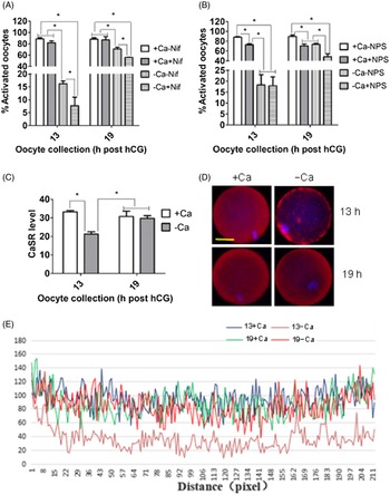
The DOs recovered 13 or 19 h post hCG injection were aged for 6 h with (+Ca) or without (−Ca) calcium before ethanol stimulus for 5 min. Immediately following the ethanol stimulus, the oocytes were incubated for 6 h with or without 400 μM of nifedipine, an L-type calcium channel blocker. While the EIA rates in both 13-h and 19-h +Ca oocytes were unaffected by the presence of nifedipine, the activation rates in both 13-h and 19-h −Ca oocytes were significantly decreased by treatment with nifedipine (Figure 3A). The results suggest that, although oocytes after ageing with calcium do not require calcium influx for EIA, calcium influx is essential for EIA of the oocytes after CFA.
Roles of extracellular calcium and calcium-sensing receptor (CaSR) in ethanol-induced activation of mouse oocytes with different levels of calcium stores. The DOs recovered 13 or 19 h post hCG injection were aged for 6 h with (+Ca) or without (−Ca) calcium before ethanol stimulus for 5 min or immunocytochemistry for CaSR expression. Immediately following the ethanol stimulus, the oocytes were incubated for 6 h with or without 400 μM of nifedipine (Nif) or 2 mM NPS-2143 (NPS). Graphs (A) and (B) show percentages of activated oocytes after 13-h or 19-h +Ca or −Ca oocytes were treated with Nif and NPS, respectively. Each treatment was repeated three or four times with each replicate including 20–30 oocytes. Graph (C) shows levels of CaSR in 13-h and 19-h +Ca or −Ca oocytes. Each treatment was repeated three times with each replicated containing 25 oocytes. *Indicates significant difference (P < 0.05). Panel (D) shows confocal images showing CaSR distribution in 13-h or 19-h +Ca or −Ca oocytes. These are merged pictures with chromosomes and CaSR puncta pseudo coloured blue and red, respectively. The bar is 20 μm and applies to all images. Panel (E) shows fluorescence intensity curves for CaSR in 13-h or 19-h +Ca or −Ca representative oocytes.

Role of CaSR in EIA of oocytes following CFA
The DOs recovered 13 or 19 h post hCG injection were aged for 6 h with (+Ca) or without (−Ca) calcium before ethanol stimulus for 5 min. Immediately following the ethanol stimulus, the oocytes were incubated for 6 h with or without 2 mM of NPS-2143, a CaSR inhibitor. While treatment with NPS-2143 had no significant effects on the activation rates of the 13-h −Ca oocytes, it significantly decreased the activation rates of the 13-h +Ca and 19-h +Ca and −Ca oocytes (Figure 3B). Our quantification of CaSR expression demonstrated that, while the 13-h +Ca oocytes and 19-h +Ca and −Ca oocytes expressed similarly high levels of CaSR, the CaSR level in 13-h −Ca oocytes was decreased significantly (Figure 3C–E). The results suggested that CFA impaired CaSR expression in the 13-h oocytes and that inhibiting CaSR decreased EIA only in oocytes with a full complement of CaSR. Therefore, our results confirmed that CaSR played a significant role in the EIA of mouse oocytes.
Effects of CFA on cortical granules (CGs) distribution of oocytes
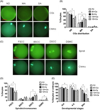
The DOs collected at 13 h post hCG were treated immediately after collection (FC) or aged for 6 or 12 h with (+Ca) or without (−Ca) calcium before immunocytochemistry for observation of CG distribution. We classified the CG distribution into normal distribution (ND) and mildly abnormal (MA) or severely abnormal (SA) distribution (Figure 4A). Percentages of oocytes with ND CG distribution were significantly lower in 12 h-aged than in 6 h-aged +Ca or −Ca oocytes (Figure 4B). The percentages of oocytes with ND distribution of CGs did not differ between +Ca and −Ca oocytes of either the 6 h-aged or 12 h-aged groups. The results suggest that the ageing time was the main factor that impaired CG distribution, while the presence of calcium during ageing had a mild effect on CG distribution in ageing oocytes.
Effects of calcium stores and ageing time on cortical granules (CGs) distribution, spindle/chromosomes morphology and developmental potential of ageing oocytes. The DOs collected at 13 h post hCG were treated immediately after collection (FC) or aged for 6 or 12 h with (+Ca) or without (−Ca) calcium before further treatment. The FC and the +Ca or −Ca aged oocytes were either processed for immunocytochemistry for detection of CG distribution and spindle/chromosomes morphology, or activated with SrCl2 for embryo development. Panels (A) and (C) are micrographs showing CG distribution and spindle/chromosomes morphology, respectively. The bar is 24 μm and applies to all images. The oocytes were observed under a fluorescence microscope after staining with Hoechst 33342 for chromosomes (Chrms) and with antibodies for CGs or spindle (Spndl) detection. The CG distribution was classified into normal distribution (ND), mildly abnormal (MA) or severely abnormal (SA) distribution. The spindle/chromosome morphology was classified into focused pole spindle (FS), barrel-shaped pole spindle (BS) or disintegrated spindle (DS) with congressed chromosomes (CC) or misaligned chromosomes (MC). While graph (B) shows percentages of oocytes with ND, MA and SA distributions of CGs, graph (D) shows percentages of oocytes with FSCC, BSCC, DSCC and DSMC spindle/chromosomes in FC oocytes and oocytes aged +Ca or −Ca for 6 or 12 h. Each treatment was repeated three or four times with each replicate containing 20–25 oocytes. Graph (E) shows percentages of activated oocytes (Act), 4-cell embryos (4-C) and blastocysts (Blst) following activation of different groups of oocytes. Each treatment was repeated three or four times with each replicate including 25–30 oocytes. Percentages of Act oocytes, 4-C and Blst embryos were calculated from SrCl2 treated oocytes, Act oocytes and 4-C embryos, respectively. *Indicates significant difference (P < 0.05).

Effects of CFA on spindle/chromosomes morphology of oocytes
The DOs recovered at 13 h post hCG were treated immediately after recovery (FC) or aged for 6 or 12 h with (+Ca) or without (−Ca) calcium before immunocytochemistry for observation of spindle/chromosome morphology. We classified the spindle/chromosomes into focused pole spindle (FS), barrel-shaped pole spindle (BS) or disintegrated spindle (DS) with congressed chromosomes (CC) or misaligned chromosomes (MC; Figure 4C). We considered the FS (FSCC) or BS (BSCC) spindles with CC chromosomes as normal, while the DS spindles with CC (DSCC) or MC (DSMC) chromosomes had abnormal morphology. Percentages of the oocytes with FSCC did not differ between +Ca and −Ca ageing for either 6 or 12 h, but were significantly lower in oocytes aged for 12 h rather than for 6 h (Figure 4D). Percentages of the oocytes with DSCC did not differ between +Ca and −Ca ageing for either 6 or 12 h but were significantly higher in oocytes aged for 12 h than +Ca aged for 6 h. Furthermore, the percentage of oocytes with DSMC was significantly higher in oocytes −Ca aged for 12 h than that in other groups of oocytes. Therefore, the results suggested that, while ageing time is the major factor that affects spindle/chromosomes in ageing oocytes, the presence of calcium during ageing may also play a role in this regard.
Effects of CFA on developmental potential of oocytes
The DOs recovered at 13 h post hCG were SrCl2 activated for embryo development immediately after recovery (FC) or after ageing for 6 or 12 h with (+Ca) or without (−Ca) calcium. Blastocyst rates were significantly lower in oocytes after −Ca than +Ca ageing for either 6 or 12 h and in oocytes after +Ca or −Ca ageing for 12 h than for 6 h (Figure 4E). Therefore, the results suggested that both the ageing time and the presence of calcium during ageing affected the developmental potential of oocytes.
Discussion
The present study demonstrated that in vitro CFA of mouse oocytes significantly decreased sCa, downregulated CaSR expression, and impaired EIA, spindle/chromosomes morphology and developmental potential, while making EIA more dependent on the calcium influx. It is known that calcium influx across the plasma membrane is an essential calcium source for the repletion of the calcium stores and, therefore, it is essential to sustain prolonged calcium signals within the oocyte (Miao et al., Reference Miao, Stein, Jefferson, Padilla-Banks and Williams2012; Wang and Macháty, Reference Wang and Macháty2013). Most cells use a mechanism called store-operated calcium entry (SOCE) to regulate calcium influx (Soboloff et al., Reference Soboloff, Rothberg, Madesh and Gill2012). In this mechanism, stromal interacting molecule 1 (STIM1) molecules sense calcium depletion in the internal calcium store and migrate to plasma membrane contact sites to trigger Ca2+ influx through Orai1 channels. However, Bernhardt et al. (Reference Bernhardt, Padilla-Banks, Stein, Zhang and Williams2017) found that mouse oocytes used the TRPM7-like channels instead of SOCE for fertilization-induced Ca2+ signalling.
The current results suggest a positive correlation between the levels of sCa and EIA of mouse oocytes. The importance of sCa in oocyte activation/fertilization has been extensively reported. For example, according to Stricker (Reference Stricker1999), fertilization causes most oocytes to produce multiple wave-like calcium oscillations that arise, at least in part, from the release of internal calcium stores that are sensitive to IP3. In Xenopus oocytes, Ilyin and Parker (Reference Ilyin and Parker1992) observed that exposure to ethanol triggered a rapid calcium release from intracellular stores by promoting the generation of IP3, which facilitates calcium release from ER through interactions with IP3 receptors (Hofer and Brown, Reference Hofer and Brown2003). Furthermore, in human oocytes, Yeste et al. (Reference Yeste, Jones, Amdani, Patel and Coward2016) reported that abnormalities in the molecular machinery within the oocyte, such as IP3 receptors, might affect the pulsatile release of calcium within the ooplasm, and result in activation failure.
The present results suggest a positive correlation between levels of CaSR expression/activity and EIA of mouse oocytes. Therefore, the extremely low EIA rate was closely associated with the low level of CaSR expression following CFA of oocytes recovered 13 h post hCG injection. While showing no effect on the EIA of the 13-h −Ca oocytes with significantly downregulated levels of CaSR, inhibiting CaSR significantly decreased the EIA rates of the 13-h +Ca and 19-h +Ca or −Ca oocytes that had a full complement of CaSR. Yang et al. (Reference Yang, Sun, Ji, Zhang, Yuan, Luo, Liu and Tan2018) observed that the level of CaSR functional dimer protein was positively correlated with the spontaneous activation susceptibility of postovulatory rat oocytes. As Yang (Reference Yang2017) observed no significant change in the CaSR levels of mouse oocytes from 13 to 25 h post hCG injection, the present results suggest that CFA for 6 h significantly downregulated the CaSR expression in mouse oocytes recovered 13 h post hCG injection, but had no effect on that of oocytes recovered 19 h after hCG injection. Furthermore, Liu et al. (Reference Liu, Liu, Luo, Lu, Fu, Cui, Zhu and Hou2020) demonstrated that CaSR promoted the activation of pig oocytes with its expression markedly upregulated in activated oocytes.
This study demonstrated that, although oocytes after ageing with calcium do not require calcium influx for EIA, calcium influx is essential for the EIA of the oocytes after CFA. Because the CFA oocytes also suffered from sCa insufficiency, the results suggest a marked negative correlation between the levels of sCa and the dependence on calcium influx for EIA of mouse oocytes. It is known that oocytes or eggs can use a combination of external calcium influx and internal calcium release to generate multiple wave-like calcium oscillations during activation (Stricker, Reference Stricker1999). There have been some reports that oocytes with a full complement of sCa can be activated without calcium influx. For example, Wang et al. (Reference Wang, Macháty, Ruddock, Abeydeera, Boquest, Prather and Day1999) observed that A23187 could induce pig oocyte activation in calcium-free medium without a typical increase in the intracellular calcium. Yang (Reference Yang2017) showed that treatment with T-type or L-type calcium channel blockers had no effect on the EIA of mouse oocytes. Therefore, the present results suggest that CFA of mouse oocytes increases their dependence on calcium influx for EIA through reducing sCa.
The present results indicated that, although CFA significantly impaired EIA in both the 13-h and 19-h oocytes, the impairment was more severe in 13-h than in 19-h oocytes, suggesting that EIA in the 13-h oocytes was more sensitive to CFA than was that in 19-h oocytes. The factors causing the different sensitivity of EIA to CFA between the 13-h and 19-h oocytes were therefore analyzed. Although the 13-h and 19-h CFA oocytes showed a similar level of calcium stores, the EIA rate in the latter was much higher than that in the former, excluding the involvement of calcium store levels in the EIA impairment of the 13-h CFA oocytes. The EIA rates in both 13-h and 19-h CFA oocytes were similarly decreased by blocking the L-type calcium channel with nifedipine, suggesting that the two groups of oocytes shared a similar dependence on the calcium influx for EIA. However, the expression of CaSR in 13-h oocytes was significantly lower than that in 19-h oocytes following CFA, suggesting that the impaired EIA in the 13-h oocytes after CFA was due mainly to the impaired expression of CaSR in this group of oocytes.
This study showed that CFA significantly decreased the rates of EIA in both oocytes recovered 13 h and 19 h post hCG injection. Although this suggests that CFA might be used to control the increase in oocyte susceptibility to activating stimuli, our observation on oocyte developmental potential showed that CFA significantly impaired the blastocyst rates of both oocytes recovered 13 h and 19 h post hCG injection. Furthermore, our observation also demonstrated that CFA significantly increased the proportion of oocytes with abnormal spindle/chromosomes. Therefore, our results have excluded the feasibility of using CFA for inhibition of the oocyte’s increase in the susceptibility to activating stimuli.
In summary, we have studied the role of calcium in oocyte ageing and the effects of intracellular calcium stores, calcium influx and CaSR on the EIA of mouse oocytes. The results showed that CFA significantly decreased sCa, downregulated CaSR expression, and impaired EIA, spindle/chromosomes morphology and developmental potential of mouse oocytes. Although EIA in oocytes with full sCa after ageing with calcium does not require calcium influx, calcium influx is essential for the EIA of oocytes with reduced sCa after CFA. Furthermore, the fact that the oocytes with downregulated CaSR expression following CFA showed an extremely low EIA rate, and that inhibiting CaSR significantly decreased the EIA of oocytes with a full complement of CaSR, suggested that CaSR plays a significant role in the EIA of ageing oocytes. Because the activation rates of mouse oocytes recovered 18 h post hCG were much higher than those of oocytes recovered 13 h post hCG (Kubiak, Reference Kubiak1989; Lan et al., Reference Lan, Ma, Wang, Luo, Chang and Tan2004; Ma et al., Reference Ma, Liu, Miao, Han, Zhang, Miao, Yanagimachi and Tan2005) and the present results showed that the 19-h mouse oocytes were equipped with a full complement of sCa and CaSR, it is suggested that while calcium influx is not essential, CaSR is required for EIA of mouse oocytes. The data obtained have provided important evidence that will facilitate our understanding of the mechanisms for EIA and postovulatory oocyte ageing.
Conflict of interest
The author(s) declared no potential conflicts of interest with respect to the research, authorship, and/or publication of this article.
Funding
This study was supported by grants from the China National Natural Science Foundation (Nos. 32072738, 31902160, 31772599 and 31702114), and the Natural Science Foundation of Shandong Province (Nos. ZR2022MC036).