Giant cell myocarditis is a rare form of severe myocarditis with an average survival without immunosuppressive treatment of 3 months. Reference Cooper, Berry and Shabetai1 Although most cases occur in middle-aged adults, Reference Cooper, Berry and Shabetai1,Reference Kandolin, Lehtonen, Salmenkivi, Räisänen-Sokolowski, Lommi and Kupari2 paediatric cases have been described. Reference Cooper, Berry and Shabetai1,Reference Cooper3–Reference Lind-Ayres, Abramowsky and Mahle5 Aetiology is generally considered idiopathic or autoimmune, but some reports suggest other aetiologies, including infectious agents. Reference Blauwet and Cooper6,Reference Vaideeswar and Cooper7 Patients usually present with recent-onset heart failure, rhythm disturbances (ventricular tachyarrhythmias or complete atrioventricular block) and/or chest pain. Reference Cooper, Berry and Shabetai1,Reference Kandolin, Lehtonen, Salmenkivi, Räisänen-Sokolowski, Lommi and Kupari2 Diagnosis requires a histology sample. Reference Blauwet and Cooper6 Cyclosporin-based immunosuppressive regimens are the most consensual therapy. Reference Kandolin, Lehtonen, Salmenkivi, Räisänen-Sokolowski, Lommi and Kupari2
Case presentation
A previously healthy 15-year-old teenage boy was referred to our Paediatric Cardiology Department with a 1-month history of fever and worsening heart failure symptoms. In the first week of symptoms, he also experienced chest pain, which later disappeared. His medical history included frequent consumption of unsanitised water (patient lives in the countryside) and a febrile episode about 2 weeks before symptom onset, treated with amoxicillin/clavulanate.
On presentation, he had a blood pressure of 89/54 mmHg and heart rate of 111 bpm, was tachypnoeic on light exertion, and had low-grade fever (38.6°C). The electrocardiogram showed sinus rhythm and T wave inversion in the inferior and left precordial leads. Chest radiograph was normal. Echocardiogram demonstrated a non-dilated left ventricle with systolic dysfunction, ejection fraction of 40% and global longitudinal strain of −7.3%; normal right ventricle, fractional area change of 51%, Tricuspid annular plane systolic excursion 22.4 mm; mildly dilated left atrium and an estimated pulmonary artery systolic pressure of 50 mmHg. Blood work exhibited elevated C-reactive protein, erythrocyte sedimentation rate and fibrinogen, with low procalcitonin. Cardiac biomarkers were elevated: high sensitivity troponin I 7620 pg/mL (normal < 34), B-natriuretic peptide 1903 pg/mL (normal < 100). Renal and hepatic function were normal. The patient was diagnosed with myocarditis, put on bed rest and started on carvedilol and enalapril.
There was a transient symptomatic improvement and biomarker decrease, followed by severe clinical worsening with progression to cardiogenic shock. Chest radiograph showed pulmonary oedema and bilateral pleural effusions, echocardiogram showed worsened systolic function with an ejection fraction of 25%, global longitudinal strain of −4.9% and a new increase in troponin and B-natriuretic peptide levels. The patient was transferred to our ICU and started on IV furosemide, dopamine and milrinone. Pleural effusions were drained and intravenous immunoglobulin were administered.
At this point, aetiologic investigation was highly indicative of immune phase Leptospira spp. infection – positive direct observation in urine, inconclusive serology, negative polymerase chain reaction – and penicillin treatment was administered. All other serologies, polymerase chain reaction and auto-immunity tests were negative. Hypothyroidism was identified and treated, probably related to the patient’s critical condition; anti-thyroid autoantibodies were negative. Anti-cardiac muscle autoantibodies were positive. A summary of investigations is presented in Supplementary Table.
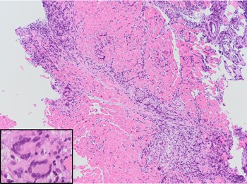
Right ventricular endomyocardial biopsy was performed. Histologic examination showed significant interstitial chronic inflammatory infiltrate composed of lymphocytes and eosinophils, with associated myocyte damage, and giant multi-nucleated cells with no granulomas – giant cell myocarditis was diagnosed (Fig 1). Biopsy viral screen was negative and bacterial DNA polymerase chain reaction was negative. The patient was started on triple immunosuppression with cyclosporine, azathioprine and corticosteroids.
Endomyocardial biopsy histology: diffuse myocardial infiltration by a chronic infiltrate composed of lymphocytes, eosinophils and scattered prominent multi-nucleated giant cells without granuloma formation (inset), with associated cardiomyocyte degeneration. (100× and 400× magnification).

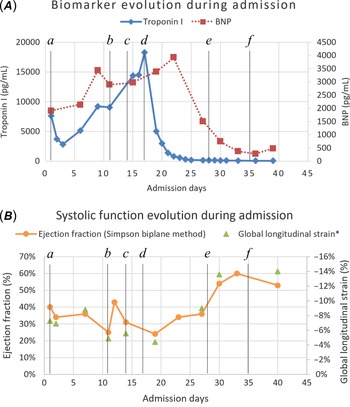
After initiation of immunosuppression, there was rapid and impressive clinical and biomarker improvement (Fig 2). Left ventricular systolic function improved and, after one levosimendan cycle, inotropes were weaned uneventfully. On discharge, both echocardiographic and laboratory parameters showed only mild abnormalities – ejection fraction 53%, global longitudinal strain −14%; troponin 55 pg/mL, B-natriuretic peptide 305 pg/mL; the electrocardiogram was normal. Immunosuppression was maintained, with slow reduction in corticosteroids.
Evolution of ( A ) laboratory and ( B ) echocardiographic parameters during admission, related to therapeutic interventions: a, oral anti-congestive medication; b, inotropic support and IV furosemide; c, intravenous immunoglobulin (2 g/kg total, divided in 5 days); d, cyclosporine-based triple immunosuppression; e, levosimendan; f, discontinuation of dopamine and milrinone. *Global longitudinal strain axis inverted for ease of reading, more negative values are better.

After 18 months of follow-up, the patient was asymptomatic, with negative cardiac biomarkers. On serial magnetic resonance imaging, there was evidence of progressive left ventricular dilation (120 mL/m2) with preserved systolic function, focal subepicardial fibrosis consistent with previous myocarditis overlapping on repeat exams, and there was no myocardial oedema. No active inflammatory process was seen.
Discussion
In our case, giant cell myocarditis presentation was severe acute heart failure (the most common presentation), associated with transient chest pain. Reference Cooper, Berry and Shabetai1,Reference Kandolin, Lehtonen, Salmenkivi, Räisänen-Sokolowski, Lommi and Kupari2 There was no associated autoimmune disorder identified, but that is the case in only ≈20% of patients. Reference Cooper, Berry and Shabetai1,Reference Kandolin, Lehtonen, Salmenkivi, Räisänen-Sokolowski, Lommi and Kupari2 However, a Leptospira infection may have been the trigger to the development of giant cell myocarditis. Our patient had epidemiological context, was treated with an adequate antibiotic during his first febrile episode (which can blunt serological response) and had positive direct observation on darkfield microscopy. Reference Levett8 Moreover, classic polymerase chain reaction has only modest sensitivity on the immune phase. A possible connection between giant cell myocarditis and Leptospira has been reported before by Vaideeswar and Cooper. Reference Vaideeswar and Cooper7
The diagnosis requires a histologic specimen fulfilling the Dallas criteria, and demonstrating the presence of giant multi-nucleated cells, without the presence of granulomas. Reference Blauwet and Cooper6 It is therefore essential to perform an endomyocardial biopsy to be able to diagnose giant cell myocarditis timely. Its use in patients with a clinical presentation of acute severe myocarditis is uncontroversial. Endomyocardial biopsy is a safe procedure, and is also indicated in children. Sensitivity is not perfect but increases with multiple samples, left/biventricular biopsy and magnetic resonance imaging guidance. Reference Kandolin, Lehtonen, Salmenkivi, Räisänen-Sokolowski, Lommi and Kupari2,Reference Kalra, Kneeland, Samara and Cooper9 Endomyocardial biopsy was not performed sooner due to the patient’s initial improvement.
Cyclosporin-based immunosuppressive regimens have demonstrated benefit for giant cell myocarditis. Reference Cooper, Berry and Shabetai1,Reference Kandolin, Lehtonen, Salmenkivi, Räisänen-Sokolowski, Lommi and Kupari2,Reference Maleszewski, Orellana, Hodge, Kuhl, Schultheiss and Cooper10 Our patient responded well to this therapy, with its value clearly demonstrated by the swift clinical, biomarker and echocardiographic improvement. As serial magnetic resonance imaging evaluation on follow-up also shows that there is no current myocardial inflammatory process, repeat biopsies were not performed.
Discontinuation of immunosuppression is not advised and reduction should be undertaken carefully, as recurrence is possible and can occur years after the diagnosis. Reference Maleszewski, Orellana, Hodge, Kuhl, Schultheiss and Cooper10 Monitoring of renal function and infectious complications related to immunosuppression is also paramount, as these are frequent in the adult giant cell myocarditis population. Reference Maleszewski, Orellana, Hodge, Kuhl, Schultheiss and Cooper10 Arrhythmic events are also frequent on follow-up, Reference Kandolin, Lehtonen, Salmenkivi, Räisänen-Sokolowski, Lommi and Kupari2,Reference Maleszewski, Orellana, Hodge, Kuhl, Schultheiss and Cooper10 and can be life-threatening. Until now our patient has had no late complications.
Children appear to have poorer outcomes than adults. Our patient, to our knowledge, is only the second-reported paediatric case in which the outcome was not death, heart transplant or listing for heart transplant. Reference Cooper3–Reference Lind-Ayres, Abramowsky and Mahle5 Generally, giant cell myocarditis patients who were transplanted were listed within 9 months of diagnosis, Reference Kandolin, Lehtonen, Salmenkivi, Räisänen-Sokolowski, Lommi and Kupari2 and our patient has surpassed that threshold by a large margin. Even though the disease is controlled, progression to dilated cardiomyopathy is possible in the future.
Supplementary material
To view supplementary material for this article, please visit https://doi.org/10.1017/S1047951121004327
Acknowledgements
The authors want to acknowledge Eugénia Pinto, MD from the Anatomic Pathology Department, the Paediatric Infectious Disease Unit of Hospital Dona Estefânia and the Leptospirosis Laboratory of the Instituto de Higiene e Medicina Tropical for their contributions.
Financial support
This research received no specific grant from any funding agency, commercial or not-for-profit sectors.
Conflicts of interest
None.