Introduction
Epidemiological and experimental studies published since 1980 have linked the influence of environmental factors to the development of diseases in adulthood. Reference Silveira, Portella, Goldani and Barbieri1 The DOHaD concept points out that insults occurring during periods of organ and system development can have lasting effects on the individual, altering the structure and/or function of these organs and systems and thus programming them for the occurrence of diseases in adulthood. Reference Khazipov and Luhmann2,Reference Barker3 According to the literature, the main insults that can program reproductive dysfunctions are those related to diet, exposure to pollutants and food contaminants, dysfunctions in the parents’ reproductive system, intrauterine growth restriction, and stress. Reference Silveira, Portella, Goldani and Barbieri1,Reference Ibáñez, Erthal and Ogo4 The main phases susceptible to programming diseases in adult life are the periods prior to conception, pregnancy, lactation, childhood and adolescence. Reference Barker3,Reference Barker, Eriksson, Forsén and Osmond5
In this context, studies using experimental models show that adolescence is also considered a critical stage of development Reference De Oliveira, Lisboa and De Moura6,Reference De Oliveira, De Moura and Miranda7 and show that protein restriction or a high-fat diet (HFD) during adolescence promote changes in the metabolism in adulthood. Reference De Oliveira, De Moura and Miranda7 As for the reproductive system, there are few studies in the literature using these models during adolescence, which is a period characterized by intense neuroendocrine and reproductive changes. Reference Salam, Bhutta, Koletzko, Koletzko, Bhatia, Bhutta, Cooper, Makrides, Uauy and Wang8 Proteins are essential for male fertility, contributing to processes such as sperm differentiation, motility, and function of ion channels. Reference Beeram, Suman and Divya9,Reference Ajuogu, Al-Aqbi, Hart and Wolden10 On the other hand, HFDs negatively impact male fertility by reducing testicular size, semen quality, and mating and fertilization success. Reference Crean and Senior11 These dietary patterns can disrupt reproductive health, leading to compromised male fertility outcomes. A previous study by our laboratory Reference Ibáñez, Erthal and Ogo4 showed that animals that consumed a HFD in adolescence showed an abnormal enlargement of the seminiferous tubule in adulthood, paired with a reduction in both the height of the seminiferous epithelium and the diameter of the seminiferous tubule, concluding that HFD can damage spermatogenesis and may be harmful in the long term.
All the programmed dysfunctions may depend on functional and structural alterations that are already in place during the pubertal period. However, no studies are known to have specifically evaluated, within the same experiment, the impact of both low-protein and HFDs on the testes of males during this critical developmental stage. Nutrition is known to play a fundamental role in human development, significantly impacting various aspects of health and well-being. During childhood and adolescence, a balanced diet rich in essential nutrients supports physical growth, bone development and the maturation of the reproductive system. 12 In addition, nutritional choices during these years can have long-term effects on adult health. Understanding the importance of a healthy and balanced diet at every stage of life is crucial to promoting development and well-being in individuals of all ages.
Therefore, the aim of the present study was to evaluate the effects of high-fat (HFD) and low-protein (LP) diets during the juvenile and peripubertal stages on testicular, epididymal, and sperm parameters in pubertal rats.
Materials and methods
Animals and experimental conditions and experimental design
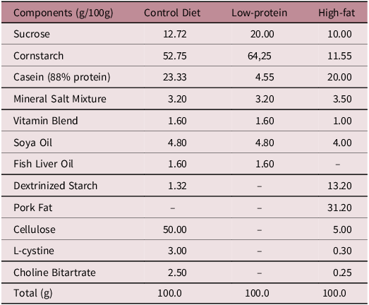
Male Wistar rats (n = 30) at postnatal day (PND) 25 were retrieved from the Central Animal Facility of the State University of Maringa and kept in the Sectorial Vivarium of the Department of Biotechnology, Genetics and Cell Biology for adaptation for 5 days. Animals were housed in a cage remaining under controlled temperature (22 ± 2°C) and photoperiod (07:00 AM – 07:00 PM, light cycle) conditions, with ad libitum access to water and food. At PND 30 the animals were assigned into three experimental groups; LP group: animals fed an isocaloric low-protein palletized (4% protein; LP; Table 1); HFD group: animals exposed to a HFD (35% protein; HFD; Table 1) or Control group: animals fed a balanced commercial diet (23% protein; Nuvital®, Curitiba/PR, Brazil; Control) until PND 60 with 10 animals each group, choice based on previous studies by the research group, randomly. At DPN 61, the animals were submitted to euthanasia by decapitation with a guillotine, without anesthesia. The HFD and LP groups were compared to the control group.
Composition of low-protein, high-fat and control diets

Testis, epididymis and sperm collection
The right testis and epididymis were removed, fixed in methacarn solution (10% acetic acid, 60% methanol and 30% chloroform) and destined to histological process (n = 5/group) and assessment of oxidative stress (n = 5/group). The left testis and epididymis were frozen and stored in −20°C freezer for posterior analysis of spermatic count (n = 5/group). The vas deferens were retrieved and had its content washed with formaldehyde (10%) and its sperm content stored in the refrigerator for posterior analysis.
Daily sperm production per testis and transit time in the epididymis
Decapsulated left testis and entire epididymis (n = 5/group) were weighed and homogenized as described previously by Robb et al., Reference Robb, Amann and Killian13 with the adaptations previously described. Reference Siervo, Vieira and Ogo14 After the dilution of homogenates, a small aliquot of sample was transferred to a Neubauer chamber (five fields per animal) for counting homogenization-resistant spermatids (that correspond to stage 19 of spermatogenesis) in the testis and spermatozoa in epididymis. To calculate the daily sperm production (DSP), the concentration of homogenization-resistant spermatids per testis was divided by 6.1, which corresponds to the number of days during which mature spermatids remain in the seminiferous epithelium. To calculate the sperm transit time through the epididymis, the number of sperm in each epididymal portion was divided by DSP.
Sperm morphology
The contents of the vas deferens (n = 5/group) were removed via internal rinsing with 0.5 mL of 10% formol-saline. Histological slides were prepared from this solution and observed using an Opton photomicroscope (400x magnification). One hundred spermatozoa were analyzed per animal. Morphological analysis was classified into three general categories: normal morphology, head abnormalities (without characteristic curvature or isolated form, i.e., no tail attached), and tails abnormalities (broken, rolled into a spiral and isolated, i.e., no head attached). This analysis was performed as described by Fernandes et al. Reference Fernandes, Arena and Fernandez15
Mitochondrial activity
The mitochondrial activity of the sperm (n = 5/group) was determined as described by Silva et al. Reference Silva, Vendramini and Restelli16 with adaptations. Spermatozoa obtained from the vas deferens by internal rinsing with PBS enriched with 0.75% BSA were added in microtubes containing 200 µL of 3–30-diaminobenzidine (DAB) dissolved in PBS in a 1:3 (v/v) ratio and incubated at 37°C for 1 h in the dark. Smears were prepared on histological slides and fixed with 10% formaldehyde for 10 min. Two hundred cells were evaluated with a phase contrast microscope and visually classified as: DAB 1 (100% stained intermediate piece, indicating that the cells maintain a complete mitochondrial activity or little loss of mitochondrial activity, which may not lead to severe impairment of motility and fertilization capacity); DAB 2 (more than 50% staining in the intermediate part, indicating moderate loss in the mitochondrial activity, which might lead to impairment of motility and fertilization capacity); and DAB 3 (less than 50% staining in the intermediate part, indicating cells that maintain minimal energy production through oxidative phosphorylation, which may lead to impairment of motility and fertilization capacity).
Histopathological analysis
Testis and epididymis (n = 5/group) were fixed in methacarn solution (10% acetic acid, 60% methanol and 30% chloroform) and then put in alcohol 70%. Then, they were embedded in Paraplast® wax and cut into 5 um sections and stained with hematoxylin and eosin (H&E). The tissue was evaluated using a Motic microscope (Motic®, Richmond, Canada) (100 and 400x magnification). For the testes, random seminiferous tubular sections (100/animal) in three nonconsecutive testis cross sections were analyzed to detect the presence of abnormalities in these tubules: immature germ cells in the lumen, acidophilic cells, vacuoles, and tubular degeneration. In the epididymis, histopathological inspection was performed qualitatively in the caput-corpus and cauda of each animal as described by Favareto et al. Reference Favareto, De Toledo and Kempinas17
Number of sertoli and leydig cells
The number of Sertoli cell nuclei was determined in 20 cross sections of the seminiferous tubules (stages VII-VIII of spermatogenesis) per testis (n = 5/group) in each rat, under a light microscope at 400x magnification, as described by Nassr et al. Reference Nassr, Arena and Toledo18 The number of Leydig cells nuclei was counted in 10 random fields of interstitial tissue in each testis section per rat under a light microscope at 400x magnification. Reference Guerra, Sanabria and Leite19 The Leydig cell morphology was identified as described by Teerds and Huhtaniemi. Reference Teerds and Huhtaniemi20 The differentiation of cells is due to the following characteristics, according to the previous references: Sertoli cells feature a clear, triangular nucleus with a prominent nucleolus located in the epithelium of the seminiferous tubule, while Leydig cells display a centrally located, rounded nucleus, present in the testicular interstitial.
Seminiferous tubule diameters and seminiferous epithelium height
These assays were described by Favareto et al. Reference Favareto, De Toledo and Kempinas17 For evaluation of the seminiferous tubule diameter and epithelium height, 40 random testicular cross sections per animal (n = 5/group) in stage IX of the seminiferous epithelium cycle were examined. Seminiferous tubule diameters were measured using an Opton photomicroscope (400x magnification) and BELView software (version for Windows). Likewise, the seminiferous epithelium height was measured using the same tubules and methodology. The mean of four measures for the diameters and the mean of four measures for the heights were calculated and used in the statistical analysis.
Stereology analyses of the epididymis
For stereological analysis of the epididymis, 10 random epididymal cross sections per animal were captured and analyzed (n = 5/group). This analysis was performed by means of Weibel’s multipurpose graticulate, with 168 points superimposed on the images. By counting the overlapping dots in the seminiferous cord, lumen or interstitial compartment, it was possible to establish the respective proportions of each of these components in the epididymis for each experimental group, as described by Favareto et al. Reference Favareto, De Toledo and Kempinas17
Oxidative stress of the testis, epididymis and sperm cells
Tissue samples from the testis and epididymis caput and cauda were homogenized in 500 uL of PBS (pH 7.2) and centrifuged at 9500 g for 10 min at 4°C (n = 5/group). Protein quantification of the samples, derived from the centrifuged supernatant, was performed using the Bradford method with BSA as standard. Reference Bradford21 Samples were then normalized to 1 mg of protein per mL of solution, as described by Erthal et al., Reference Erthal, Siervo and Frigoli22 and used for the following analyses: lipid peroxidation and reduced glutathione (GSH).
Lipid peroxidation
Lipid hydroperoxides levels as thiobarbituric acid reactive substances (TBARS) were measured through lipid hydroperoxide, which is an intermediate product of fatty acids oxidation. Reference Aviram and Eias23,Reference Parfitt, Desomeaux, Bolton and Hartog24 Hydroperoxide was determined through ferric oxidation (Fe2+; ammonium ferric sulfate). The Fe3+ ion that is formed reacts with xylenol orange, forming a colorful compound detected through its absorbance at 560 nm. Reference Jiang, Woollard and Wolff25 The results are expressed as nanomoles of TBARS/mg of protein.
Reduced glutathione concentration
Concentration of GSH was determined as described by Rahman et al., Reference Rahman, Kode and Biswas26 with some adaptations made by Erthal et al. Reference Erthal, Siervo and Frigoli22 GSH levels were measured by adding 5,5-dithiobis-2-nitrobenzoic acid (DTNB) into the normalized samples, resulting in an yellow solution, having its absorbance read at 412 nm. The results are expressed as micromoles/mg of protein.
Statistical analysis
The statistical analyses were performed using software Prism 10.1.0 (Graphpad, 2023). All parameters were submitted to the Shapiro–Wilk test for normality and thus classified into parametric or nonparametric data. Data were compared using unpaired t-test for analysis of the epididymis and ANOVA for testicular analysis with Tukey’s a posteriori test, or the nonparametric Kruskal–Wallis test for testicular analysis and Dunnett test for analysis of the epididymis. Differences were considered significant when p < 0.05.
Results
Counting daily sperm production and per testicle
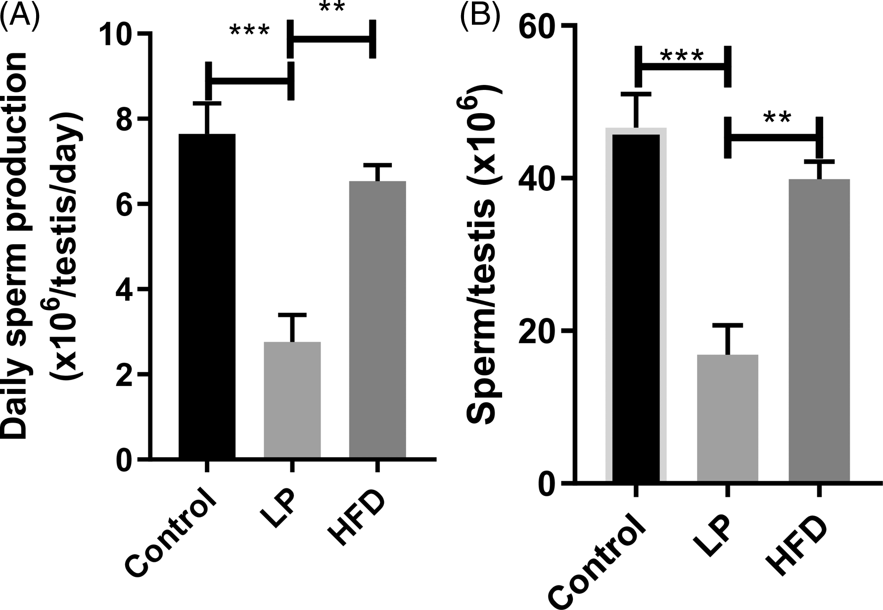
The daily sperm production (Fig. 1A) showed changes when comparing the experimental groups. Thus, the LP diet introduced to the animals showed a statistically significant difference, with a marked reduction in the DSP of these animals when compared to the Control and LP groups. In relation to sperm production per organ (Fig. 1B), the LP experimental group, as shown above, showed a significant reduction when compared to the other two groups analyzed.
Daily sperm production (A) and production of sperm per testicle (B) daily sperm production and sperm production per testicle in the animals of each group: the control group, the LP group fed a low-protein diet and the HFD group fed a high-fat diet. Values expressed as mean ± S.E.M. ANOVA with Tukey’s a posteriori test. **p < 0.005; ***p < 0.001.

Sperm transit time through the epididymis
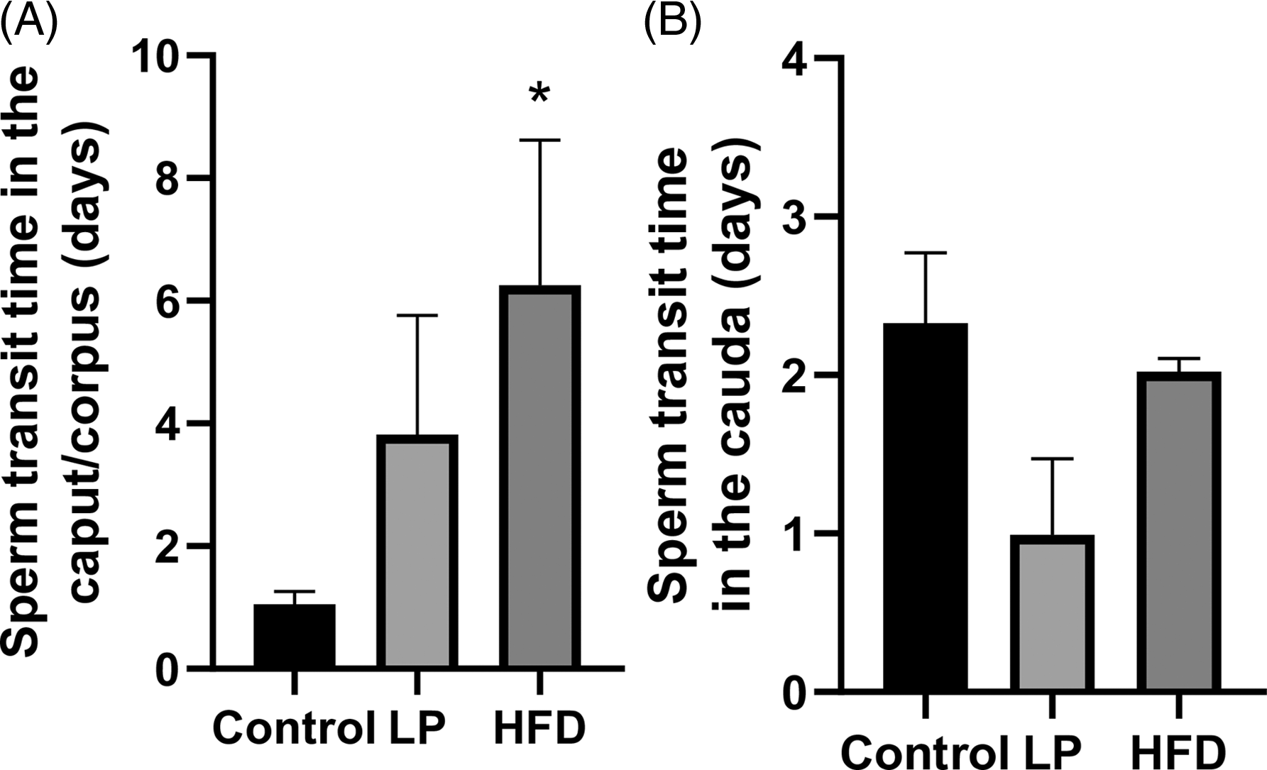
Regarding sperm transit time, in the caput region (Fig. 2A), there was a significant increase in the HFD group when compared to the control group, while in the cauda region (Fig. 2B), there were no significant differences in the diet groups when compared to the control group. Thus, we can say that the HFD was able to alter sperm transit time in the epididymal regions.
Sperm transit time through the caput (A), and cauda epididymal (B). Analysis of sperm transit through the epididymis in the animals of each group: the control group, the LP group maintained on a low-protein diet and the HFD group maintained on a high-fat diet. Results presented as mean ± S.E.M. ANOVA with Dunnett’s a posteriori test. *p < 0.05.

Sperm morphology
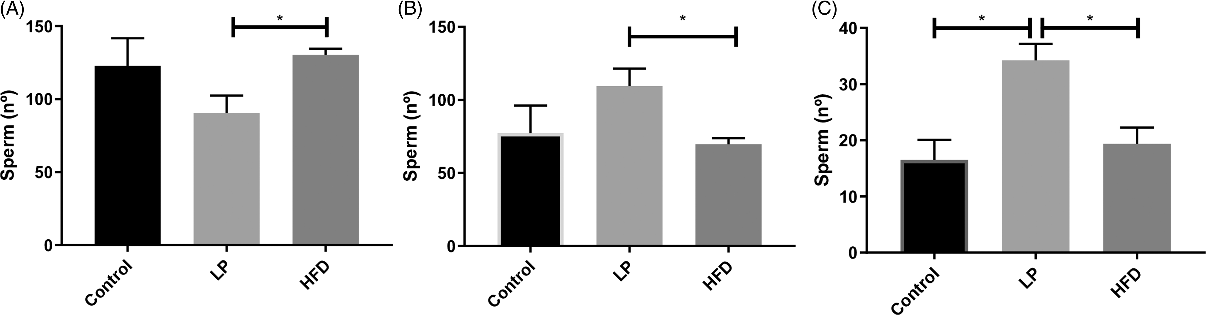
The sperm morphology parameters are highlighted in Fig 3. Compared to the control group, the results evidenced LP diet or HFD did not cause damage to the sperm morphology, regarding the proportions of normal and abnormal sperm. However, when the groups submitted to the diets were compared, there was a reduction in the number of normal sperm in the LP group compared to the HFD group (Fig. 3A). Therefore, the opposite result was observed in the number of abnormal sperm (Fig. 3B).
Normal (A), abnormal (B) sperm number morphology and abnormal sperm morphology with broken tail (C) parameters of normal (head and tail intact) and abnormal (head without characteristic curvature or isolated form, no tail attached, and tails broken, rolled into a spiral and isolated) sperm morphology in the animals of each group: the control group, the LP group maintained on a low-protein diet and the HFD group maintained on a high-fat diet. Values expressed in sperm number as mean ± S.E.M. ANOVA with Tukey’s a posteriori test. *p < 0.05.

Regarding the data shown in Fig. 3, the classification category of the abnormalities that stood out among the three groups was investigated separately. Thus, the broken tail abnormality (Fig. 3C) was the characteristic where the LP experimental group showed a significant increase in the number of sperm with this aspect, differing from the other experimental groups.
Mitochondrial activity
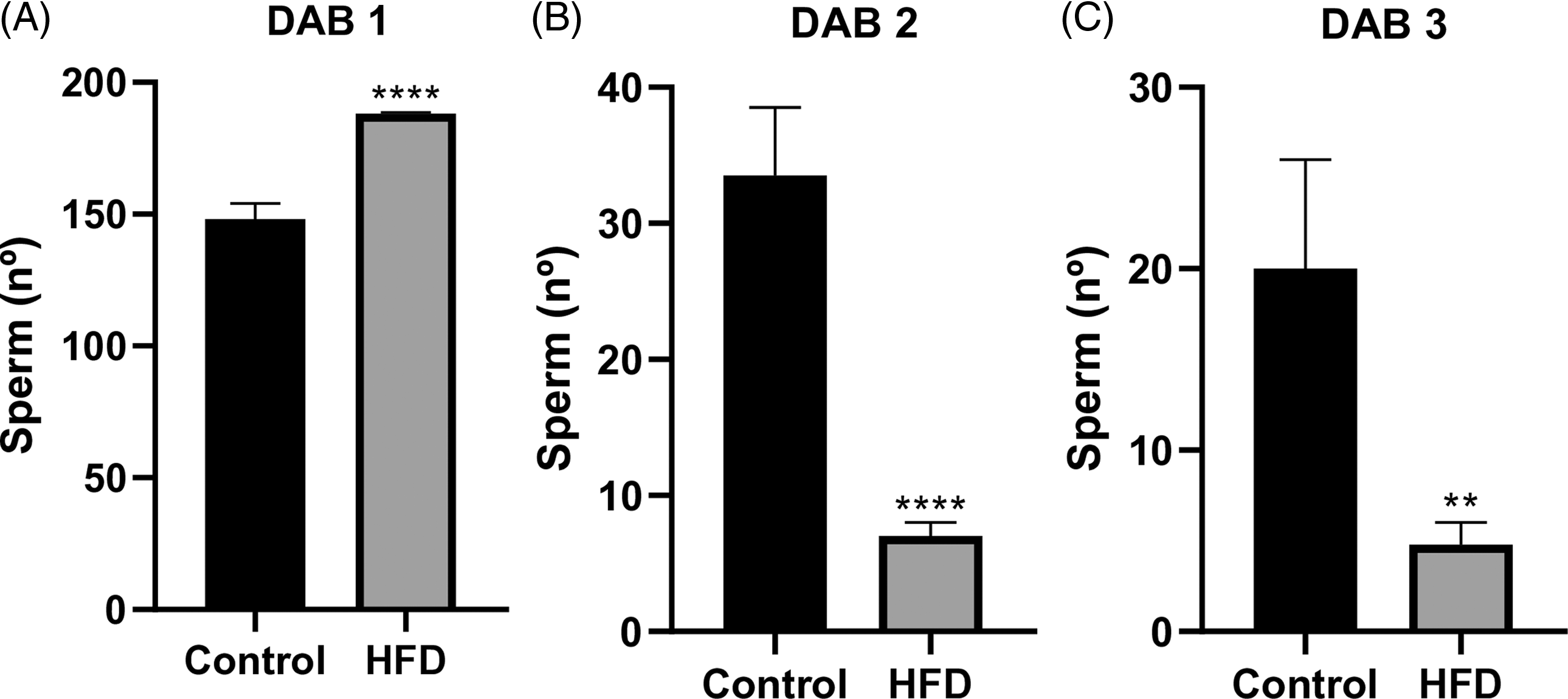
Compared to the control group, the analysis of mitochondrial activity (Fig. 4) revealed an increase in the percentage of sperm classified as DAB I because of a decrease in the percentage of sperm classified as DAB II and DAB III in the HFD group. Thus, there was a greater number of sperm with possible increase in mitochondrial activity in the animals exposed to the HFD compared to the control group. Thus, sperm classified in the other categories showed a significant reduction in the number of gametes with lower mitochondrial activity.
Sperm mitochondrial activity. Analysis of mitochondrial activity in the animals of the experimental groups, with the control and HFD groups. DAB 1 - number of sperm with a fully stained mitochondrial sheath; DAB 2 - number of sperm with a partially stained mitochondrial sheath; DAB 3 - number of sperm with an unstained mitochondrial sheath. Values expressed as mean ± S.E.M. Student’s t-test. **p < 0.005; ****p < 0.00001.

Histopathology of the testicles and epididymis
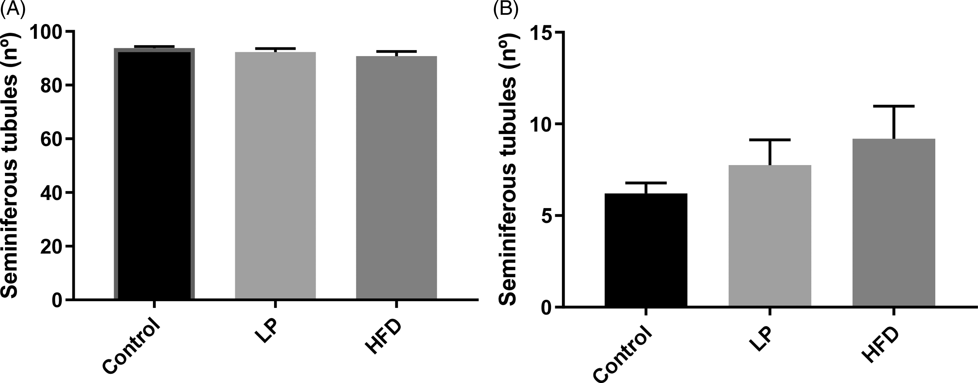
The histopathological analysis of the testicles of the animals used in the study did not show any statistically significant difference in relation to the seminiferous tubules, either normal (Fig. 5A) or abnormal (Fig. 5B) when comparing the three experimental groups.
Histopathology of normal (A) and abnormal (B) seminiferous tubules. Histopathological parameters of the testicles of the animals in each group: the control group, the LP group fed a low-protein diet and the HFD group fed a high-fat diet. Values expressed as mean ± S.E.M. ANOVA. p < 0.05.

In relation to epididymal histopathology assay, no significant differences were observed in the experimental groups when compared to the control group.
Counting the number of sertoli and leydig cells
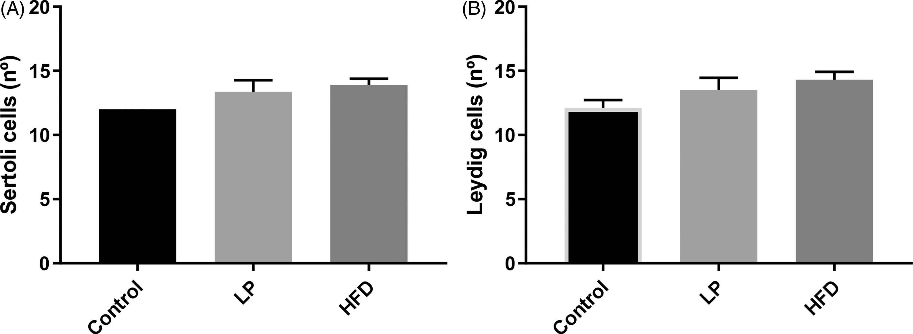
The number of Sertoli cells (Fig. 6A) and the number of Leydig cells (Fig. 6B) remained unchanged when comparing the three experimental groups.
Number of sertoli cells (A) and leydig cells (B) number of sertoli cells and number of leydig cells for each experimental group: the control group, the LP group maintained on a low-protein diet and the HFD group maintained on a high-fat diet. Values expressed as mean ± S.E.M. ANOVA. p < 0.05.

Diameter of the seminiferous tubules and height of the germinal epithelium
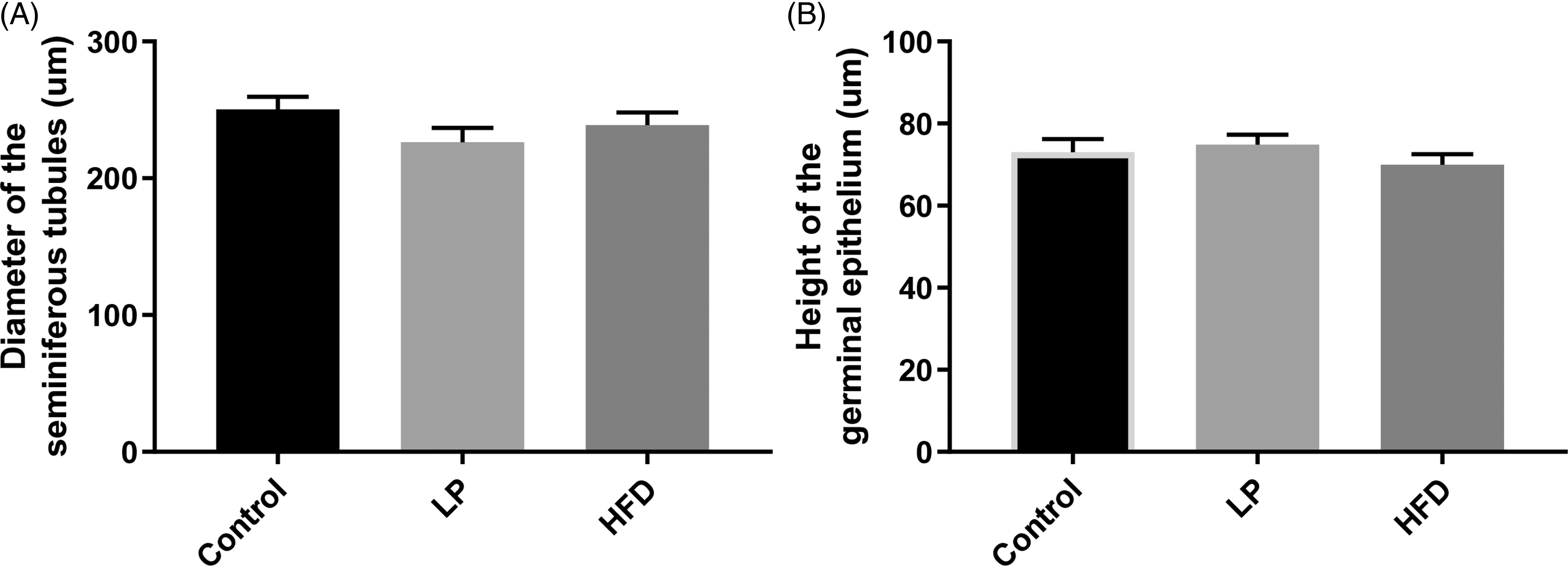
The analysis in which the diameter of the seminiferous tubule and the height of the germinal epithelium are measured. These analyses showed no change in the diameter of the seminiferous tubule (Fig. 7A) and the same occurred when comparing the height of the germinal epithelium (Fig. 7B).
Diameter of the seminiferous tubules (A) and height of the germinal epithelium (B) diameter of the seminiferous tubules and height of the germinal epithelium of the animals in each experimental group: the control group, the LP group fed a low-protein diet and the HFD group fed a high-fat diet. Values expressed in micrometers by mean ± S.E.M. ANOVA. p < 0.05.

Stereology of the epididymis
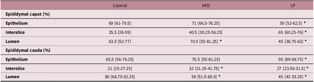
In the stereological analysis (Table 2) of the epididymal caput region, a significant decrease in the epithelial and luminal compartments was observed, as well as a significant increase in the interstitial compartment in the LP group when compared to the control group. In addition, a significant increase in the luminal region was observed in the HFD group when compared to the control group. As for the stereological analysis of the epididymis cauda, a significant increase in the epithelial compartment and a significant decrease in the luminal region were observed in the LP group when compared to the control, as well as a significant increase in the interstitial compartment and a significant decrease in the luminal compartment in the HFD group when compared to the control group. These results indicate that tissue remodeling occurred in the epididymis.
Epididymal stereology

Effect of low-protein and high-fat diets on the epididymal stereology of the animals in each experimental group: the control group, the LP group maintained on a low-protein diet and the HFD group maintained on a high-fat diet. Results are expressed as a percentage, representing the proportion of each compartment analyzed. Values expressed as median [Q1 - Q3]. Mann-Whitney test. *p < 0.05.
Lipid peroxidation
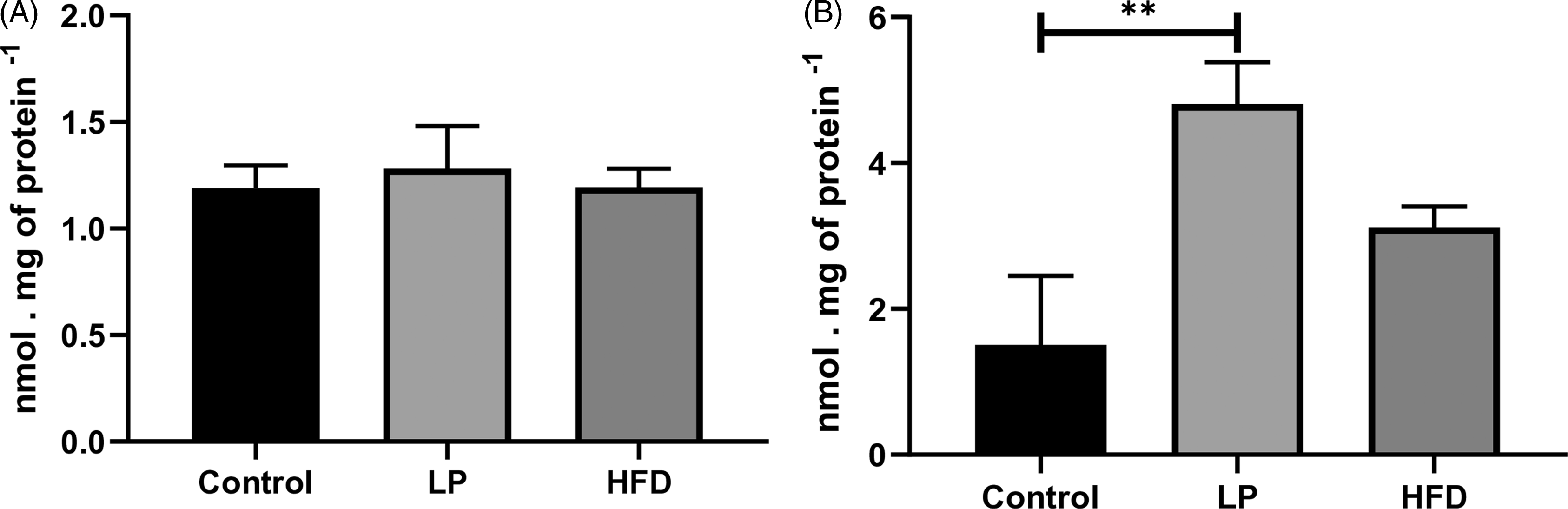
In the analysis of lipid peroxidation in the testicle (Fig. 8A), no statistically significant changes were seen when comparing the experimental groups with the control group, which differs from what was seen in the same analysis carried out on the epididymis (Fig. 8B). In the epididymis, the LP group showed an increase in lipoperoxidation when compared to the control group, demonstrating greater damage to the cell membrane in this organ.
Lipid peroxidation in the testicle (A) and lipid peroxidation on the epididymis (B) lipid peroxidation in the testicle and epididymis in each experimental group: the control group, the LP group fed a low-protein diet and the HFD group fed a high-fat diet. Values expressed in nanomol/mg of protein by mean ± S.E.M. ANOVA with Tukey’s a posteriori test. p < 0.05; **p < 0.005.

Reduced glutathione concentration
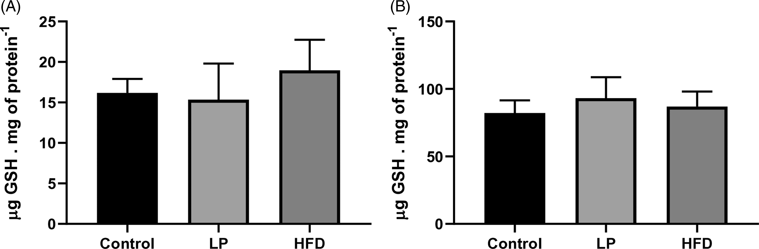
The concentration of reduced glutathione in the testes (Fig. 9A) showed no significant difference when comparing the experimental groups with the control group. The same occurred when the analysis was carried out on the epididymis (Fig. 9B) of the animals, showing no change in the concentration of this antioxidant.
Reduced glutathione concentration in the testicle (A) and epididymis (B) reduced glutathione concentration in the testicle and epididymis in each experimental group: the control group, the LP group fed a low-protein diet and the HFD group fed a high-fat diet. Values expressed in micromoles/mg of protein by mean ± S.E.M. ANOVA. p < 0.05.

Discussion
This study provides important information showing that protein restriction or a HFD administered during the peripubertal period causes significant changes in the sperm, testicles and epididymis of pubertal rats.
The present results did not show a reduction in the number of Leydig and Sertoli cells in the groups submitted to the HFD and LP diet, indicating no damage to spermatogenesis. Thus, Leydig cells, which are responsible for testosterone synthesis, play a crucial role in testicular maturation and in stimulating spermatogenesis. Reference Martin27 In turn, Sertoli cells, known for their morphophysiological support in spermatogenesis, provide an essential environment for germ cells throughout the process, and are coordinated by testosterone levels, with no change in these processes. Reference Martin27
In addition to their role in spermatogenesis, Sertoli cells play a fundamental role in spermiogenesis. This is a complex process which includes the elongation of spermatids, the condensation of nuclei, the development of the tail and acrosome, and the reduction of cytoplasmic excess, culminating in the formation of mature spermatozoa. Reference Griswold28 Given this, the morphology of the sperm in the LP group was impaired, and it can be concluded that the origin of these alterations was not related to the Sertoli and Leydig cells, which did not show any alterations. However, we believe that the notorious broken tail alteration observed in the protein-restricted diet groups may be related to the alterations presented in the analysis of the stereology of the epididymal cauda and caput, considering the possible influence of sperm on the differentiation of the epididymal epithelium, Reference Sofikitis and Giotitsas29 and the organ’s dependence on protein for its development and the capacitation and storage of sperm. Reference Sun and Flickinger30
As for the findings on the epididymis, sperm transit time depends on the contractile activity of the smooth muscle cells in this organ, influenced by androgens and autonomic neurotransmitters. Reference Robaire and Viger31 The epididymal epithelium, responsible for secreting proteins and factors essential for sperm maturation, can be affected by changes in the secretion pattern, impairing sperm quality. Reference Elfgen, Mietens, Mewe, Hau and Middendorff32,Reference Cornwall33 The lack of progression in the epididymis development observed in the cauda, where the spermatozoa are stored, Reference Cornwall33 suggests a possible stagnation in the proliferation of epithelial cells in this portion of the epididymal duct, which could compromise the environment for the spermatozoa. In addition, the reduction in the luminal compartment in LP and HFD groups, both in the caput and cauda, reflects the reduction in sperm production in the testes of these cells in the epididymis.
The stereological analysis of the epididymis shows that the protein restriction altered its proper postnatal development, showing a regressed histology in relation to the animal’s age, which may be due to the lack of adequate proliferation of the epithelial cells. It is important to evidenced that in the caput portion, the HFD group showed an increase in the luminal compartment, demonstrating an above-normal proliferation of these cells, while the LP group showed a reduction in the epithelial and luminal compartments, with an increase in the interstitial tissue. In the cauda portion of the epididymis, we infer that the diets prevented adequate postnatal development since the LP diet and HFD caused an increase in the interstitial compartment and a reduction in the lumen of the epididymal ducts, while only in the LP group there was a significant increase in the percentage related to the epithelial compartment. As there were no histopathological differences in the organ, it is understood that there is a possibility of hormonal interference in this process, as plasma androgens may be responsible for the growth of the epididymis during the undifferentiated period, as well as being necessary to maintain the structure and function, mainly epithelial, in the epididymis in the adult animal. Reference Dacheux and Dacheux34
Sperm transit time is crucial for the development of parameters such as the acquisition of motility. Reference Fernandez, Porto, Arena and Kempinas35,Reference Souza, Schorr-Lenz, Lucca and Bustamante36 For proper motility, it is vital that mitochondrial activation occurs in the middle part of the sperm, ensuring the availability of ATP. Reference Devi, Kushwaha, Maikhuri, Singh and Gupta37 Mitochondria are essential for oxygen processing and are both the source and target of oxidative stress. Lipids can behave as uncouplers and inhibitors of oxidative phosphorylation, but the detour of fatty acids to lipid droplets in the cytoplasm can prevent lipotoxicity. Direct delivery of lipids to the mitochondria ensures energy efficiency and redox control, preventing excess lipid storage. Reference Fernandez, Porto, Arena and Kempinas35 Thus, the increase in mitochondrial activity in the animals in the HFD group, exposed to a greater amount of lipids in the diet, was evidenced by the increase in cells classified as DAB I, indicating greater availability of energy for the sperm in this experimental group, but not guaranteeing its effects on sperm motility.
The oxidative stress test measured damage to cell membranes by assessing thiobarbituric acid reactive substances (TBARS) such as malondialdehyde and hydroxynonenal levels. GSH repair mechanism was also evaluated, showing no change in GSH concentration in either organ. Reference Piner and Üner39 Despite the increase in lipoperoxidation in the epididymis of the LP group, spermatogenesis or spermiogenesis were not affected. Reference Salem, Salem and Maarouf40 The epididymis is a site with high oxidative metabolism, essential for reproductive functions, so oxidative stress, measured by TBARS, can compromise sperm maturation and reduce sperm quality. Reference O’Flaherty and Scarlata41 These data are in line with the results of the epididymis stereology, which showed compromised tissue compartments and altered sperm morphology in the LP group.
Based on the information provided, we conclude that the influence of low-protein, HFDs during peripubertal age had no adverse effects on postnatal testicular development in Wistar rats. However, these diets had a negative impact on sperm quality, suggesting possible implications for fertility and the development of the male reproductive system. The results indicate that both protein restriction and a HFD during peripuberty affected epididymal development and sperm quality, influencing sperm morphology. Also, proper nutritional monitoring during windows of developmental plasticity, emphasizing the adolescent period, is extremely important.
Acknowledgments
The authors are grateful to State University of Londrina and ProPPG-UEL (Office of the Dean of Research and Postgraduate Studies) for providing a Scientific Initiation scholarship to L. P. Bomfim and M. S. S. Silva. The authors also thank the Academic Writing Center of the State University of Londrina for assistance with English language developmental editing.
Funding statement
This research did not receive any specific grant from funding agencies in the public, commercial, or not-for-profit.
Competing interests
The authors declare no competing interests.
Ethical standard
The experimental procedures described in this study were performed in accordance with the Ethical Principles in Animal Research adopted by the Brazilian College of Animal Experimentation and were approved by the Ethics Committee on Animal Use of the State University of Maringá (CEUA/UEM protocol no. 2910011021).