Introduction
Local drug delivery system has been accepted as an alternative to conventional oral dosage to address challenges such as poor targeting and efficacy, severe toxicity in nontarget tissues, and uncontrolled pharmacokinetics [Reference Alkhraisat, Rueda, Cabrejos-Azama, Lucas-Aparicio, Mariño, Garcia-Denche, Blanco Jerez, Gbureck and Cabarcos1, Reference Otto, de Villiers, de Villiers, Aramwit and Kwon2]. Calcium phosphate (CaP)–based implants, especially HA, have been proven as excellent materials for tissue integration [Reference Bandyopadhyay, Bernard, Xue and Bose3, Reference Rey4, Reference Dasgupta, Banerjee, Bandyopadhyay and Bose5, Reference Nandi, Fielding, Banerjee, Bandyopadhyay and Bose6] and also as reservoirs for slow drug elution for extended time periods with a variety of surface modifications [Reference Vallet-Regí, Balas and Arcos7, Reference Bose and Tarafder8]. Matrix degradation mechanism, drug solubility, polymer–drug interactions, and the surface microstructure have been shown to be pivotal in controlling the drug release kinetics from these CaP-based drug delivery devices [Reference Bose and Tarafder8, Reference Reginster and Burlet9, Reference Pastorino, Canal and Ginebra10].
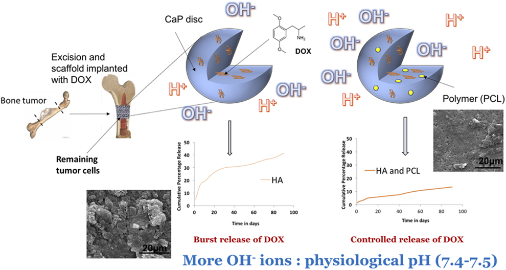
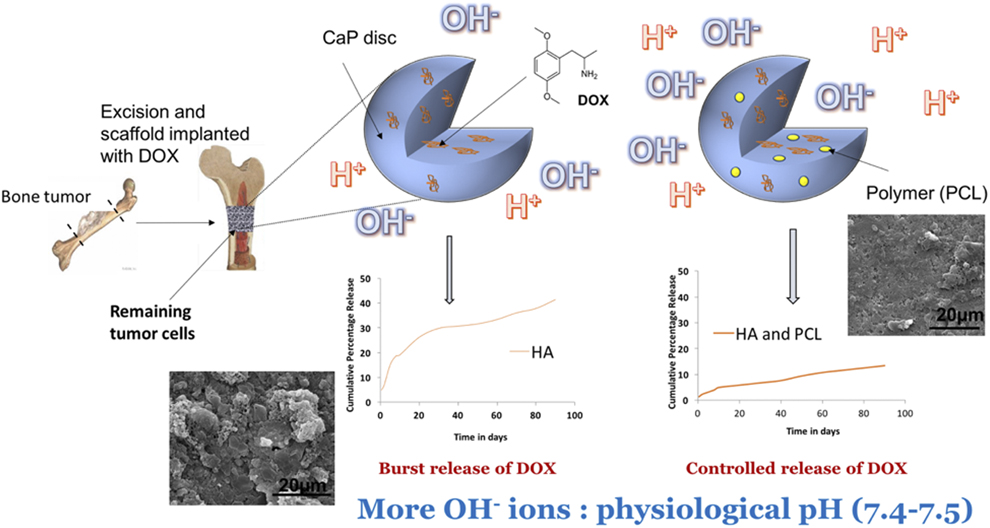
Localized drug delivery still poses an important challenge in the treatment of bone cancer. Much research has been dedicated in the quest of developing technologies for efficient local drug delivery to help in selectively eradicating the remnant cancer cells after excision of bone tumor with negligible effects on the healthy bone cells. Although drugs consumed orally are delivered to all body cells by getting absorbed in the blood stream, however, cancer drugs have adverse effects on the noncancerous bystander cells [Reference Kantoff, Higano, Shore, Roy Berger, Small, Penson, Redfern, Ferrari, Dreicer, Sims, Xu, Frohlich and Schellhammer11, Reference Lee, Barrett, Mackall, Orentas and Grupp12, Reference Vasir and Labhasetwar13]. Also, cellular uptake of the drugs through the plasma membrane of the tumor in appropriate and efficient dosage is still a difficult work. Hence, this study intends in investigating the effects of incorporation of biocompatible polymers in controlling and sustaining the local release of a commercial cancer drug doxorubicin with minimal effects on healthy bone cells. Figure 1 shows the application of a CaP-based controlled doxorubicin delivery vehicle used for the eradication of remaining tumor cells after excision [Reference Banerjee and Bose14].
The application and utility of a calcium phosphate based controlled doxorubicin delivery vehicle which can be used for eradication of residual bone tumor cells after excision [Reference Banerjee and Bose14].
Doxorubicin is a chemotherapeutic drug used commonly in cancer treatment [Reference Banerjee and Bose14]. It is classified as a Class I anthracycline antibiotic and deemed effective in the treatment of leukemia and other kinds of tumors, especially breast tumors. Studies have shown metastases of breast cancers as one of the most common reasons for the bone tumors [Reference Kang, Siegel, Shu, Drobnjak, Kakonen, Cordón-Cardo, Guise and Massagué15], showing the rationale behind the usage of doxorubicin for the development of the bone tissue engineering–based delivery system. The drug causes cell death by acting on the S phase of the cell cycle by the inhibition of topoisomerase II enzyme activity [Reference Capranico, Tinelli, Zunino, Kohn and Pommier16]. After topoisomerase II breaks the DNA duplex for replication, it is intercalated and stabilized by doxorubicin, preventing the resealing of the DNA double helix. Free doxorubicin has very low therapeutic index, and the oral dosage needed to be administered for appropriate therapeutic requirement is quite high. The administered dosage through intravenous injections causes toxic effects in nontarget tissues [Reference Pigram, Fuller and Hamilton17]. The FDA approves the dosage of 50–75 μg/m2 of the drug to be administered every 3–4 weeks if used as a single agent. A person can be exposed to a cumulative dosage of 500 mg/m2 of the drug in his lifetime [18]. So, an adequate local CaP drug carrier system is required for the delivery of doxorubicin for bone tissue engineering applications.
Burst release of drug has always been a concern for CaP-based delivery vehicles [Reference Bose and Tarafder8] and hinder the efficient treatment of bone cancer. Drug delivery through bioactive and biodegradable polymers incorporated in HA implants is an effort to clinch release at the most desired dosage and rate at the site of implantation for better tumor excision [Reference Xue, Bandyopadhyay and Bose19]. Poly glycolic lactic acid (PLGA) has been used as a potential agent for controlling drug release because of the inherent properties of biodegradability and tailorable mechanical properties, along with an exhibition of a variety of degradation kinetics in the physiological system [Reference Takeuchi, Takeshita, Suzuki and Makino20] and used extensively as drug carriers in scaffolds used for hard and soft tissue repair. Polycaprolactone (PCL) is another bioactive polymer explored for potential uses as drug carrier due to its low cost, processing ease, and inherent biodegradability [Reference Coombes, Rizzi, Williamson, Barralet, Downes and Wallace21, Reference Tarafder, Nansen and Bose22].
Although a lot of research has been advocated to the mechanism of doxorubicin activity in the human body and its effects on the sarcoma cells [Reference Kang, Siegel, Shu, Drobnjak, Kakonen, Cordón-Cardo, Guise and Massagué15, Reference Capranico, Tinelli, Zunino, Kohn and Pommier16], no studies have been done till date to the best of our knowledge to analyze the release of doxorubicin from polymer and CaP matrix to be used as a treatment of bone cancer. Thus, the novelty of this study lies in the development of a polymer ceramic composite as a sustained release vehicle for the cancer drug doxorubicin. We hypothesize that the interactions of doxorubicin with the hydrophobic polymers, PCL and PLGA will be able to control the release kinetics. The report provides a comparison of how the chemistry and varying amounts of the polymer into HA-based implants alter the release kinetics of doxorubicin in the physiological system. The understanding of the ceramic polymer composite with controlled release characteristics can prove to be a potential drug carrier vehicle.
Results
Cumulative release behavior of doxorubicin
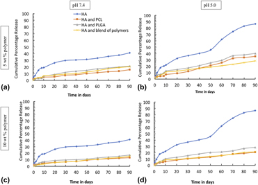
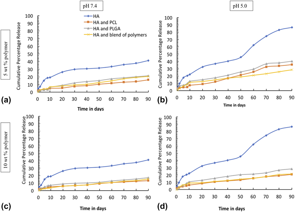
Figure 2 shows the cumulative percent release of doxorubicin at pH 7.4 phosphate buffer and pH 5.0 acetate buffer. All the compositions were stocked up with similar amount of doxorubicin, 50 µg per sample (DOX). Burst release of around 20% of DOX was noted from the bare uncoated HA samples within 10 days at pH 7.4. The cumulative percent release of doxorubicin from different concentrations of PCL has also been compared in the plots. Figure 2(a) shows the release data from 5% PCL incorporated samples, whereas Fig. 2(c) shows the release data from 10% PCL-incorporated samples at pH 7.4 and Figs. 2(b) and 2(d) show the similar release date at pH 5.0. Faster release was observed from 5% PCL-incorporated samples compared to 10% PCL-incorporated samples as seen from plots Figs. 2(a) and 2(c). The higher polymer concentration has proved to be more efficient in terms of controlled release kinetics. The release data at pH 7.4 for both the PCL concentrations show a plateau region after a burst release in the initial days. For both PCL concentrations, faster release kinetics was observed at pH 5.0 compared to pH 7.4. DOX release behavior from 5 to 10% PLGA incorporated HA samples at pH 7.4 phosphate buffer and pH 5.0 acetate buffer at pH 7.4 has been presented in Figs. 2(a) and 2(c) and at pH 5.0 in Figs. 2(b) and 2(d). The total amount of DOX release decreased at both pH 5.0 and pH 7.4, with increased PLGA concentration, as was expected. A higher initial and final DOX release was observed at pH 5.0 compared to pH 7.4 for both the concentrations of PLGA. A very similar trend of release kinetics has also been presented with HA samples incorporated with different concentrations of blend of PCL and PLGA. The blend of polymers shows an overall sustained release instead of burst release at the initial period. At pH 5.0, release is very similar in cases of 10 wt% blend of polymers and 10 wt% PCL. Overall, polymer incorporation has been able to control the release kinetics compared to the bare HA samples.
The cumulative percentage release of doxorubicin from HA, HA and PCL composite, HA and PLGA composite and HA and blend of PCL and PLGA polymer composites at (a) pH 7.4 with 5 wt% polymers (b) pH 5.0 with 5 wt% polymers (c) pH 7.4 with 10 wt% polymers (d) pH 5.0 with 10 wt% polymers (n = 3). Faster release pattern has been noted at pH 5.0 than pH 7.4. Also, lower release of the drug has been seen in polymer incorporated composites compared to the control HA discs.
Surface morphologies by SEM before release
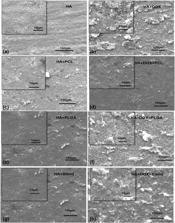
Figure 3 shows the SEM morphologies of the polymer-infused HA samples prior to the release study. Figures 3(a) and 3(b) show the SEM morphologies of the bare HA and HA incorporated with DOX. Figures 3(c) and 3(d) show surface morphologies of HA incorporated with 5% PCL solution in acetone (w/v) with and without the presence of the drug. The samples indicate a rough surface with the appearance of pores in them. Figures 3(e) and 3(f) show the SEM morphologies of HA samples coated with 5% PLGA solution in ethanol with and without the drug.
The surface morphologies of (a) bare HA samples (b) bare HA + DOX (c) HA + 5 wt% PCL (d) HA + DOX + 5 wt% PCL (e) HA + 5 wt% PLGA (f) HA + DOX + 5 wt% PLGA (g) HA + 2.5 wt% PCL + 2.5 wt% PLGA (h) HA + DOX + 2.5 wt% PCL + 2.5 wt% PLGA, before any release study was performed. All the DOX marked samples contain 50 µg of doxorubicin.

Much bigger pores are noted in the PLGA-incorporated samples, which may be a reason for the higher release kinetics observed from these samples. Figures 3(g) and 3(h) show the surface morphologies of the HA coated with 5% blend solution in acetone (w/v) with a constant DOX amount of 50 µg per sample before any drug release.
SEM morphologies after release
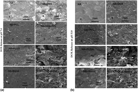
SEM surface morphology of all the compositions after DOX release from HA discs and PCL and PLGA HA discs at pH 7.4 and 5.0 have been shown in Figs. 4(a) and 4(b), respectively. A visibly porous morphology has been noted in all the compositions after release at pH 5.0 which is due to the polymer degradation in the acidic release media. The more porous surface morphology of the PLGA after release at pH 5.0 was attributed to the faster degradation of the PLGA.
The surface morphologies of (A) bare HA samples (B) bare HA + DOX (C) HA + 5 wt% PCL (D) HA + DOX + 5 wt% PCL (E) HA + 5 wt% PLGA (F) HA + DOX + 5 wt% PLGA (G) HA + 2.5 wt% PCL + 2.5 wt% PLGA (H) HA + DOX + 2.5 wt% PCL + 2.5 wt% PLGA, after release study was done at (a) pH 7.4 and (b) pH 5.0. All the DOX marked samples contain 50 µgs of doxorubicin. Faster release of DOX from HA-PLGA and HA-Blend observed in Fig. 2 can be explained from higher degradation of polymer matrix for HA-PLGA and HA-Blend composite compared to the HA-PCL composite.

Surface property analysis by contact angle measurement
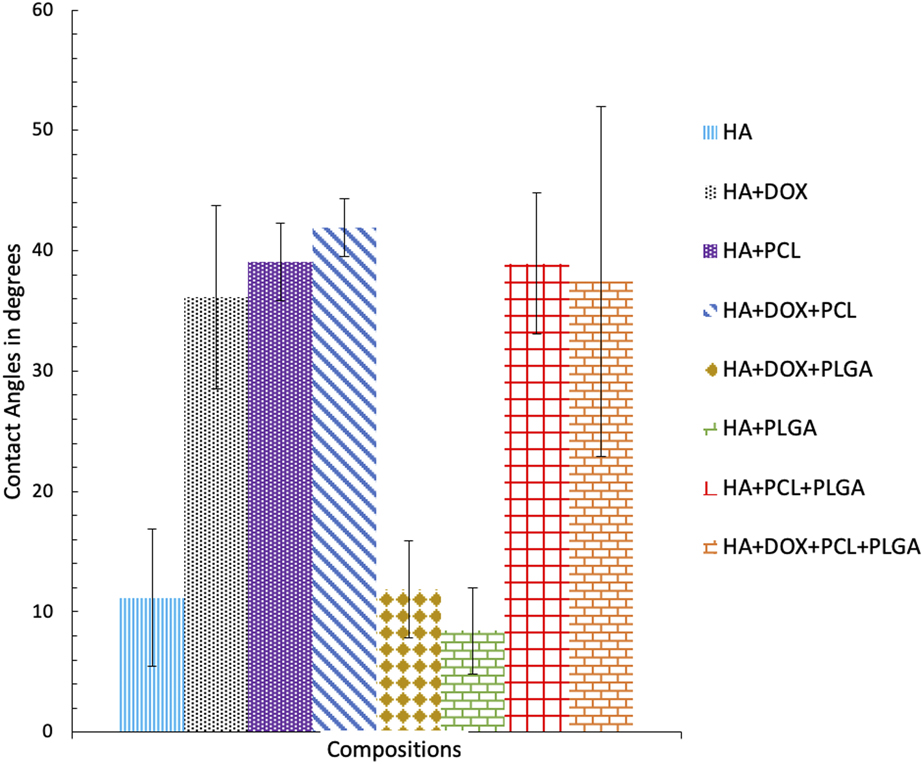
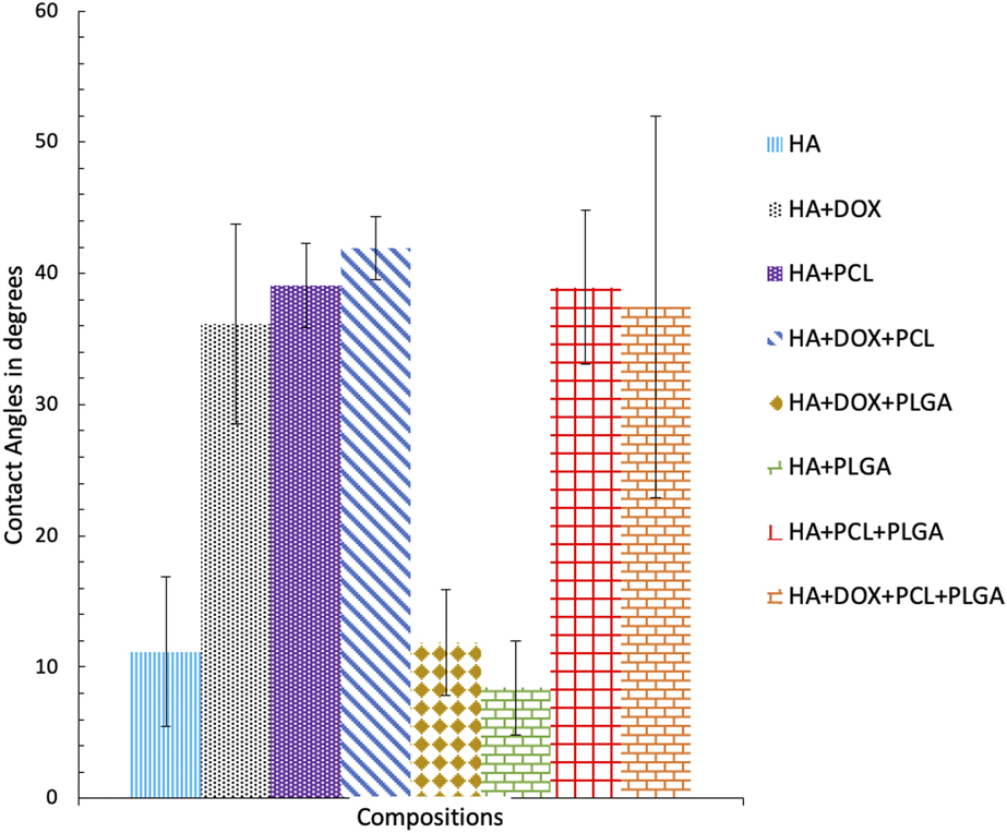
Figure 5 showing the contact angle values indicated that the surface property changes with the change in the type of polymer and its concentration. The contact angle of any surface depends on many factors such as chemistry and surface morphology of the samples like surface roughness. Increased wettability has been observed in the samples incorporated with PCL. Irrespective of the compositions, the samples with the drug in them show higher contact angles compared to their nondrug counterparts. This can be explained by the hydrophilicity of the drug in neutral or higher pH values. The samples incorporated with PLGA show the lowest contact angle, so more will be the surface energy of the samples. PCL is more hydrophobic than PLGA and thus lower contact angles have been witnessed with PLGA as the polymer. Increased wettability of the PLGA samples would be useful for better cell adhesion and proliferation. The more interaction of the PLGA-incorporated samples with the body fluid also explains the higher release kinetics from these samples. Samples with PCL show higher contact angles, which is consistent with the fact that it interacts less with the body fluid and entraps the drug better, leading to more sustained rate of release.
The contact angle values obtained with the various compositions. The contact angles are a direct reflectance of the surface energies of the samples which influences the cell attachment. Higher the contact angle shows higher the surface energies.
Bone cell materials interaction
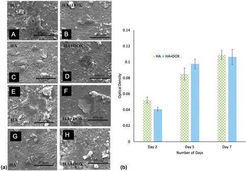
Figure 6(a) shows the growth and proliferation of human osteoblast cells on the samples after 7 days of cell seeding. This clearly indicates that the drug does not have any cytotoxic effect on the samples at the early time points. This is more clearly proven by the MTT assay shown in Fig. 6(b), which shows similar number of cells on both HA control and HA incorporated with the drug.
(a) Human osteoblast cells on (A, C, E, G) bare HA samples after 1, 2, 5, and 7 days of culture and (B, D, F, H) show the cell growth on HA samples with a constant amount (10 µg) of drug incorporated in it. (b) Presents cell viability on HA and HA + DOX samples after 2, 5, and 7 days of culture. No significant alteration in cell viability proves that the incorporated doxorubicin dosage is not toxic at the early experimental days (n = 3).
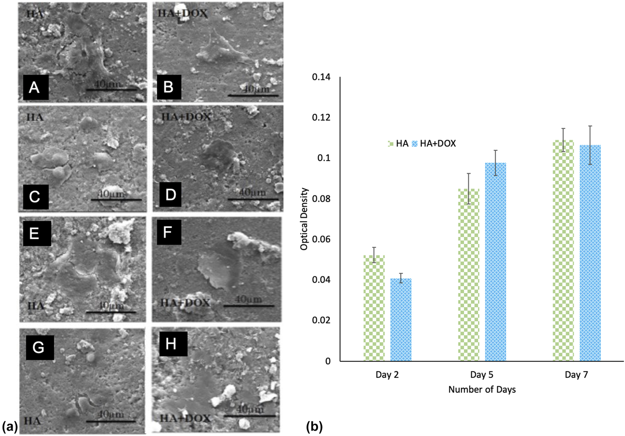
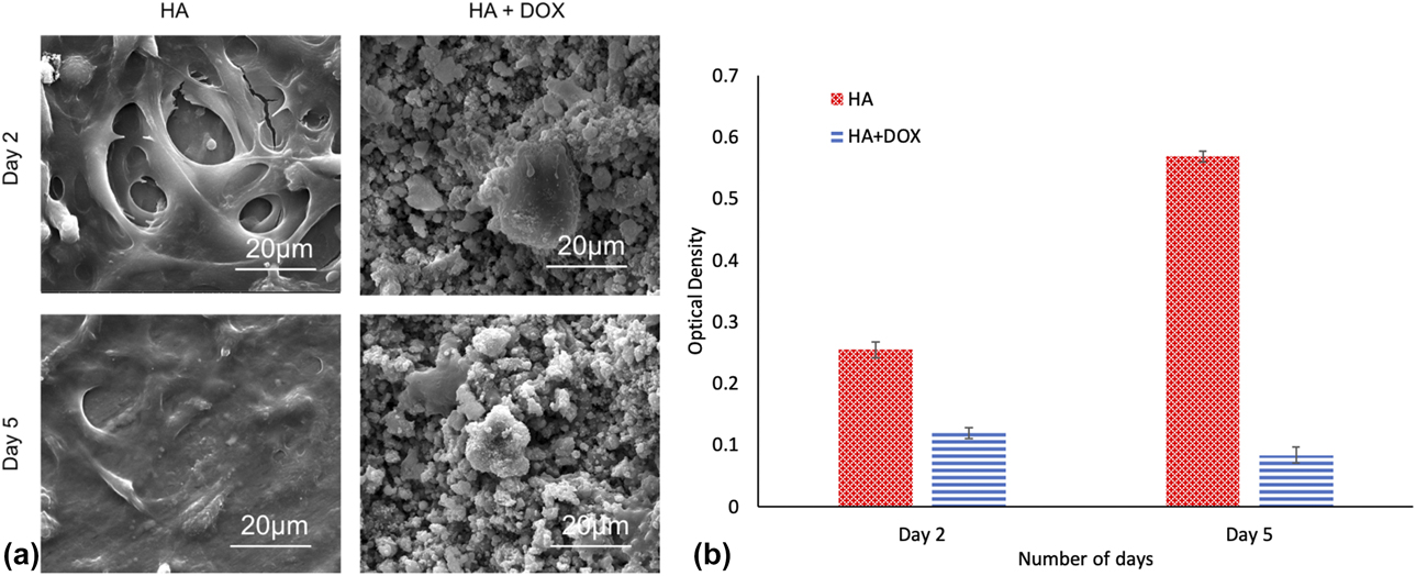
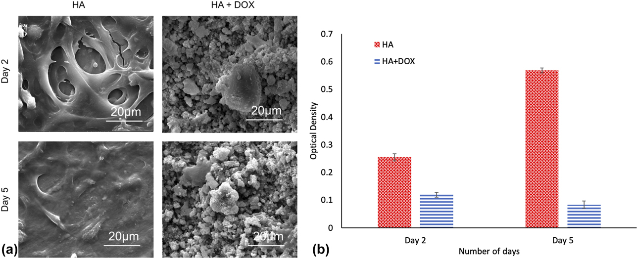
Figure 7(a) represents the osteosarcoma cell attachment and proliferation after 2 and 5 days of culture. This indicates the early stage efficacy of doxorubicin in reducing osteosarcoma cell growth. The results are in coherence with the quantitative cell viability data demonstrated in Fig. 7(b).
(a) Human osteosarcoma cells on bare HA samples and 10 µgs of DOX incorporated samples after 2 and 5 days of culture demonstrating the effectiveness of DOX in abating the osteosarcoma cell growth. (b) Presents osteosarcoma cell viability on HA and HA + DOX samples after 2 and 5 days of culture. Lower cell viability by DOX incorporation after both 2 and 5 days of culture demonstrates the potency of doxorubicin to be used for bone cancer treatment (n = 3).
Discussions
Strong or weak interactions attach the drug molecules on any biomaterial surface [Reference Kunieda, Seki, Nakatani, Wakabayashi, Shiro, Inoue, Sougawa, Kimura and Harada23]. Stronger interactions between the drug molecules and the body fluid or the release media often oust these weak interactions between the drug and the biomaterial surface [Reference Bose and Tarafder8, Reference Reginster and Burlet9, Reference Pastorino, Canal and Ginebra10, Reference Tarafder, Nansen and Bose22, Reference Tarafder, Banerjee, Bandyopadhyay and Bose24]. Burst release characteristics is observed as soon as the drug molecules loaded on a biomaterial surface locally come in contact with the release media [Reference Alkhraisat, Rueda, Cabrejos-Azama, Lucas-Aparicio, Mariño, Garcia-Denche, Blanco Jerez, Gbureck and Cabarcos1, Reference Otto, de Villiers, de Villiers, Aramwit and Kwon2] or the body fluid [Reference Bose and Tarafder8]. Burst release from very high dose of drug can have adverse effects on the target cells. Owing to the weak electrostatic interactions with drug molecules, burst release accompanied by controlled release has also been noticed from CaP matrix [Reference Tarafder, Banerjee, Bandyopadhyay and Bose24] or CaP coatings on metallic samples [Reference Nyan, Miyahara, Noritake, Hao, Rodriguez, Kuroda and Kasugai25]. A similar burst doxorubicin release trend had been observed from the bare HA shown in Fig. 2 [Reference Tarafder, Nansen and Bose22], and thus, it called for the necessity to develop a controlled drug release carrier vehicle for biomedical applications. Over some time now, experiments have presented the usage of biodegradable polymer coating in controlling the release kinetics of a drug on an implant surface [Reference Radin, Campbell, Ducheyne and Cuckler26, Reference Kim, Knowles and Kim27]. The pH of the release media, drug concentration, polymer concentration, and the drug interaction with the polymer affect release kinetics of the drug molecules from an implant surface. It is very crucial to have a proper understanding of these interactions to be able to successfully design a polymer-based controlled release CaP drug delivery vehicle [Reference Vallet-Regí, Balas and Arcos7, Reference Bose and Tarafder8]. For our experimental purposes, we solubilized the drug and the HA powder in water and stirred vigorously, ensuring a homogeneous mixture of the powder along with the drug molecules. Doxorubicin and PCL, and doxorubicin and PLGA were soluble in acetone. For the blend of the polymers, the drug and the polymers were made soluble in acetone. To rule out any chances of deviation in the release behavior, cropping in from the variation in polymer-intercalated microstructure and difference in processing parameters, environmental conditions and the processing time were kept uniform for all sample preparations.
Doxorubicin release has been investigated in two different pH values. PH 7.4 is the physiological pH of the body, whereas the pH drops significantly to around 5.0 in case of infection and inflammation. Higher release of doxorubicin has been observed at lower pH of 5.0 irrespective of the compositions. This specifies a favorable interaction of the dissociated doxorubicin hydrochloride with the release media at lower pHs. Release kinetics of drug molecules from the polymer incorporation is highly influenced by the polymer solubility in the release media [Reference Li, Hardy and Gu28]. Usually diffusion is a much faster process compared to the degradation of the polymer or the calcium phosphate matrix and release kinetics is mostly dominated by the diffusion process. In this study, diffusion dominated release behavior at pH 7.4 and polymer and calcium phosphate matrix degradation dominated release at pH 5.0 has been noted [Reference Tarafder and Bose29].
Doxorubicin release from the 5 wt% PCL and PLGA increased linearly for around 70 days before reaching a plateau. Thus, early on it was a diffusion-controlled release, after which polymer degradation and interactions with the drug and polymer influenced the release kinetics. Solubility of HA is substantially higher at acidic pH as compared to basic pH. Physicochemical interactions, electrostatic interactions, diffusion of drug molecules, matrix degradation, or external processes are deemed to have an impact on the doxorubicin release behavior. The porous structure observed after release in the microstructures of all the compositions was due to the matrix degradation in acidic pHs, shown in Figs. 3 and 4. The visible changes of degradation in the higher pH of 7.4 after 90 days of release were mainly seen due to the polymer degradation after such a long course of study. Whereas in the acidic pH, with the considerable degradation of the ceramic–polymer matrix, more and more doxorubicin came in contact with the acidic environment, leading to a hike in the release kinetics 50 days from the release was started.
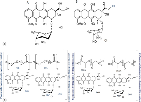
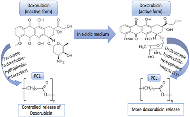
The favorable and unfavorable hydrophilic–hydrophobic interactions between the drug molecule and the polymer can be explained from the chemical structures of doxorubicin hydrochloride and PCL and PLGA. The chemical structures of the active and the inactive form of the drug have been shown in Fig. 8(a). In the lower pH, in an abundance of hydrogen ions, the doxorubicin accepts a proton and forms a cation. This cationic form of the drug is more hydrophilic in nature and readily interacts with the release media. Thus, we see higher release kinetics of the drug at the lower pHs. In the higher pH, the drug remains in an undissociated form which is more hydrophobic, and the deprotonated form of the drug remains more stable in the release media in the presence of the hydrophobic polymer. The favorable hydrophobic–hydrophobic interactions of the polymer with the drug molecule help in the sustained release kinetics. With a fixed amount of the drug, more favorable hydrophobic interactions are witnessed with increase in polymer concentration. Thus, a more sustained release is seen with 10 wt% of polymer concentration compared to 5 wt% of the polymer concentration.
(a) (A) Unprotonated inactive and (B) protonated active form of doxorubicin. (b) Presents a schematic representation of the steps involved in the release of DOX from the HA surface in the presence of PCL and PLGA in acidic or basic release medium, primarily due to the shift driven equilibrium change and/or rate limiting HA dissolution.
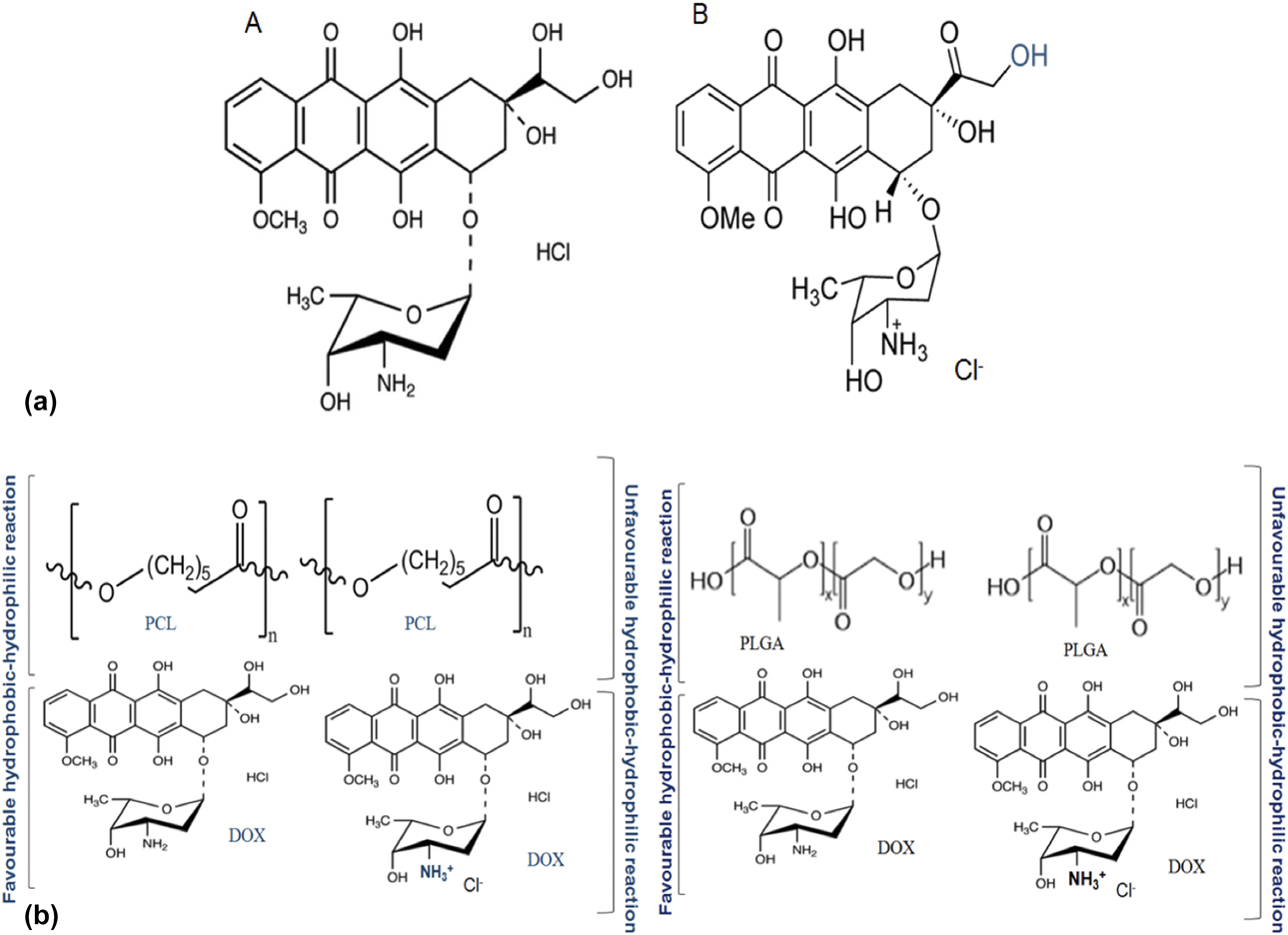
Polymer solubility also affects the release behavior significantly [Reference Saylor, Kim, Patwardhan and Warren30]. Even with identical processing parameters, environmental conditions and drug loading, much higher release is witnessed from soluble polymer intercalation compared to an insoluble or less soluble polymer matrix. The higher release kinetics from the PLGA polymer compared to the blend and the PCL alone can be attributed to the higher dissolution of the PLGA polymer. In line with our findings, a very similar release trend was also observed with the sodium salt of alendronate from PCL coating on 3D printed tricalcium phosphate scaffolds [Reference Tarafder and Bose29]. A schematic of the unfavorable hydrophilic–hydrophobic interactions at lower pH and the favorable hydrophobic–hydrophobic interaction has been presented in Fig. 8(b).
As indicated from these experimental results, the release behavior of the doxorubicin, from the polymer-intercalated HA discs, was primarily diffusion dominated. The complex polymer drug molecule interaction also influenced the release rate to a significant level. This release kinetics graph can be best fitted using the Higuchi and zero-order release kinetics model which demonstrates the release from a drug from an insoluble matrix [Reference Siepmann, Lecomte and Bodmeier31]. A variety of factor influenced the release rate, including the higher molecular weight, size, chemistry, and drug interaction with the calcium phosphate matrix. The number of hydroxyl groups and positive charged amino group in the doxorubicin molecule interacted with the calcium phosphate matrix and the surrounding media in different ways including electrostatic interactions and hydrophobic–hydrophobic and hydrophilic–hydrophobic interactions [Reference Hongquan, Yuhua, Youfa and Shipu32]. These interactions caused slower release of the drug molecule from the calcium phosphate and polymer matrix. Thus, the understanding of the complex hydrophobic polymer interactions with the hydrophobic deprotonated form or the hydrophilic active form of the drug gave us considerable insight into controlling the release kinetics. Since, following of zero-order kinetics is an ideal pattern for any pharmaceutical dosage [Reference Siepmann, Lecomte and Bodmeier31], these results show that calcium phosphate with polymers can be employed as a successful drug delivery platform for the local release of doxorubicin to combat remaining tumor cells after excision of the bone tumor and implantation with DOX-containing discs.
The absorption of doxorubicin with HA using a freeze-drying method also showed a sustained release for a period of 30 days [Reference Kunieda, Seki, Nakatani, Wakabayashi, Shiro, Inoue, Sougawa, Kimura and Harada23]. Improvement of hepatocellular carcinoma in a mouse model with the administration of 100 mg/kg was observed. Studies also revealed induced apoptosis by doxorubicin by functionalizing with hyaluronic acid and HA [Reference Xiong, Du, Ni, Zhou and Yao33]. Encapsulation of doxorubicin in HA particles with poly-lactic-glycolic acid also showed sustained release with reduced epithelial carcinoma cell viability [Reference Zheng, Wang, Shen, Zhu and Shi34]. The efficacy of doxorubicin was demonstrated on the early stage osteosarcoma cell culture after 2 and 5 days of culture. Cell viability was found to diminish significantly by the incorporation of doxorubicin. Layered cells, somewhat resembling the morphology of tumor cells, were noticed in the control HA samples under the SEM, whereas only couple cells were witnessed in the doxorubicin-containing samples. Also, human osteoblastic cell growth and proliferation were investigated on the HA discs with doxorubicin to check the cytotoxicity of the dosage of the drug used in the discs on bone cells. The cell adhesion was compared to the control bare HA discs without any drug. Very similar count of the cells from the MTT assay in Fig. 6(b), with or without the drug, showed no cytotoxicity of the drug up to 7 days of culture. SEM images indicated well-adhered flattened cells on the surface of the HA discs even with the doxorubicin drug which was again in consistence with the MTT assay results. Thus, the dosage of the drug (50 μg per discs) used in the HA discs was observed to be efficient to inhibit osteosarcoma cell culture, but not found to be cytotoxic on osteoblast cells in the early time points.
Conclusions
Doxorubicin release kinetics from polymer incorporated HA discs at physiological pH of 7.4 and acidic pH of 5.0 was studied to develop an appropriate delivery vehicle for local drug release. The role of chemistry of the polymer and the interaction of the drug with the polymer, based on the pH, was accessed to have a better understanding on the development of the drug carrier vehicle. An overall lower release kinetics (40% compared to 80% from bare HA) was noted at higher pH of 7.4 compared to pH 5.0 which was attributed to the higher matrix degradation at acidic pH. Primarily, the release of doxorubicin was controlled by the hydrophobic polymer interaction with the drug molecule at the physiological pH. Cytotoxicity analysis showed inhibition of osteosarcoma cell viability with the 50 μg of doxorubicin per disc at the early time points, which had no adverse effects on the bone cells at the early time points as well. A mechanistic understanding of doxorubicin release from HA discs has been proposed involving hydrophobic and hydrophilic interaction based on pH. This insight into the chemistry and the physicochemical behavior of the drug will be beneficial toward the future development of drug delivery vehicle for controlled and local drug delivery from polymer incorporated calcium phosphate matrix.
Materials and methods
Disc fabrication
0.2 gm of commercially available HA powder, average particle size of 100 nm (Berkeley Advanced Biomaterials Inc., Berkeley, California), was uniaxially pressed to fabricate disc samples [10 mm (Ф) × 0.5 mm (h)]. These discs were further compressed by cold isostatic press (CIP) at 50 psi for 5 min to achieve higher mechanical properties.
PCL and PLGA incorporation
Two different concentrations of PCL (M w = 14,000) (Sigma, St. Louis, Missouri) and PLGA (M w = 190,000–240,000) (Sigma, St. Louis, Missouri), 5 and 10 wt% were used to study the effects of PCL and PLGA concentration on doxorubicin release kinetics. To make the 5% and 10% (w/v) solution, measured amounts of PCL and PLGA were dissolved in 10 mL of acetone and kept in water bath at 60 °C for 15–20 min 5 gm of HA powder was mixed with PCL solution and stirred vigorously at room temperature. The samples were further dried overnight and then kept in a desiccator.
PCL and PLGA blend incorporation
2.5% PCL and 2.5% PLGA were dissolved in 5 mL of acetone each and used as 5% polymer blend. Likewise, 5% PCL and 5% PLGA were dissolved in 5 mL of acetone each and referred to as 10% polymer blend. The polymers were further mixed together along with similar DOX amount of 50 µgs per sample for the release and cell culture studies.
Doxorubicin incorporation
50 µg of doxorubicin (DOX) (Sigma Aldrich, St. Louis, Missouri) (lower dosage approved by FDA [18]) was mixed vigorously with 0.2 gm s of HA or HA and polymer composite powder in 10 mL of water. The solubility of the DOX was not affected by the presence of HA. The powder was dried for 72 h at room temperature in dark containers to inhibit light penetration and pressed as described before for further usage.
Surface energy determination by contact angle measurement
Experimentally obtained values of contact angles were utilized in investigating surface energy analysis of the samples. A sessile drop method with a face contact angle setup was utilized in the measurement of the contact angles. The setup was further equipped with a camera and a microscope to improve further accuracy of the measurement. As the images were captured with the connected camera, the face contact angle between the liquid droplet and the substrate was calculated from the magnified image from the microscope. Approximately 6 measurements were taken for each combinations of the samples and the liquid to minimize the statistical chances of error. Surface energy is inversely related with contact angles, and hence, higher the contact angle, less the surface energy.
DOX release study
Release of doxorubicin was measured by using 0.1 M pH buffers. A pH 7.4 PBS (phosphate buffer saline) was utilized to imitate the body’s physiological pH, whereas a pH 5.0 acetate buffer was used to imitate the slightly acidic pH in the vicinity of the surgical site right after inflammation or the execution of surgery [Reference Tarafder, Nansen and Bose22]. Three samples from each concentration (5 and 10%) of PCL, PLGA, and blend of PCL and PLGA with 50 µgs of DOX incorporated were placed in 4 mL of pH 7.4 phosphate buffer and pH 5.0 acetate buffer in individual vials. The vials were placed at 37 °C under constant shaking conditions of 150 rotations per minute. For the study, buffer solutions were changed at 0.5, 1, 2, 5, 8, 12, 15, 20, 30, 40, 50, 60, 70, 80, and 90 days. At each time point, 4 mL of the buffer solution was pipetted out and was restored with a new 4 mL of the buffer solution for volume make-up. The drug concentration was investigated at 479 nm wavelength using a BioTek Synergy 2 SLFPTAD microplate reader (BioTek, Wiooski, Vermont). Standard curve was utilized to find the final cumulative percentage release of the drug.
Surface morphology before and after release of drug
Field emission scanning electron microscope (FEI Inc., Hillsboro, Oregon), endowed with an ETD (Everhart-Thornley Detector) was utilized to observe the surface morphologies of the disc samples of all compositions following gold sputter-coating (Technics Hummer V, San Jose, California) before conducting the release study. After the release of the drug was completed for the 90 days study period, discs were dried carefully to ensure the absence of moisture in an oven at 37 °C for a week before the investigation of their surface morphologies.
In vitro cell materials interaction
Human fetal osteoblast cells (hFOB) (ATCC, Manassas, Virginia) and human osteosarcoma cells (ATCC, Manassas, Virginia) derived from the human bone tissue were incubated for a maximum of 7 days with the aim of investigating the in vitro materials and bone cell interactions and analyzing the efficacy of doxorubicin on bone cancer treatment. After sterilization, each sample was seeded directly with 2.5 × 10⁵ osteoblast or osteosarcoma cells and added with 1 mL of Dulbecco’s modified Eagle’s medium (DMEM) or Eagle’s Minimum Essential Medium (EMEM), respectively, enriched with 10% fetal bovine serum following ATCC’s hFOB cell culture protocols. The cell media was altered every second day throughout the span of the experiment to provide fresh media to aid in enhancing cell growth and proliferation. The osteoblast cultures were incubated in a sterilized environment at 34 °C under 5% CO2, whereas the osteosarcoma cell cultures were incubated at 37 °C under 5% CO2 and 95% humidified air atmosphere.
Cell morphology
The seeded samples were cultured after a maximum of 7 days to observe their growth and proliferation. To observe under the SEM, the samples were fixed with 1 mL of 2% paraformaldehyde and 2% glutaraldehyde in 0.1 M phosphate buffer and refrigerated overnight at 4 °C. The samples were further treated with 2% osmium tetroxide (OsO4) and maintained for 2 h at room temperature. Following this, the samples were dehydrated by rinsing them in a consecutive series of ethanol drying and further by a hexamethyldisilane (HMDS) drying procedure. The dehydrated samples then were gold coated to ensure elimination of any kind of scanning faults arising from electrostatic charges on the sample surface and observed under an FESEM under high vacuum at 30 kV acceleration voltages.
Cell proliferation using MTT assay
The MTT assay (Sigma, ST. Louis, Missouri) was executed for 2, 5, and 7 days of incubation to assess osteoblast and osteosarcoma cell proliferation on the discs and check the cytotoxicity of the drug on the cell proliferation. 5 mg/mL of MTT solution was made by 3-(4,5-Dimethylthiazol-2-yl)-2,5-Diphenyltetrazolium Bromide in phosphate buffer and sterilized by filter sterilization. 100 μL of MTT solution was then added on the surface of each sample in the 24-well plates and covered with 900 μL of the DMEM culture media. These were incubated for 2 h in the respective culture conditions. Following incubation, the samples were transferred to a new well plate and added with 600 μL of MTT solubilizer composed of 10% Triton X-100, 0.1 N HCl, and isopropanol to dissolve the formazan crystals, responsible for the purple color on the sample surface. 100 μL aliquots of this resulting solution was taken from each group in triplicates and measured at 570 nm by a microplate reader.
Acknowledgments
The authors would like to acknowledge financial support from the National Institutes of Health under grant numbers R01 AR066361. The content is solely the responsibility of the authors and does not necessarily represent the official views of the National Institutes of Health.