Implications
Supplementing the diet of the ewe with excess iodine, for the final 4 weeks of gestation, altered the tri-iodothyronine concentration, ileal gene expression and serum IgG concentration of the lamb postpartum.
Introduction
Altered maternal nutrition during gestation influences foetal development with adaptions to the structural, physiological and metabolic functions of the offspring (van der Linden et al., Reference van der Linden, Kenyon, Blair, Lopez-Villalobas, Jenkinson, Peterson and Mackenzie2009). In ruminants, the neonate is born hypoimmunocompetent (O’Dohert and Crosby, Reference O’Doherty and Crosby1997; Boland et al., Reference Boland, Hayes, Sweeney, Callan, Baird, Keely and Crosby2008) due to the presence of a placental barrier which prevents the in utero transfer of immunoglobulins from mother to offspring (Brambell, Reference Brambell1970). Neonatal lambs are therefore dependent upon passively acquiring maternal immunoglobulins from colostrum to confer disease resistance in early life. However, due to the onset of gut maturation, the ability of the newborn lamb to absorb macromolecules diminishes rapidly in the first 24 to 48 h of life (Gilbert et al., Reference Gilbert, Gaskins, Hillers, Parker and McGuire1988).
It is common practice for high producing ruminant livestock to be supplemented with minerals especially during the late gestational phase (Rose et al., Reference Rose, Wolf and Haresign2007). Previous studies have shown that a high level of iodine supplementation to the pregnant ewe produces progeny with decreased serum IgG concentrations and a reduced efficiency of colostral IgG absorption at 24 h postpartum (Boland et al., Reference Boland, Guinan, Brophy, Callan, Quinn, Nowakowski and Crosby2005a). While this occurs independently of colostrum intake, colostral IgG concentration (Boland et al., Reference Boland, Brophy, Callan, Quinn, Nowakowski and Crosby2004) or colostrum source (Boland et al., Reference Boland, Callan, Brophy, Quinn and Crosby2006); the influence of iodine carrier source material has not been investigated. Boland et al. (Reference Boland, Keane, Nowakowski, Brophy and Crosby2005b) indicated the presence of a ‘pre-programming’ effect of iodine supplementation on intestinal absorptive capacity in the newborn. The primary role of iodine in the animal’s body is as a constituent of the thyroid hormones (Mayer et al., Reference Mayer, Zolnai, Frenyo, Jancsik, Szentirmay, Hammarstrom and Kacskovics2002); therefore, the inhibition of IgG transfer from colostrum to the newborn may be mediated via the thyroid hormones, tri-iodothyronine (T3) and thyroxine (T4).
The major site of IgG absorption in the lamb is the ileum (Yvon et al., Reference Yvon, Levieux, Valluy, Pélissier and Mirand1993), where the IgG is transported across the intestinal epithelium via non-specific transcytosis (Mayer et al., Reference Mayer, Zolnai, Frenyo, Jancsik, Szentirmay, Hammarstrom and Kacskovics2002). The Fc receptor for IgG (FcRn) is suggested as the primary receptor involved in this process (Praeter et al., Reference Praetor, Ellinger, Fuchs and Hunziker2000); however the exact control of IgG transfer and the potential mechanisms through which an elevated iodine intake inhibits the transfer of immunoglobulins from colostrum across the intestinal epithelial layer in the newborn remain unclear. The objectives of this experiment were (1) to determine the effect of iodine supplementation to the dam on the expression of ileal located genes, purported to influence IgG transfer in the newborn lamb before colostrum consumption; (2) to determine the impact of iodine supplementation on the plasma T3 and T4 concentrations in the offspring at 1 h postpartum and (3) to examine the effect of iodine source material (calcium iodate (CaIO3) or potassium iodide (KI)) on lamb serum IgG concentration.
Material and methods
Ethical approval
All procedures involving ewes and lambs in this study were conducted under experimental licence in accordance with the European Community Directive, 86-609-EC (EC, 2002). Ethical approval (AREC-P-09-12-Boland) was obtained from the Animal Research Ethics Subcommittee, University College Dublin. This study was conducted at University College Dublin, Lyons Research Farm, Newcastle, Co. Dublin.
Pre-experimental animal management
Four weeks before the mean predicted lambing date 45 mature, twin bearing ewes of mixed breed were selected and randomly allocated to one of three nutritional treatments (n=15). Before selection the ewes were blocked on the basis of BW (80.0±1.81 kg) and balanced for body condition score (BCS) (3.17±0.07 BCS units). BCS assessments were made by a trained technician and ewes were scored on a scale of 0 to 5 according to Russel (Reference Russel1984).
Four months before initiation of the experiment, the ewes were oestrus synchronised using intervaginal progestagen pessaries (Chronogest and Folligon, Intervet Ireland, Ltd) followed by an intramuscular injection of 500 international units PMSG (Chronogest and Folligon, Intervet Ireland, Ltd), before being individually inseminated with fresh diluted semen, at a rate of 20 million spermatozoa per uterine horn, using laparoscopic artificial insemination (AI). Rams from a range of breeds including Belclare, Charolais, Rouge L’ouest, Suffolk, Texel, Vendeen were used. Post insemination the ewes remained outdoors where they were offered a grazed grass only diet until they were group housed on day 42 of gestation. From the initial time of housing until day 119 of gestation the ewes were offered a diet of grass silage ad libitum only. Ewes were pregnancy scanned and shorn on day 63 and 72 of gestation, respectively.
Nutritional treatments
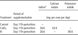
On day 113 of gestation, ewes were allocated to one of three nutritional treatments and transferred to individual, wooden slatted, pens measuring 1.1×1.4 m. The animals were allowed a 7 day adaption period before experimental feeding began on day 119 of gestation. All ewes were fed a basal diet of grass silage ad libitum supplemented with 750 g (fresh weight) of a 190 g CP/kg DM concentrate. Ewes received either the basal diet with no supplementary iodine (Control) or the basal diet plus 26.6 mg/day of iodine supplied as either CaIO3 or KI (Table 1). This level of supplementary iodine equates to the level offered to ewes in a previously reported study (Boland et al., Reference Boland, Hayes, Sweeney, Callan, Baird, Keely and Crosby2008) and is reflective of the level of intake achieved when ewes are offered free access mineral supplements indoors (Crosby et al., Reference Crosby, Boland, Brophy, Quinn, Callan and Joyce2004). In accordance with a previous study, carried out at this institute, the dietary intake of iodine on the control diet can be estimated at ~0.57 mg iodine per ewe per day (Boland et al., Reference Boland, Hayes, Sweeney, Callan, Baird, Keely and Crosby2008).
Iodine inclusion levels, expressed in mg (DM) per ewe per day of active ingredient and in mg (DM) per ewe per day of source material

1 The 26.6 mg level of iodine equates to a mineral intake of 34 g which we have previously shown to have a negative effect on IgG and vitamin E absorption (Boland et al., 2008).
Feeding
Fresh silage was offered to ewes each morning while ewes had free access to water at all times throughout the study. At 0730 h each day, uneaten silage was removed and fresh silage offered at 1.1 times the previous day’s intake. Concentrate was offered as a coarse ration. Total daily concentrate allowance was split fed in two equal portions offered at 0900 and 1730 h; with no concentrate refusals recorded. The iodine supplement was manually mixed into the concentrate on the day before feeding. Before mixing, the daily iodine allowance was first added to 20 g (fresh weight) of the concentrate which was used as a carrier for the supplement. Before the mineral inclusion, this carrier was dried using forced air circulation at 55°C for 72 h, following which it was then ground through a 0.8 mm screen using a Christy and Norris hammer mill (Christy and Norris Ltd, Ipswich, UK). The daily concentrate allowance was then adjusted to take into account the carrier contribution.
Ewe measurements and lambing data
Ewes were assessed for BCS on day 114 and day 140 of gestation and at 24 h postpartum as described above. Ewe BW was recorded electronically (Tru-Test Group, Auckland, New Zealand) at each BCS measurement. All ewes lambed down in their individual pens and remained there until 24 h postpartum. Immediately after the birth of the first lamb an udder cover was placed on the ewe to prevent suckling. Under careful supervision, the udder covers were entirely successful. Within the first hour after parturition, the navel of each lamb was dipped in a 10% iodine solution, to aid in the control of Erysipelas Polyarthritis (joint-ill). Lambs were weighed and time of birth, birth weight and sex were recorded for each lamb. The time of birth of the first lamb was used to calculate the gestation length of the ewe with 1200 h on the day of AI taken as hour zero.
Milking
All ewes were hand milked at 1, 10 and 18 h postpartum as previously described by (Crosby et al., Reference Crosby, Boland, Brophy, Quinn, Callan and Joyce2004). Briefly, each ewe received an intramuscular injection of 10 i.u. of oxytocin (Oxytocin-S, Intervet Ltd) immediately before each milking to ensure total milk let down (Doney et al., Reference Doney, Peart, Smith and Louda1979). Ewes were milked out completely by hand. Total volume was recorded after each milking and the total volume produced up to 18 h postpartum was calculated. At 24 h postpartum the udder cover was removed.
Following each milking, the lambs were fed measured quantities of colostrum from their dam via stomach tube. Depending on the yield of colostrum collected, lambs received the maximum amount of colostrum available, within the range of 20 to 50 ml colostrum per kg of birth weight. The quantity of colostrum fed to each lamb was recorded. When colostrum production was insufficient to provide a minimum of 20 ml/kg birth weight, lambs received substitute pooled colostrum up to the maximum allowance of 50 ml/kg birth weight.
Tissue collection
At 1 h postpartum 10 ewes from each of the Control and KI treatments were randomly selected and one of their progeny euthanised by the intravenous administration of Euthatal (pentobarbitone sodium BP; Merial Animal Limited); at a dose rate of 1 ml/kg BW. Following euthanasia the intestinal tissue from the middle section of the ileum, ~10 cm from the ileo-cecal junction, was aseptically isolated and flushed using phosphate buffered saline (PBS). Approximately 1 to 2 g of tissue was then harvested and placed into 15 ml tubes (Sarstedt, Wexford, Ireland), containing 5 ml of RNAlater (Life Technologies Ltd, Dublin, Ireland), for 24 h. Following this, the preservative was removed and tissue samples stored at −80°C until used for total RNA extraction.
Lamb blood sample collection
Before euthanasia, at 1 h postpartum, a 10 ml plasma blood sample was collected via jugular venepuncture and heparinised VacutainersTM (Becton-Dickson Ltd, Plymouth, UK) from all lambs. All blood samples were immediately placed on ice and centrifuged at 4°C and 1800 g for 15 min after which the plasma was pipetted into separate pour off tubes and frozen at −20°C until further analysis. At 24 h postpartum a serum blood sample was collected, from all remaining offspring, via jugular venepuncture and non-heparinised vacutainers (Becton-Dickson Ltd). Blood samples were stored at room temperature for 1 h post collection before being transferred to a refrigerator where they remained at 4°C for 24 h. Samples were subsequently centrifuged at 4°C and 1800×g for 15 min to obtain the serum, which was then frozen at −20°C until required for further analysis.
Laboratory analysis
Blood sample analysis
Total serum immunoglobulin concentration (g/l) was determined, from samples collected at 24 h postpartum, using the zinc sulphate turbidity test (McEwan et al., Reference McEwan, Fisher and Selman1970). These results were then multiplied by 0.91 to provide the IgG only content of the serum (Larson et al., Reference Larson, Ward, Frederiksen, Ardrey and Frank1974; Boland et al., Reference Boland, Brophy, Callan, Quinn, Nowakowski and Crosby2005c). Plasma free T3 and total T4 concentrations were determined by solid phase time-resolved fluoroimmunoassay using AutoDelfia kits (Wallac Oy, Turku, Finland), as described previously (Boland et al., Reference Boland, Hayes, Sweeney, Callan, Baird, Keely and Crosby2008).
Extraction and assessment of RNA integrity
Total RNA was isolated using TRIzol reagent (Molecular Research Center, Cincinnati, USA) as per the manufacturer’s instructions followed by a clean-up using the Qiagen RNeasy mini kit (Qiagen Ltd, Manchester, England). Briefly, ~25 mg of tissue was homogenised in 1.0 ml of Trizol using a sterile stainless steel bead and the Qiagen tissue lyser II (2×120 s max. speed; (Qiagen Ltd). After homogenisation, 200 µl of chloroform was added to each sample and centrifuged (12 000×g, 15 min). Following centrifugation, the upper aqueous phase was transferred directly onto a RNeasy column and RNA was purified using an on-column DNase digestion step. Total RNA was quantified using a NanoDrop-ND1000 Spectrophotometer (Thermo Fisher Scientific Inc, Boston, MA, USA) and all samples were shown to a 260/280 nm ratio >1.8. RNA quality was determined using an RNA 6000 chip on the Agilent 2100 Bioanalyzer (Agilent Technologies Inc., Santa Clara, CA, USA); all samples were shown to have a RNA Integrity Number (RIN) >7.5.
Complementary cDNA synthesis and quantitative real-time PCR (qPCR)
For each sample cDNA was prepared from 1.0 µg of total RNA using the High Capacity cDNA Reverse Transcription Kit (Life Technologies Ltd) according to the manufacturer’s instructions. The purified cDNA was then diluted in RNase- and DNase-free water up to a volume of 250 µl and stored at −20°C until required for subsequent use.
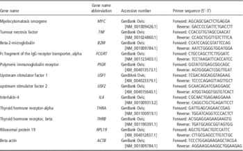
Ten mammalian genes (Table 2) shown to be involved in the process of immunoglobulin transcytosis and absorption were chosen for investigation based on data from the literature and interaction network data contained in the Ingenuity Pathway Analysis knowledgebase (Laegreid et al., Reference Laegreid, Heaton, Keen, Grosse, Chitko-McKown, Smith, Keele, Bennett and Besser2002; Spiekermann et al., Reference Spiekermann, Finn, Ward, Dumont, Dickinson, Blumberg and Lencer2002; Cervenak and Kacskovics, Reference Cervenak and Kacskovics2009). Oligonucleotide primers were designed using Primer ExpressTM Software, version 2.0 package (Life Technologies Ltd) and synthesised by MWG Biotech (Ebersberg, Germany). Primers were also designed and synthesised for two reference genes, ACTB and RPL19; these genes were selected from a panel of four candidates via GeNorm analysis using the qbase+ plus software package (Biogazelle, Ghent, Belgium) and the cDNA samples generated in the current study.
Ovine oligonucleotide primers used for quantitative real-time PCR (qPCR)

Duplicate qPCR assays were performed in a total volume of 20 µl, containing 1× Fast SYBR PCR Master Mix (Life Technologies Ltd), 300 nM of each forward and reverse primer, and 1ng cDNA. qPCR was performed in duplicate for each sample on a 7500 ABI Prism Sequence Detection System (Life Technologies Ltd) together with non-template PCR and non-reverse transcriptase control samples. Thermocycling conditions were as follows: 95°C for 2 min for one cycle, followed by 95°C for 15 s, and 60°C for 1 min for 40 cycles. Finally, calibrated normalised relative quantities were determined for each gene using the qbase+ package (Biogazelle) based on normalisation factors generated for the two reference genes.
Statistical analysis
Data was analysed as a completely randomised block design using the mixed model procedure (PROC MIXED) in SAS (SAS, 2013). Individual ewe was the experimental unit for all parameters analysed. Descriptive statistics were performed to identify continuous and categorical variables. All models were analysed to fit the assumptions of normality using the UNIVARIATE procedure in SAS. Data was fitted to the following statistical model for analysis:
 $$Y_{{ijklmnp}} {\rm \,{=}\,}\mu {\plus}\alpha _{i} {\rm {\plus}}\beta _{j} {\rm {\plus}KL}_{p} {\rm {\plus}}{\varepsilon}_{{ijklmnp}} $$
where Y is the observation, µ is the overall mean, α i the fixed effect of the ith treatment (i=control, CaIO3 or KI), β j the fixed effect of the jth block, KL p the random effect of the pth ewe and ε the associated error. The fixed effect of ram breed and ewe age were included in the model however, effects were removed from the final model if their P value was >0.25. Rearing rank (single or twin) was included in the model for all parameters analysed following euthanasia.
A repeated measure analysis was performed on ewe DM intake, BW, BCS and colostrum production data using PROC MIXED. The repeated measures were fit using variance-covariance structures; the most appropriate (lowest Bayesian information criterion values) used for each analysis. Data was fitted to the following model for repeated measures analysis:
 $$Y_{{ijklmnp}} {\rm \,{=}\,}\mu {\rm {\plus}}\alpha _{{_{i} }} {\rm {\plus}}\beta _{j} {\rm {\plus}}\rho _{m} {\rm {\plus}}\beta \rho _{{jm}} {\rm {\plus}KL}_{p} {\rm {\plus}} {\varepsilon}_{{_{{ijklmnp}} }} $$
where Y is the observation, µ the overall mean, α i the fixed effect of the ith treatment (i=control, CaIO3 or KI), β j the fixed effect of the jth block, ρ m the repeated effect of day, βρ jm the interaction between the jth treatment and the mth day, KL p the random effect of the pth ewe and ε the associated error. All data presented in the tables are expressed as least squares means±SEM. The probability value, which denotes statistical significance, was P⩽0.05, while values tending towards significance were 0.05<P<0.10.
Results
Ewe BW and BCS
Dietary treatment had no effect (P>0.05) on the BW of the ewes on day 114 and day 140 of gestation or at 24 h postpartum when ewes on the Control, CaIO3 and KI treatments weighed 74.0, 71.8 and 73.5±1.87 kg (mean±SEM). Similarly, there was no difference (P>0.05) in BCS or BCS change among ewes on the Control, CaIO3 or KI treatments on day 114 (3.16 v. 3.16 v. 3.20±0.070 BCS units) and day 140 (2.65 v. 2.68 v. 2.82±0.074 BCS units) of gestation.
Lambing parameters
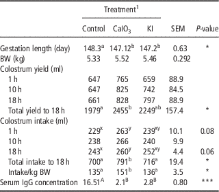
There was an effect of treatment (P<0.05) on ewe gestation length (Table 3). Ewes offered the iodine supplemented diets (CaIO3 or KI) had a shorter gestation length, regardless of iodine source. Dam nutritional treatment had no effect on individual lamb birth weight (P>0.05), with treatment means ranging from 5.33 to 5.52±0.292 kg.
The effect of prepartum dam nutritional treatment on ewe gestation length, individual lamb BW, colostrum yield and colostrum intake per lamb, at 1, 10 and 18 h, total yield intake to 18 h, lamb intake per kg BW and lamb serum IgG concentration at 24 h postpartum. (least square means±SEM)

Values represent least square means±SEM (n=15 per treatment for gestation length and colostrum yield; n=30 per treatment for BW; n=20 per treatment for Control and CaIO3, and n=30 for KI treatment for colostrum intake and serum IgG concentration.
a,bWithin a row, means without a common superscript differ (P⩽0.05).
A,BWithin a row, means without a common superscript differ (P<0.01).
x,yWithin a row, means without a common superscript differ (P<0.10).
1 C=control, basal diet, CaIO3=basal diet plus 26.6 mg of iodine as calcium iodate, KI=basal diet plus 26.6 mg of iodine potassium iodide.
*P < 0.05; **P < 0.01; ***P < 0.001.
Colostrum production and intake
The effect of iodine supplementation on colostrum production, intake and serum IgG concentration at 24 h postpartum is presented in Table 3. Dam nutritional treatment had no effect (P>0.05) on colostrum yield at 1, 10 or 18 h postpartum. Ewes on the CaIO3 diet had a higher (P<0.05) total colostrum yield to 18 h postpartum than Control ewes.
Progeny of CaIO3 ewes tended to have a higher intake of colostrum at 1 h (P=0.08) and 18 h (P=0.06) postpartum (Table 3), when compared with progeny of the Control group. The progeny of ewes offered the Control and the KI diets had lower colostrum intakes (P<0.05), on an absolute basis to 18 h postpartum when compared with CaIO3 progeny. Similarly, supplementation with calcium iodate significantly increased the amount of colostrum fed, per kg of lamb birth weight, to lambs in the first 18 h postpartum reflecting higher colostrum production. Progeny of both the CaIO3 and KI treatment groups had significantly lower serum IgG concentrations (P<0.01) when compared with the progeny of ewes offered the control diet.
Thyroid hormone concentration
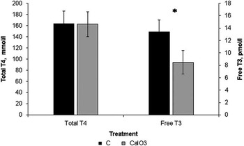
Iodine supplementation to the dam had no effect (P<0.05) on the total concentration of T4 quantified in the progeny (Figure 1) however there was an effect (P<0.01) of dam nutritional treatment on free T3 concentration at 1 h postpartum. Lower free T3 concentrations were observed in the CaIO3 progeny relative to the control progeny (13.39 v. 8.48 pmol/l; P<0.01).
The effect of prepartum dam nutritional treatment on the concentration of total T4 (mmol/l) and free T3 (pmol/l) in the lamb at 1 h postpartum. Treatments: Control=basal diet, CaIO3=basal diet plus 26.6 mg of iodine as calcium iodate. Asterisk (*) indicates a significant difference between Control and CaIO3 for free T3 concentration (P<0.01). Least square means±SEM are presented; n=10 lambs per treatment group.

Immunoglobulin associated gene expression
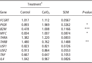
Iodine supplementation of the dam significantly affected (P<0.05) the mRNA expression of B2M, PIGR, THRB and MYC in the ileum of the lamb at 1 h postpartum (Table 4). Progeny of the CaIO3 ewes had greater (P<0.05) levels of B2M, PIGR and MYC mRNA expression when compared with those born to ewes fed the control diet. However, the level of THRB mRNA expression was lower (P<0.01) in the progeny of the CaIO3 supplemented ewes. Supplementation of the dam with iodine in late gestation had no effect (P>0.05) on the mRNA expression of FCGRT, THRA, USF1, USF2 and the cytokine genes TNF and IL4.
The effect of prepartum dam nutritional treatment on the calibrated normalised relative quantity of immunoglobulin associated genes in ileal tissue of the lamb at 1 h postpartum

Values represent least square means of the normalised relative quantities±SEM (n=10 per treatment).
1 Control=basal diet, CaIO3=basal diet plus 26.6 mg of iodine as calcium iodate.
*P < 0.05; **P < 0.01; ***P < 0.001.
Discussion
In ruminant livestock the sole source of immunoglobulins to the newborn is via postnatal absorption from colostrum (Laegreid et al., Reference Laegreid, Heaton, Keen, Grosse, Chitko-McKown, Smith, Keele, Bennett and Besser2002). In specific livestock species, low immunoglobulin levels in the newborn have been associated with impaired health in later life, thus emphasising the importance of colostrum for the growth and well-being of the newborn (Speikermann et al., Reference Spiekermann, Finn, Ward, Dumont, Dickinson, Blumberg and Lencer2002). Previous studies have, however, recently demonstrated a negative relationship between iodine supplementation of the pregnant ewe in late gestation and the serum IgG concentration of her progeny at 24 h postpartum (Boland et al., Reference Boland, Callan, Brophy, Quinn and Crosby2006 and Reference Boland, Hayes, Sweeney, Callan, Baird, Keely and Crosby2008; Rose et al., Reference Rose, Wolf and Haresign2007).
In the present study the expression patterns of a range of genes associated with colostral IgG absorption in the newborn lamb were examined. Maternal iodine supplementation altered the expression of B2M, PIGR and MYC in the neonatal lamb. Previous physiological studies have associated these genes with intestinal immunoglobulin (Ig) transfer (Bruno et al., Reference Bruno, West, Schneeman, Bresnick and Kaetzel2004; Baumrucker and Bruckmaier, Reference Baumrucker and Bruckmaier2014). It is evident also that THRB expression at the newborn intestinal level is diminished following maternal prepartum supplementation (CaIO3) in conjunction with reduced free T3 concentrations in these animals. The failure of passive transfer observed is therefore potentially mediated via the altered thyroid hormone status of the newborn and reduced THRB expression of these progeny. This represents new and significant information pertaining to the control of intestinal IgG transfer in ruminant species.
Dietary iodine requirements of 0.7 mg/kg DM have been reported for sheep (Boland et al., Reference Boland, Brophy, Callan, Quinn, Nowakowski and Crosby2005c). Thus, in the current study, ewes in both the CaIO3 and KI treatments received iodine in excess of requirements; however the quantity fed remained below the published toxicity level of 50 mg per ewe per day (Cervenak and Kacskovics, Reference Cervenak and Kacskovics2009). The level of iodine offered was lower than that consumed by pregnant ewes when offered ad-libitum access to a mineral block under group housing conditions, and thus replicating normal commercial farm practice (Crosby et al., Reference Crosby, Boland, Brophy, Quinn, Callan and Joyce2004).
Previous studies have examined the effect of offering supplementary iodine at levels ranging from 8.9 to 40 mg per ewe per day and identified a negative linear relationship between iodine supplementation of the dam and the serum IgG concentration of her offspring at 24 h postpartum (Boland et al., Reference Boland, Guinan, Brophy, Callan, Quinn, Nowakowski and Crosby2005a, Reference Boland, Callan, Brophy, Quinn and Crosby2006 and Reference Boland, Hayes, Sweeney, Callan, Baird, Keely and Crosby2008); provided the mineral supplement is offered within the final 7 to 14 days of gestation (Boland et al., Reference Boland, Hayes, Sweeney, Callan, Baird, Keely and Crosby2008; Rose et al., Reference Rose, Pearson and Cratchley2012). This is in agreement with the present study; where ewes offered either the CaIO3 or KI treatments had progeny with significantly reduced serum IgG concentrations.
Mineral supplementation has been shown to have an inconsistent effect on colostrum production (Boland et al., Reference Boland, Brophy, Callan, Quinn, Nowakowski and Crosby2004, Reference Boland, Brophy, Callan, Quinn, Nowakowski and Crosby2005c and Reference Boland, Hayes, Sweeney, Callan, Baird, Keely and Crosby2008). In the present study, a wide variation in colostrum yield was observed within all treatments with ewes on the CaIO3 diet producing a higher total yield to 18 h in comparison to those on the control diet. It is unlikely that this increase in production can be fully attributable to iodine supplementation as ewes on the KI treatment produced an intermediate yield. As the colostrum intake per lamb was dictated by colostrum yield the higher total intake to 18 h by the CaIO3 lambs is a reflection of the elevated total colostrum production by these ewes. All lambs in this study had colostrum intakes in excess of the 112 ml per kg birth weight which has previously been described as sufficient to meet both the lamb’s immunoglobulin and energy requirements to prevent hypothermia in an indoor lambing situation, during the first 24 h of life (O’Doherty and Crosby, Reference O’Doherty and Crosby1997). Previous research identified a positive linear relationship between total colostrum produced and total IgG produced up to 18 h postpartum (O’Doherty and Crosby, Reference O’Doherty and Crosby1997) however, despite the fact that CaIO3 ewes had a significantly higher total colostrum yield to 18 h in the current study; the serum IgG concentrations of the progeny were significantly lower at 24 h postpartum. This further emphasises the presence of a pre-programming effect from the iodine supplementation on the lamb in utero (Boland et al., Reference Boland, Keane, Nowakowski, Brophy and Crosby2005b).
In the current study, the gestation length of the non-supplemented ewes was significantly longer than that of the supplemented ewes. This is somewhat inconsistent with earlier work which saw no influence of either mineral or iodine supplementation on the gestation length of the ewe (Boland et al., Reference Boland, Keane, Nowakowski, Brophy and Crosby2005b). The birth of lambs between 88% and 95% gestation has been linked to the birth of offspring with an immature gastrointestinal tract (Sangild, Reference Sangild2006) thus decreasing Ig absorption. However, despite the observed difference in gestation length lamb birth weight remained unaffected and gestation length was within the standard accepted range for sheep (Boland et al., Reference Boland, Keane, Nowakowski, Brophy and Crosby2005b), thus indicating the adequacy of nutrition during the final weeks of gestation (Agricultural and Food Research Council, 1993).
Taking into account the findings of earlier work carried out by our group (Boland et al., Reference Boland, Keane, Nowakowski, Brophy and Crosby2005b) the negative effect of mineral and specifically iodine supplementation on the serum IgG concentration of the lamb is a consequence of a failure of the consumed IgG being transferred into circulation and is not due to the IgG contained in the colostrum being unavailable for absorption.
The lower ileum has been identified as the primary site of Ig absorption in the lamb (Yvon et al., Reference Yvon, Levieux, Valluy, Pélissier and Mirand1993). Immunoglobulins are absorbed across the epithelial lining of the ileum via non-specific transcytosis (Praeter et al., Reference Praetor, Ellinger, Fuchs and Hunziker2000). Following absorption, immunoglobulins are transported through the mucosal layer, into the lymphatic system and then into the bloodstream (Quigley et al., Reference Quigley, Hammer, Russel and Polo2005). Previously, authors have shown that transcytosis is a receptor-mediated vesicular process, to which the neonatal Fc receptor (FcRn) is primarily associated (Praeter et al., Reference Praetor, Ellinger, Fuchs and Hunziker2000). FcRn was identified in rodents as the predominant receptor involved in the transfer of maternal immunoglobulins from the mother to the newborn via the neonatal intestine (Jones and Waldmann, Reference Jones and Waldmann1972). FcRn molecules, which are located in the intestinal brush border, bind ingested maternal immunoglobulins and transport them through enterocytes to the systemic circulatory system of the ungulate (Yvon et al., Reference Yvon, Levieux, Valluy, Pélissier and Mirand1993). Previous authors stated that the FcRn molecule is composed of two proteins: the Fc fragment of IgG receptor transporter alpha (FCGRT) and beta-2-microglobulin (B2M) (Rodewald, Reference Rodewald1976). Therefore, the neonatal expression of both the FCGRT and B2M genes were investigated. The lack of difference in the expression of the FCGRT gene identified in the ileum of newborn lambs, before colostrum consumption, is in agreement with previous work stating that the FcRn receptor, while present in the intestine, is not directly involved in the absorption of IgG1, the most abundant form of IgG found in colostrum (Simister and Rees, Reference Simister and Rees1985). Further investigation has hypothesised that perhaps the primary function of the FcRn receptor is its involvement in the recycling of IgG back into the intestinal lumen where it contributes to the protection of the gastrointestinal tract against infection (Rojas and Apodaca, Reference Rojas and Apodaca2002). At birth, the ruminant intestinal tract is undergoing a maturational process in preparation for colostrum ingestion and absorption before gut closure. Due to the requirement for B2M in the maturation and functioning of FcRn (Baumrucker and Bruckmaier, Reference Baumrucker and Bruckmaier2014) and the increased relative expression of B2M in the ileum of CaIO3 lambs at 1 h postpartum it was concluded that iodine supplementation of the dam has no negative influence on the functioning of the FcRn receptor. Additionally, the lower expression of PIGR and increased serum IgG in non-supplemented progeny is in agreement with Johansen et al. (Reference Johansen, Pekna, Norderhaug, Haneberg, Hietala, Krajci, Betsholtz and Brandtzaeg1999) who found that PIGR knockout mice had increased levels of serum IgG in comparison to PIGR abundant mice. The authors hypothesised that this was due to an enhancement of their systemic immune system following a reduction in IgA absorption.
The only known physiological role of iodine is as a constituent of the thyroid hormones T4 and T3 (Mayer et al., Reference Mayer, Zolnai, Frenyo, Jancsik, Szentirmay, Hammarstrom and Kacskovics2002; Zhu et al., Reference Zhu, Peng, Raychowdhury, Nakajima, Lencer and Blumberg2002). In the foetus and newborn the thyroid hormones have been shown to exert a major influence on cellular differentiation, growth and development (Underwood and Suttle, Reference Underwood and Suttle1999). Altered thyroid hormone concentrations may, therefore, have an influential impact on the maturation of the gastrointestinal tract (Pácha, Reference Pácha2000) however further histological examination is warranted in order to investigate the effect of thyroid hormone concentration on neonatal intestinal morphology and subsequent passive immunity.
Studies have shown that thyroid gland development in the sheep takes place primarily during the latter two-thirds of gestation (Fowden et al., Reference Fowden, Li and Forhead1998), at which time the placental transfer of T3 and T4 is absent or at least minimal in the ovine, when compared with both the human and rat (Cabello and Levieux, Reference Cabello and Levieux1982). However, the foetal production of thyroid hormones remains dependent upon iodide uptake from maternal circulation by the follicular cells of the foetal thyroid gland (Choksi et al., Reference Choksi, Jahnke, St Hilaire and Shelby2003). While T4 is the most predominant thyroid hormone in circulation (Piosik et al., Reference Piosik, Van Groenigen, Van Doorn, Baas and De Vijlder1997), with concentrations increasing in the lamb before parturition and declining rapidly within the first few days of life (Forhead and Fowden, Reference Forhead and Fowden2014), T3 is deemed the most biologically active (Choksi et al., Reference Choksi, Jahnke, St Hilaire and Shelby2003; Rose et al., Reference Rose, Wolf and Haresign2007). There is a strong relationship between both total the free thyroid hormone fractions with circulating levels of T3 arising from the de-iodination of T4 by a selenium containing enzyme, type 1 iodothyronine 5’-deiodinase (Barry et al., Reference Barry, Duncan, Sadler, Millar and Sheppard1983; Camacho et al., Reference Camacho, Meyer, Neville, Hammer, Redmer, Reynolds, Caton and Vonnahme2012). However, in the current study a lower concentration of free T3 was observed in lambs born to CaIO3 ewes. This is in agreement with Boland et al. (Reference Boland, Hayes, Sweeney, Callan, Baird, Keely and Crosby2008) who observed a positive correlation between plasma T3 concentrations at 1 h and serum IgG absorption from colostrum at 24 h postpartum in the lamb. However, in contrast, Rose et al. (Reference Rose, Wolf and Haresign2007) reported an increase in T4 concentrations with no effect observed on T3 concentrations at 1 h and 24 h postpartum in the lamb. According to Boland et al. (Reference Boland, Hayes, Sweeney, Callan, Baird, Keely and Crosby2008), the decrease in free T3 concentrations may be attributed to an increase in the conversion of T4 to the biologically inactive rT3 or an inability of the animal to degrade rT3. Furthermore, the functions of T3 are mediated by the thyroid receptors and in-turn it has been stated that the expression of the thyroid hormone receptors is reflective of the animal’s thyroid hormone status (Gauthier et al., Reference Gauthier, Chassande, Plateroti, Roux, Legrand, Pain, Rousset, Weiss, Trouillas and Samarut1999). Consequently, the decreased concentration of T3 in the present study was combined with a decline in the expression of the THRB gene in the ileum of CaIO3 lambs.
In conclusion, this study provides evidence of the link between iodine supplementation of the dam and a failure of IgG absorption in the ileum of the newborn lamb; a mechanism potentially mediated through a decline in the thyroid hormone status of the newborn. However, it remains to be seen whether this decline in thyroid hormone status affects gut maturation in the newborn and whether or not the decrease in serum IgG absorption impairs the animal’s response to infection and ultimately their immune function in later life. Additionally, the negative impacts of iodine on lamb serum IgG concentration are independent of the carrier material used indicating it is the inclusion of excess iodine in the diet of the ewe that is negatively effecting the in utero development of the lamb.
Acknowledgements
The authors wish to thank Mr David Hogan for his assistance throughout the running of the experiment and Mr Stephen Lott for his assistance with animal management. Thanks are also extended to Dr Karina Pierce, Dr Bridget Lynch and Dr Alan Kelly for their assistance with sample collection.