Hypoplastic left heart syndrome comprises 4–9% of children born with congenital heart disease and has an incidence of about 0.162/1000 live births.Reference Morris, Outcalt and Menashe 1 In typical hypoplastic left heart syndrome, oxygenated blood from the left atrium drains through an interatrial communication to the right atrium. If there is obstruction to the pulmonary venous drainage during intrauterine life, the embryologic pulmonary–systemic venous connections can reopen or remain patent leading to an anomalous drainage. The levoatriocardinal vein is a form of pulmonary–systemic connection that connects the left atrium (62%) or one of the pulmonary veins (32%) to the cardinal venous system.Reference Bernstein, Moore, Stanger and Silverman 2 Hypoplastic left heart syndrome can also have associated aberrant subclavian artery in 10% of cases.
Defining the head and neck vessels before Norwood palliation is important, especially if Blalock–Taussig shunt is being considered. Echocardiography typically provides sufficient pre-operative data in most cases of hypoplastic left heart syndrome. However, it can be a challenge to optimally define aortic arch vessels in atypical cases.
This case report describes a newborn with hypoplastic left heart syndrome, intact atrial septum, levoatriocardinal vein, and right aberrant subclavian artery in which a cardiac magnetic resonance imaging/angiography proved invaluable in the diagnosis and surgical preparation.
Case report
A full-term newborn prenatally diagnosed with hypoplastic left heart syndrome was transferred on prostaglandin infusion to the intensive care unit at our centre for further management. Pre-operative echocardiogram showed evidence of aortic and mitral atresia with intact atrial septum and levoatriocardinal vein draining the left atrium to the left innominate vein (as shown in Supplementary Video S1), patent ductus arteriosus with flow from pulmonary artery to the descending aorta, and hypoplastic ascending aorta. The branching of the aortic arch was poorly visualised, likely because of the presence of levoatriocardinal vein and dilated left innominate vein. Cardiac magnetic resonance imaging/angiography was obtained to further define the arch anatomy/branches. The echocardiogram findings were confirmed and an aberrant right subclavian artery was identified. The patient underwent Norwood palliation with Sano shunt on the fifth day of life; the post-operative period was unremarkable.
Discussion
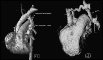
During foetal life, the pulmonary venous plexus, a part of the splanchnic plexus, has a connection to the pre-cardinal, post-cardinal and umbilicovitteline venous systems. These connections are lost after the pulmonary venous plexus establishes a connection to the common pulmonary vein. Anomalous pulmonary venous connections are felt to result from proximal obstruction to the pulmonary venous drainage, leading to persistence of these vestigial connections. If the obstruction to the pulmonary venous drainage is at the level of the heart, then persistence of these connections leads to egress of blood from the left atrium, thus forming the levoatriocardinal vein. The term levoatriocardinal vein was first recommended by Edwards and Dushane.Reference Edwards, Dushane, Alcott and Burchell 3 It typically runs posterior to the left pulmonary artery (as shown in Supplementary Figure S2) as it drains the left atrium to the left innominate vein. This is in contrast to the persistent left superior vena cava, which courses anterior to the left pulmonary artery and usually drains into the coronary sinus (Fig 1).
Three-dimentional reconstruction of the magnetic resonance imaging/angiography images showing the course of the levoatriocardinal vein connecting the left atrium to the left innominate vein and passing posterior to the left pulmonary artery (LPA). Compare it with another image where the left superior vena cava (LSVC) runs anterior to the left pulmonary artery (image taken from a different patient).

Levoatriocardinal vein has been described in association with hypoplastic left heart syndrome and intact atrial septum.Reference Bernstein, Moore, Stanger and Silverman 2 An intact atrial septum is a rare finding in hypoplastic left heart syndrome, occurring only in 1% of the pathology specimens.Reference Rychik, Rome, Collins, DeCampli and Spray 4 In all, 10% of the patients with hypoplastic left heart syndrome were also noted to have aberrant subclavian artery with left aortic arch in previous studies.Reference Ramaswamy, Lytrivi and Thanjan 5 To our knowledge, no prior reports have identified a case with combination of the lesions as described in our case. Finding individually rare lesions in the same patient makes our case rare and unique.
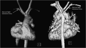
A cardiac magnetic resonance imaging/angiography was obtained to confirm the venous anatomy and identify aortic arch branching, which could not be determined by echocardiogram. The magnetic resonance imaging confirmed the echocardiogram findings and identified previously unknown aberrant right subclavian artery (Figs 1, 2). The presence of a levoatriocardinal vein and dilated left innominate vein most likely obscured the aortic arch and its branches. Pre-operative identification of aortic arch sidedness and presence of aberrant subclavian arteries is critical in surgical planning, especially if a Blalock–Taussig shunt is incorporated into the palliation. In addition, during Norwood repair, it is prudent to identify and clamp the aberrant right subclavian artery to improve visualisation of the surgical field. From a monitoring standpoint during surgery, previous reports have suggested that a radial artery catheter be placed in the arm opposite the side of the aberrant subclavian artery to avoid it being compressed by a trans-oesophageal probe.Reference Bensky, O’Brien and Hammon 6 Prior knowledge of arch anatomy will enable the surgeon to optimally plan surgery. In addition, during follow-up after surgery, right arm to leg blood pressure gradient can no longer be used to identify the persistence or development of a recoarctation in a patient who has right aberrant subclavian artery.
Three-dimentional reconstruction from magnetic resonance imaging/angiography images showing the aortic arch with its branches; the aberrant right subclavian artery can be visualised coming distal to the left subclavian from the descending aorta.

Conclusion
Unusual venous pathways – levoatriocardinal vein – in infants with hypoplastic left heart syndrome and intact atrial septum could provide a challenge in echocardiographic identification of aortic arch and its branches. Known association of aberrant subclavian artery in hypoplastic left heart syndrome should prompt consideration of a cardiac magnetic resonance imaging/angiography for better delineation of venous and arterial anatomy if echocardiogram is not definitive.
Acknowledgement
None.
Financial Support
This research received no specific grant from any funding agency, commercial or not-for-profit sectors.
Conflicts of Interest
None.
Supplementary material
To view supplementary material for this article, please visit http://dx.doi.org/10.1017/S1047951113002370.