Adults with congenitally malformed hearts suffering with Eisenmenger’s syndrome can present with haemoptysis, which often does not settle with conservative treatment. Embolisation of appropriate systemic-to-pulmonary collateral arteries may represent another potential therapeutic strategy. We present our experience of this approach in 2 such patients.
Case reports
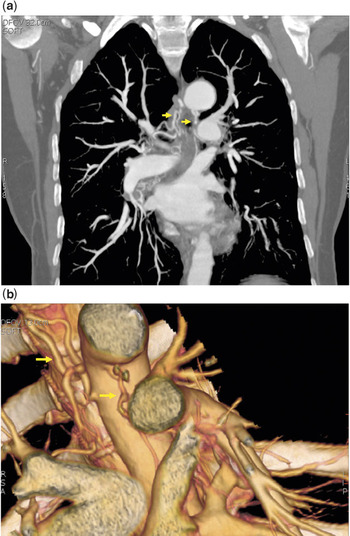
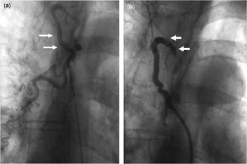
Our first patient was a 59 year old female, who had never smoked, with Eisenmenger’s syndrome developed in the setting of an unrepaired aortopulmonary window. She presented to her local hospital with an initial episode of haemoptysis. The markers of her inflammatory state were normal, and she was systemically well. She continued to have haemoptysis, and her concentration of haemoglobin dropped by 2 gm/dl. A computed tomographic pulmonary angiogram revealed confluent pulmonary arteries, with no evidence of pulmonary embolism. The scan revealed 2 collateral arteries arising from the descending aorta and joining the right and left pulmonary arteries (Fig. 1), The right-sided collateral artery was larger, and there was evidence of blood in the parenchyma of the lower lobe of the right lung. The pulmonary circulation, however, was not dependent on the collateral artery. Angiography using the right femoral artery for access confirmed the presence of these two aortopulmonary collateral channels. In view of evidence of bleeding in the right lung, we elected to embolise the right-sided collateral artery, deeming this vessel to be the most likely source of her continuing haemoptysis. We used a Cordis MPB3 (Cordis Corp., Miami, FL) catheter of 6 French dimension selectively to enter the collateral vessel, and delivered 1 coil of 5 mm diameter, and 2 of 3 mm diameter, using MREYE coils® (William Cook Europe, Bjaeverskov, Denmark). Repeated angiography demonstrated complete cessation of flow through the collateral artery (Fig. 2). The patient was discharged 3 days later, and remains free from haemoptysis after follow-up of 1 year.
Multiplanar reconstruction (a) showing a coronal view of the thorax. Two anatomically ectopic vessels (yellow arrows) are observed arising from the descending aorta producing ectactic collaterals to the right and left pulmonary arteries, respectively. Volume rendering reconstruction of descending thoracic aorta (b) shows the origin of the two aortopulmonary collateral arteries (yellow arrows).

The angiogram of the aortopulmonary collateral artery (yellow arrows) to the right pulmonary artery (a) was taken prior to embolisation. Insertion of coils (yellow arrows) in the collateral artery produced cessation of flow (b).

Our second patient was a 31 year old male with pulmonary atresia, intact interventricular septum, and non-confluent pulmonary arteries. He was admitted with haemoptysis that had lasted for longer than 1 week. He had previously undergone construction of a Waterston anastomosis to the right pulmonary artery, and a modified left Blalock-Taussig shunt to the left pulmonary artery. He was known to have severe pulmonary hypertension in his right lung due to the non-restrictive nature of the shunt, and normal pressures in the left pulmonary artery due to a significant stenosis at the insertion of the Blalock-Taussig shunt into the left pulmonary artery. Computed tomographic pulmonary angiography revealed no evidence of pulmonary embolisation, but showed evidence of bronchiectasis in the right lung. He had ongoing haemoptysis, and hence we performed cardiac catheterization. Angiograms obtained in the descending thoracic aorta demonstrated one significant collateral artery, which arose from the aorta and communicated with the distal right pulmonary artery. The scan also showed several other smaller collateral vessels. The flow of blood to the lung was shown to be dependent on the shunt rather than the collateral arteries. The larger collateral vessel was entered using an MPB3® (Cordis Corp., Miami, FL) 6Frenchcatheter, and a 2 mm Vortex® coil (Boston Scientific, Cork) was deployed through a Progreat® (Terumo Corp., Piscataway, NJ) system. Additional platinum coils of 2 mm and 3 mm diameter were deployed, resulting in cessation of flow through the collateral channel. At follow-up after I year, it emerged that he had suffered only 1 further small episode of haemoptysis, related to an infection of the respiratory tract.
Discussion
Haemoptysis is a common feature of Eisenmenger’s syndrome, and can occur in a significant proportion of patients suffering this reaction during the course of their life. In the classic description provided by Wood of 127 patients with the syndrome, all reported haemoptysis by the age of 40.Reference Wood1 In a similar study of a large cohort of patients with the syndrome, almost two-fifths reported at least one episode of haemoptysis.Reference Daliento, Somerville and Presbitero2 In the series reported by Wood, haemoptysis was responsible for deaths in two-fifths of his cohort, whereas othersReference Daliento, Somerville and Presbitero2, Reference Saha, Balakrishnan and Jaiswal3 have reported mortality in up to one-sixth of patients due to this problem.
In patients with Eisenmenger’s syndrome, haemoptysis can occur due to pulmonary infarction secondary to thrombosis, or due to neovascularisation. Studies have previously demonstrated that both neovascularity, and hilar intercostal collateral arteries, are much more common in patients with Eisenmenger’s syndrome than in those with acyanotic pulmonary hypertension.Reference Sheehan, Perloff, Fishbein, Gjertson and Aberle4
Pulmonary thrombus can be visualised on computed tomographic pulmonary angiography in up to three-tenths of patients with Eisenmenger’s syndrome,Reference Perloff, Hart, Greaves, Miner and Child5 with similar observations of massive pulmonary arterial thrombosis in patients with the syndrome having been reported elsewhere.Reference Broberg, Ujita and Babu-Narayan6 Our 2 patients both had Eisenmenger physiology, but with different anatomic substrates, one having an unrepaired aortopulmonary window with confluent pulmonary arteries, and the other pulmonary atresia with an intact ventricular septum, but non-confluent pulmonary arteries and shunt-dependant pulmonary arterial circulations. Both presented with haemoptysis.
In neither of these patients did we find evidence of pulmonary thrombosis on the computed tomographic pulmonary angiography. In both, we also identified more than one systemic–to-pulmonary collateral artery. When multiple collateral arteries are identified, as in these cases, attempts should be made to determine potential anatomical sites of bleeding, which may guide the decision of which collateral vessel to embolise. This can be determined anatomically through evidence of parenchymal bleeding on the computed tomographic scan with collateral supply to this area, or infrequently by identification of active bleeding from the collateral vessel during cardiac catheterization. Occasionally as in our second patient, there is no radiological evidence of bleeding, making difficult identification of the causative systemic-to-pulmonary collateral artery. In these situations, a careful analysis has to be made based on the collateral circulation to ensure that the aortopulmonary collateral artery targeted for embolisation is not the sole source of flow of blood to a parenchymal segment. With this is mind, in the absence of clear evidence of a culprit vessel, the most prominent collateral vessel may initially be considered for embolisation. In both our cases, the pulmonary circulation was not dependant on the collateral arteries, although the flow to the affected lung was dependant on a shunt in our second case.
Catheter embolisation of these systemic-to-pulmonary collateral vessels produced a marked improvement in symptoms in both patients, with cessation of haemoptysis. Although large series of embolisation, and its efficacy and outcomes in patients with Eisenmenger’s syndrome, have not previously been published, many published series did include small numbers of patients with congenital cardiac disease and haemoptysis within their reports,Reference Hartnell7 albeit without offering separate analyses of these patients. Isolated case reports have previously demonstrated successful treatment of haemoptysis in a patient with tetralogy of Fallot and pulmonary atresia by embolising a collateral channel arising from the left internal thoracic artery,Reference Greaves, Bye, Parker and Celermajer8 and embolisation of bronchial arteries in an adult with uncorrected common arterial trunk.Reference Haskal9 Embolisation has been shown to produce immediate control of bleeding in up to nine-tenths of patients,Reference Uflacker, Kaemmerer and Picon10 but also carries significant risks, such as ischaemia of the spinal cord due to occlusion of the anterior spinal artery. Benefit may be transient, as one-fifth of patients will bleed again within 6 months, and up to half may experience further significant haemoptysis on longer term follow up.Reference Uflacker, Kaemmerer and Picon10
Our experience, nonetheless, shows that embolisation of systemic-to-pulmonary collateral arteries in suitable patients with Eisenmenger’s syndrome is an effective method of treating significant haemoptysis. Cardiac catheterisation should be performed in these patients with a view to identifying and treating these vessels if they have continuing symptoms with conservative treatment. Efforts should be made to identify the anatomical site of bleeding to guide embolisation of the causative vessel, since multiple collateral vessels may be present. Limitations of this approach would include patients in whom the responsible collateral arteries supply directly a large area of pulmonary parenchyma, when embolisation could result in infarction of the lung parenchyma. Embolisation, nonetheless, may be the only potential option left in the presence of life-threatening haemoptysis.
Acknowledgements
We acknowledge Dr Alberto Alonso, who provided the computed tomographic images.