

Introduction
Geriatric depression is marked by an onset of depressive episodes lasting for 2 weeks or more in adults above 60 or 65 years as the typical cutoff point. It has a prevalence rate of approximately 9 to 15% which spirals with advancing age (Del Sole et al., Reference Del Sole2008; Kohler et al., Reference Kohler, Thomas, Barnett and O’Brien2010) and is generally accompanied by varying severity of cognitive impairment. Amnestic mild cognitive impairment (aMCI), an intermediary stage between normal aging and dementia, is a type of cognitive dysfunction characterized by memory deficit. Numerous studies have demonstrated that geriatric depression and aMCI are closely related to the development of Alzheimer’s disease (AD) (Mahgoub and Alexopoulos, Reference Mahgoub and Alexopoulos2016; Osorio et al., Reference Osorio, Gumb and Pomara2014; Panza et al., Reference Panza2010). In contrast to healthy older adults, patients with geriatric depression have a 2- to 5-fold higher risk of developing AD (Saczynski et al., Reference Saczynski, Beiser, Seshadri, Auerbach, Wolf and Au2010) while those with aMCI had 8.6 times increased risk of conversion to AD (Lehrner et al., Reference Lehrner2005). In addition, comorbid depressive disorders and aMCI are more than twice as likely to convert to AD than aMCI alone (Modrego and Ferrandez, Reference Modrego and Ferrandez2004). Therefore, it is purported that geriatric depression and aMCI could potentially share some common pathophysiological pathways in the development of AD.
A growing amount of evidence from previous functional magnetic resonance imaging (fMRI) studies supported the hypothesis that AD is a disconnection syndrome (Filippi et al., Reference Filippi, Spinelli, Cividini and Agosta2019; Mondragon et al., Reference Mondragon, Maurits and De Deyn2019). Resting-state functional magnetic resonance imaging (rs-fMRI), which is noninvasive and reliable, could be utilized as a potential approach for exploring abnormal functional networks associated with impaired cognitive functions (Zhang and Raichle, Reference Zhang and Raichle2010). In previous fMRI studies, altered resting-state functional connectivity (rsFC) patterns of the default mode network (DMN) in aMCI patients, which were potentially induced by cerebral amyloid-beta (A beta) depositions, were putatively associated with the risk of progression to AD (Koch et al., Reference Koch2015; Myers et al., Reference Myers2014). Moreover, the altered rsFC in the DMN developed before the onset of significant clinical symptoms of AD (Koch et al., Reference Koch2015). Therefore, it can be inferred that abnormal (FC) of the DMN might be one of the pathological mechanisms influencing the conversion of aMCI to AD.
Previous studies have suggested that altered connectivity in the DMN could be a potential biomarker of acute geriatric depression (Aizenstein et al., Reference Aizenstein, Khalaf, Walker and Andreescu2014; Liu et al., Reference Liu, Yue, Hou, Yuan and Wang2018a; Liu et al., Reference Liu, Jiang and Yuan2018b; Lui et al., Reference Lui2011; Wang et al., Reference Wang, Yuan, You and Zhang2020b). However, to date, remarkably little is known about the changes of DMN FC in remitted geriatric depression (RGD) and their substantive association with cognitive impairment. It might be possible that there is a concurrent incidence of aMCI and RGD during convergent changes in FC of the DMN, which could lead to cognitive deficits. Nonetheless, the impact of interaction between the two disease states on AD development is yet to be established. In the past, no studies have clarified the difference between the changes in DMN and cognitive impairment when RGD and aMCI are independent or comorbid. Further studies are needed to compare the convergent and divergent rsFC alterations of DMN alterations between RGD and aMCI patients and determine whether the abnormal FC of the DMN, associated with cognitive dysfunctions in RGD subjects, could serve as a potential biomarker for the conversion of RGD to AD. In this context, we conducted a cross-sectional study to examine the similarity in alteration of FC in the DMN between RGD and aMCI patients.
In the present study, we hypothesized that RGD patients would exhibit abnormal FC within the DMN related to cognitive impairment, similar to aMCI patients. We also evaluated the correlations between altered DMN rsFC and impairments of cognitive function. It is crucial to investigate the convergence of potential neuropathological mechanisms between geriatric depression and aMCI as well as their relationships with cognitive deficits to provide new insights for the development of early interventions and prevention strategies for AD in geriatric depression patients.
Methods
Participants
A total of 154 participants, comprising 39 RGD patients, 45 aMCI patients, 27 RGD and aMCI comorbidity patients (RGD-aMCI), and 43 healthy comparisons , were included in this cross-sectional study. All participants were native Han Chinese, right-handed, and aged 60 to 80 years. The patients were recruited from the outpatient department of Geriatric Psychiatry at the Affiliated Brain Hospital of Nanjing Medical University from January 2017 to December 2018. HCs were enrolled from the local community through word-of-mouth advertisements during the same time period.
The diagnosis of depression was made according to the criteria of the Diagnostic and Statistical Manual of Mental Disorders, Fourth Edition, Text Revision (DSM-IV-TR) and validated independently by at least two attending psychiatrists using the Mini-International Neuropsychiatric Interview (MINI, Chinese version). In RGD and RGD-aMCI patients, the 17-items Hamilton Depression Rating Scale (HAMD-17) scores were < 7 for 2 consecutive months. aMCI was defined by subjective reports of memory impairment, which were corroborated by objective memory performance as documented by the Auditory Verbal Learning Test (AVLT), with long-delayed recall scores less than or equal to 1.5 standard deviations (SDs) of age- and education-adjusted norms (cutoff of ≤ 4 correct responses on 12 items for ≥ 8 years of education) (Petersen et al., Reference Petersen, Smith, Waring, Ivnik, Tangalos and Kokmen1999; Winblad et al., Reference Winblad2004).
All patients met the following inclusion criteria: (1) level of education ≥ 8 years; (2) Mini-Mental Status Examination (MMSE) scores > 24; (3) Mattis Dementia Rating Scale (MDRS) scores > 120; and (4) none or minimal impairments in activities of daily living. Patients with any of the following criteria were excluded from the study: (1) unable to undergo MRI scanning; (2) having endocrine diseases including hyperthyroidism or hypothyroidism, which might affect cognitive function performance (Quinlan et al., Reference Quinlan, Nordlund, Lind, Gustafson, Edman and Wallin2010); and (3) unstable chronic medical conditions, such as organic brain disorders and severe somatic disease, confirmed by past medical history or laboratory analyses. All HC met the following inclusion criteria: (1) AVLT long-delayed recall score > 4; (2) MMSE score ≥ 26; (3) MDRS scores > 120; and (4) no lifetime history of psychiatric disorders and no history of neurological disorders. Similar exclusion criteria were adopted for the HC. Additionally, they were assessed using the Family History Screen for Epidemiological Studies to confirm that their first-degree relatives had no history of psychiatric illness. HC were matched for age, gender, and level of education.
All subjects completed clinical evaluations, comprehensive neuropsychological test, and underwent a resting-state fMRI scan. Seventeen subjects were excluded from the study due to excessive head movements (RGD: n = 1, RGD-aMCI: n = 1, aMCI: n = 2, HC: n = 2), lacunar infarction (RGD-aMCI: n = 2, aMCI: n = 2, HC: n = 3), demyelinating encephalopathy (RGD: n = 1), intracranial cyst (RGD: n = 1, aMCI: n = 1), and poor image quality (RGD: n = 1). Ultimately, 24 RGD-aMCI, 35 RGD, 40 aMCI, and 38 HC were included in the final analyses. The fMRI scan was performed within two days of the neuropsychological examination.
Ethical statement
The Local Medical Ethics Committee of the Affiliated Brain Hospital of Nanjing Medical University approved the study in accordance with the ethical guidelines of the World Medical Association Declaration of Helsinki (Morris, Reference Morris2013). All participants signed a written informed consent form.
Demographic and clinical characteristics
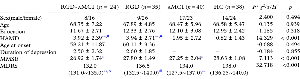
Demographic characteristics and clinical data comprised gender, age, years of education, age at onset of major depressive disorder, duration of depression, the severity of current depressive symptoms (using 17-item HAMD), MMSE scores, and MDRS scores. Age at onset of depression was defined as the age at which patients experienced the first episode of major depression and was ascertained from a self-administered questionnaire in addition to hospital records. The demographic and clinical characteristics of all participants are summarized in Table 1.
Demographic and clinical information on participants

Abbreviation: RGD: remitted geriatric depression; aMCI: amnestic mild cognitive impairment; RGD-aMCI: RGD comorbid with aMCI; HC: health control; HAMD: Hamilton depression rating scale for depression; MMSE: Mini-mental status examination; MDRS: mattis dementia rating scale.
* : HC, p < 0.05.
** : HC, p < 0.001.
# : aMCI, p < 0.05.
Δ : RGD, p < 0.05.
Neuropsychological examination
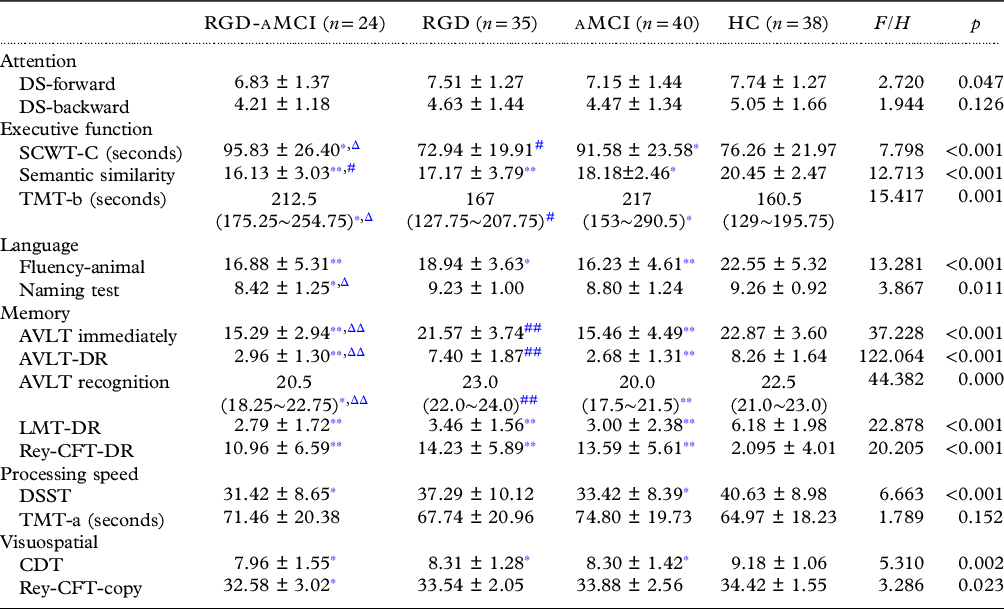
All participants were administered a comprehensive neuropsychological assessment including digit span test (DS forward; DS-backward), stroop color word test-B, C (seconds; color, word-color) (SCWT-B,C), semantic similarity, trail making test-A, B (seconds) (TMT-A,B), verbal fluency (animal), naming test, auditory verbal learning test delay recall (AVLT-DR), logical memory test-delay recall (LMT-DR), Rey–Osterrieth complex figure test-copy or delay recall (Rey-CFT-copy or DR), digit symbol substitution test (DSST), and clock drawing task (CDT). The neurocognitive measures were further categorized into six cognitive domains including attention, executive function, language, memory, processing speed, and visuospatial skill (see Table 2), based on the results of prior reports of cognitive processes which were assessed during tasks performance.
Raw neuropsychological scores in ANOVA

Abbreviation: RGD: remitted geriatric depression; aMCI: amnestic mild cognitive impairment; RGD-aMCI: RGD comorbid with aMCI; HC: health control; DS: digit span test; SCWT: stroop color word test; TMT: trail making test; AVLT: auditory verbal learning test; LMT: logical memory test; Rey-CFT: Rey–Osterrieth complex figure test; DSST: digit symbol substitution test; CDT: clock drawing task.
* : HC, p < 0.05.
** : HC, p < 0.001.
# : aMCI, p < 0.05.
## : aMCI, p < 0.001.
Δ : RGD, p < 0.05.
ΔΔ : RGD, p < 0.001.
MRI data acquisition
All participants underwent a resting-state fMRI scan using a 3.0 Tesla Siemens Verio scanner (Erlangen, Germany) with an 8-channel radiofrequency coil at the Affiliated Brain Hospital of Nanjing Medical University. They were instructed to minimize movement, keep their eyes closed, and not fall asleep. Resting-state functional images including 240 volumes were obtained using a gradient-recalled echo-planar imaging (GRE-EPI) sequence, with repetition time (TR) = 3000 ms, echo time (TE) = 40 ms, flip angle (FA) = 90°, acquisition matrix = 64 × 64, field of view (FOV) = 220 mm × 220 mm, thickness = 3.0 mm, gap = 0 mm, number of slices = 36, and voxel size = 3.75 × 3.75 × 4 mm3. High-resolution T1-weighted axial images covering the whole brain were acquired by 3D magnetization prepared rapid gradient echo (MPRAGE) sequence as described below: TR = 2000 ms, TE = 2.6 ms; FA = 8°, acquisition matrix = 256 × 256, FOV= 256 × 256 mm, thickness = 1.0 mm, gap = 0 mm, number of slices = 176, and voxel size = 1 × 1 × 1 mm3. The data acquisition lasted for 8 minutes.
MRI data preprocessing
Functional images were pre-processed via the Data Processing Assistant for Resting-State fMRI (DPARSF) Version 2.1 (http://www.restfmri.net/forum/DPARSF) based on the Statistical Parametric Mapping software (SPM8; http://www.fil.ion.ucl.ac.UK/spm). The first ten volumes were excluded to allow for signal equilibrium and enable participants to adapt to the machine’s noise. Slice timing and realignment procedures were then conducted to correct for time differences in acquisition among the slices within one volume and motion effects (6-parameter rigid body) during the scan. Subjects with excessive head motion (> 2.5 mm translation and/or >2.5° rotation) during the scan were excluded from further analyses. The resulting images were spatially normalized to the Montreal Neurological Institute template. They were then resampled into 3 × 3 × 3 mm voxels and smoothed with a 6 × 6 × 6 mm Gaussian kernel. Potential confounding signals including head motion parameters, white matter, and cerebrospinal fluid were removed as covariates of no interest. Regression of global signal time course was not executed since recent evidence indicated that it is related to major neuronal components (Chang and Glover, Reference Chang and Glover2009). Finally, linear trends removal and band-pass filtering (0.01–0.08 Hz) were performed.
Functional connectivity analyses
FC analyses were computed using a seed-based approach, whereby the posterior cingulate cortex (PCC) (Montreal Neurological Institute [MNI] space: −5, −49, 40, radius=10 mm) served as the seed region (Fransson, Reference Fransson2005; Long et al., Reference Long2008; Raichle, Reference Raichle2015; Raichle et al., Reference Raichle, MacLeod, Snyder, Powers, Gusnard and Shulman2001). The PCC was selected as the region of interest (ROI) since it is considered as a structural and functional core in the DMN. Convergent evidence from histopathologic, structural, and functional imaging suggested that while the PCC acted as a vulnerable core, it was involved in the pathophysiology of AD, aMCI, and neuropsychiatric disease (Del Sole et al., Reference Del Sole2008; Fransson and Marrelec, Reference Fransson and Marrelec2008; Liang et al., Reference Liang2008; Pengas et al., Reference Pengas, Hodges, Watson and Nestor2010; Rowe et al., Reference Rowe2007). PCC-seed FC calculations were conducted using the Resting-state fMRI data analysis Toolkit (REST; http://www.restfmri.net) (Song et al., Reference Song2011). The time series of all voxels in the ROI were averaged. A FC map of the whole brain was then obtained by conducting Pearson’s correlational analyses between averaged time series and out-seed time series at the voxel-wise with gender, age, and education as covariates. Afterward, Fisher’s z-transformation was applied to improve the normality of the correlation coefficients.
Statistical analysis
Demographic and clinical characteristics
All statistical procedures were conducted by the Statistical Product and Service Solutions (SPSS), Version 22.0 software (SPSS, Inc., Chicago, IL, USA). For RGD, RGD-aMCI, aMCI, and HC groups, between-group comparisons were made using one-way ANOVA (analysis of variance) followed by Bonferroni post hoc analysis for parametric variables (age, education, HAMD, and MMSE) and non-parametric tests (Kruskal-Wallis) for non-normally distributed data (MDRS) as well as chi-square for categorical data (gender). A two-sample t-test was used to compare the differences in age at onset and duration of depression between RGD-aMCI and RGD groups. The significance level was set at p < 0.05.
Neuropsychological data
The differences in raw scores of each cognitive test between the four groups were compared by ANOVA followed by post hoc Fisher’s least significant difference test based on z-transformed scores. The statistical significance level was set at p < 0.05. Since the variances of the TMT-b and AVLT recognition scale scores were different, non-parametric Kruskal-Wallis H test was used to determine the differences among the participant groups. Non-parametric post hoc Mann-Whitney U test was used to reveal the source of the differences between the specific group pairs (Bonferroni corrected, p < 0.05). All statistical procedures were conducted using SPSS 22.0 software.
Group-level resting-state functional connectivity analysis
To compare the rsFC in the DMN among the four groups, we utilized one-way analysis of covariance (ANCOVA) while controlling for age, gender, and level of education and correcting for multiple comparisons. Afterward, main effect contrasts were performed between groups (RGD vs. aMCI, RGD vs. HC, RGD vs. RGD-aMCI, aMCI vs. RGD-aMCI, aMCI vs HC, RGD-aMCI vs. HC). Significance after AlphaSim correction was determined at p corrected < 0.01. After that overlaps and non-overlaps between the interactive regions in RGD, aMCI, and aMCI-RGD subjects were further analyzed. The Cohen’s d, ϕ, and η 2 values, which reflected the effective size of the statistical difference among groups, were calculated by G∗Power software 3.1 (Faul et al., Reference Faul, Erdfelder, Lang and Buchner2007).
Correlation analysis between dmn functional connectivity alterations and cognitive impairment
To explore the cross-sectional relationship between abnormal DMN FC and cognitive impairment, partial correlation analysis was utilized separately with gender, age, and education as covariates in the RGD, RGD-aMCI, and aMCI groups, respectively (Goveas et al., Reference Goveas2011). Correlations were considered significant at two-tailed p < 0.05.
Results
Demographic and clinical characteristics
There were no significant differences in gender, age, and years of education between RGD-aMCI, RGD, aMCI, and HC groups. There were also no significant differences in age of onset and duration of depression between the RGD-aMCI and RGD groups. However, MMSE scores (F = 7.113, p < 0.001), MDRS scores (F = 14.993, p < 0.001), and 17-item HAMD scores (F = 14.329, p < 0.001) were significantly different among the four groups. The post hoc analysis showed that MMSE scores were significantly lower in both RGD-aMCI (p < 0.05) and aMCI (p < 0.05) compared to HC. MDRS scores were significantly reduced in both RGD-aMCI (p < 0.05) and aMCI (p < 0.05) in contrast to RGD and HC, respectively, However, there were no significant differences in MMSE and MDRS scores between RGD-aMCI and aMCI. Although the 17-item HAMD scores showed significantly higher values in RGD and RGD-aMCI compared to both HC and aMCI (p < 0.05), it was less than 7 when participants were initially enrolled in the study. Demographic and clinical characteristics of all subjects are summarized in Table 1 and Table Supplementary S1.
Neuropsychological performance in RGD, RGD-aMCI, aMCI, and HC groups
The performance on neuropsychological tests for all participants is summarized in Table 2 and Supplementary Table S2. The performance of all neuropsychological tests was statistically significantly different among the four groups, with the exception of DS forward, DS backward, and TMT-A test (in seconds). RGD-aMCI, RGD, and aMCI groups all exhibited significant impairment in one executive function measure (Semantic similarity), one language measure (Fluency-animal), two memory measures (including LMT-DR and Rey-CFT-DR), and visuospatial skill (CDT) (p < 0.05) compared to HC group. Both RGD-aMCI and aMCI had lower performances on two executive function measures (SCWT-C [seconds], TMT-b [seconds]), one processing speed measure (DSST) (p < 0.05), and three memory measures (including AVLT immediately, AVLT-DR, and AVLT recognition) (p < 0.001) in contrast to RGD and HC. Moreover, RGD-aMCI showed significant deficits in one language measure (Naming Test) and one visuospatial measure (Rey-CFT-copy) compared to the other three groups (p < 0.05). RGD-aMCI performed significantly worse in one executive function measure (Semantic similarity) than the aMCI group (p < 0.05).
Altered resting-state functional connectivity of the DMN in RGD, RGD-aMCI, aMCI, and HC groups
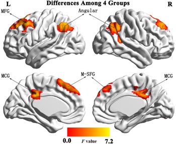
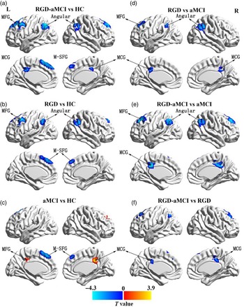
The PCC-seed DMN rsFCs were significantly different in bilateral medial superior frontal gyrus (M-SFG), bilateral angular gyrus (L-angular/R-angular), bilateral middle cingulate gyrus (MCG), and left medial frontal gyrus (L-MFG) among the four groups. The η 2 value of these brain regions ranges from 0.085 to 0.141, which signifies an intermediate to large effect (see Figure 1, Supplementary Table S3) (Tomczak M and Tomczak E, Reference Tomczak and Tomczak2014). Post hoc analysis demonstrated that the other three patient groups had similar significant reduction of DMN rsFC in bilateral M-SFG, compared with the HC group (see Figures 2a, b, and c, Supplementary Table S4). Moreover, there were no statistically significant differences in bilateral M-SFG while comparing RGD vs. aMCI, RGD vs. RGD-aMCI, and RGD-aMCI vs. aMCI subjects.
The PCC-seed DMN rsFCs comparisons among RGD, RGD-aMCI, aMCI, and HC groups. Significant differences in PCC-seed resting-state functional connectivity showed in bilateral medial superior frontal gyrus, bilateral angular gyrus, bilateral middle cingulate gyrus, and left medial frontal gyrus among RGD, RGD-aMCI, aMCI, and HC groups (p < 0.01, AlphaSim correct). Abbreviations: RGD: remitted geriatric depression; RGD-aMCI: RGD comorbid with aMCI; aMCI: amnestic mild cognitive impairment; HC: health comparisons; PCC: posterior cingulate cortex; rsFC: resting-state functional connectivity; MFG: medial frontal gyrus; MCG: middle cingulate gyrus; M-SFG: medial superior frontal gyrus.

Comparison of PCC-seed rsFCs in DMN between groups (RGD vs. aMCI, RGD vs. HC, RGD vs. RGD-aMCI, aMCI vs. RGD-aMCI, aMCI vs HC, RGD-aMCI vs. HC). Compared with HC group, the RGD-aMCI, RGD, and aMCI groups showed significantly decreased PCC-seed rsFC in the bilateral M-SFG (a, b, c), the RGD-aMCI subjects showed a significant decrease in the bilateral MCG (a), and the aMCI groups showed a significant increase in the bilateral MCG (c). Compared with the aMCI group, both the RGD (d) and RGD-aMCI (e) groups had significantly decreased PCC-seeded rsFC in the bilateral angular gyrus, MCG, and L-MFG. The PCC-seed rsFCs of the MCG and L-MFG were significantly decreased in the RGD-aMCI group than aMCI group (f) (p < 0.01, AlphaSim correct). Red is the enhanced area of functional connection; blue is the lowered area for the functional connection. Abbreviations: RGD: remitted geriatric depression; RGD-aMCI: RGD comorbid with aMCI; aMCI: amnestic mild cognitive impairment; HC: health comparisons; PCC: Posterior cingulate cortex; rsFC: resting-state functional connectivity; MFG: medial frontal gyrus; MCG: middle cingulate gyrus; M-SFG: medial superior frontal gyrus.

Furthermore, RGD-aMCI subjects displayed significantly lower rsFC in bilateral MCG compared to the HC group (see Figure 2a). In contrast, the aMCI group exhibited higher rsFC in the same regions (see Figure 2c). Compared to the aMCI group, RGD-aMCI and RGD had significant reductions in PCC-seeded rsFC of the bilateral angular gyrus, MCG, and L-MFG (see Figures 2d and e). There was a significant decline in MCG and L-MFG rsFCs of RGD-aMCI group compared to RGD patients (see Figure 2f).
Correlation analysis between DMN resting-state functional connectivity and cognitive deficit
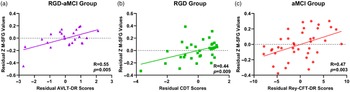
Partial correlation analysis demonstrated that the decreased rsFCs between PCC and bilateral M-SFG regions were correlated with poor neuropsychological test performance in RGD, RGD-aMCI, and aMCI groups. In particular, rsFCs which connected the PCC with bilateral M-SFG were positively correlated with a memory measure (AVLT-DR, r = 0.55, p = 0.005) in RGD-aMCI (see Figure 3a), with a visuospatial measure (CDT, r = 0.44, p = 0.009) in RGD (see Figure 3b) as well as a memory measure (Rey-CFT-DR, r = 0.47, p = 0.003) in aMCI (see Figure 3c). It is important to note that the potential outliers did not drive our findings because the results remained consistent when the outlier was excluded (Supplementary Figure S1).
Partial correlation analysis between alterations of rsFCs in the DMN and cognitive deficit in the RGD-aMCI, RGD, and aMCI group, respectively. The PCC-seed rsFC of M-SFG was positively correlated with scores of AVLT-DR in the RGD-aMCI subjects (a), positively correlated with scores of CDT in the RGD subjects (b), and positively correlated with scores of Rey-CFT-DR in the aMCI subjects (c) (two-tailed p < .05). Abbreviations: RGD: remitted geriatric depression; RGD-aMCI: RGD comorbid with aMCI; aMCI: amnestic mild cognitive impairment; HC: health comparisons; PCC: Posterior cingulate cortex; rsFC: resting-state functional connectivity; M-SFG: medial superior frontal gyrus; AVLT-DR: auditory verbal learning test-delay recall; CDT: clock drawing task; Rey-CFT-DR: Rey–Osterrieth complex figure test-delay.

Discussion
In the present study, we demonstrated that patients with RGD, aMCI, and RGD-aMCI exhibited altered FC in DMN accompanied by significant deficits in multiple domains of cognitive function. Furthermore, we observed that both shared and distinct FC alterations among RGD, RGD-aMCI, and aMCI. There was a similar significant decrease in the PCC-seed rsFC of bilateral M-SFG, which was positively correlated with the severity of cognitive impairment in all three patient groups (RGD, RGD-aMCI, and aMCI). Moreover, both RGD-aMCI and RGD participants had substantially lower rsFCs in the bilateral angular gyrus, middle cingulate gyrus, and left medial frontal gyrus compared with aMCI subjects. In our study, the differences in the rsFC of DMN among the four groups of patients were significant with a medium to large effect size. The results suggested that the abnormal FC between the PCC and bilateral M-SFG might serve as an underlying pathological mechanism for the development of AD in both depression and cognitive impairment. Altogether, this study provides novel insights into the underlying mechanisms of susceptibility conversion to AD in RGD and aMCI patients via altered rsFC of the DMN, which could have valuable clinical implications (Tomczak M and Tomczak E, Reference Tomczak and Tomczak2014).
It is widely accepted that multi-dimensional cognitive deficits, including executive function, language, memory, processing speed, attention, and visuospatial ability, are present in geriatric depression patients during an acute episode phase (Jamieson et al., Reference Jamieson, Goodwill, Termine, Campbell and Szoeke2019; Morin et al., Reference Morin, Gonzales, Bickford, Catalinotto, Nelson and Mackin2019). Our results indicated that impairments of executive function, memory, and information processing speed persisted in individuals with RGD. These findings potentially imply that the cognitive dysfunctions might be trait characteristics of geriatric depression patients. Numerous studies have confirmed that cognitive impairments persisted throughout the degeneration process from aMCI to AD (Dunn et al., Reference Dunn2014; Lehrner et al., Reference Lehrner2005; Liu et al., Reference Liu2019b; Serra et al., Reference Serra2010). Our study findings are in consistence with previous studies (Bombin et al., Reference Bombin2012; Liao et al., Reference Liao, Zhang, Shu, Wang, Liu and Zhang2017) which also demonstrated that aMCI patients had cognitive impairment across five cognitive domains, including executive function, language, memory, processing speed, and visuospatial ability, similar to RGD patients. In addition, we found that the RGD-aMCI group showed more severe impairments in both executive function and memory compared to RGD and aMCI groups. The findings of this study might collectively infer that multi-dimensional cognitive deficits, which exist in the remission phase of patients with geriatric depression, might act as the trait characteristics of RGD patients and could be implicated in the susceptibility to develop AD. It is noteworthy that there were no significant differences in the performance of attention tests between RGD and HC groups.
A growing body of evidence concerning depressive disorder has revealed that emotional processing, reward, self-representation, and external stimulus interactions are associated with structural and functional abnormalities in brain circuits, including the hippocampus, amygdala, anterior cingulate cortex, and medial prefrontal cortex (Breukelaar et al., Reference Breukelaar2020; Davidson et al., Reference Davidson, Pizzagalli, Nitschke and Putnam2002; Liu et al., Reference Liu, Jiang and Yuan2018b; Northoff et al., Reference Northoff, Wiebking, Feinberg and Panksepp2011). Interestingly, these brain regions also fall within the DMN, a brain network involved in self-referential processing, affective cognition, and emotion regulation (Andrews-Hanna et al., Reference Andrews-Hanna, Reidler, Sepulcre, Poulin and Buckner2010; Buckner et al., Reference Buckner, Andrews-Hanna and Schacter2008). Prior studies have reported that the FC of DMN with the subgenual anterior cingulate cortex (sgACC) was higher in major depressive disorder patients during an acute phase (MD et al., Reference MD2007). However, the increased FC in the DMN and sgACC was reduced in patients with remitted depression after treatment (Liston et al., Reference Liston2014; Liu et al., Reference Liu2019a). These findings suggested that the FC of DMN could be valuable in understanding the underlying mechanism of depressive disorders.
Neuroimaging evidence consistently supports the hypothesis that the AD spectrum includes geriatric depression, aMCI, and dementia due to AD stages (Delbeuck et al., Reference Delbeuck, Collette and Van der Linden2007; Vallet et al., Reference Vallet, Hudon, Simard and Versace2013). Both geriatric depression and aMCI are deemed as disconnection syndromes with altered cerebral networks, hence, leading to multi-dimensional cognitive and behavioral dysfunctions. Numerous fMRI studies have shown that the alterations within the DMN were linked to depressive symptoms and might serve as potential biomarkers of geriatric depression (Liu et al., Reference Liu2019a; Shi et al., Reference Shi2020; Wang et al., Reference Wang2020a; Yan et al., Reference Yan2019). Nonetheless, there is a paucity of studies on the incidence of abnormal FC in the DMN of RGD patients and its relationship to cognitive impairment. The present study demonstrated that RGD patients had significantly lower PCC-seed rsFC in the bilateral M-SFG than HC. Likewise, aMCI and RGD-aMCI groups showed significant correlations between decreased rsFCs and cognitive function impairment.
Previous studies have reported that geriatric depression, aMCI, and AD might exist in a clinical continuum (Mah et al., Reference Mah, Anderson, Verhoeff and Pollock2017; Panza et al., Reference Panza2010), whereby geriatric depression and aMCI are closely related to the development of AD. Geriatric depression and aMCI have several common clinical features, including impairment in cognitive function, reduction in social and occupational function along with some common risk factors, such as vascular symptoms, alterations in glucocorticoid steroids, hippocampal atrophy, and inflammatory changes (Byers and Yaffe, Reference Byers and Yaffe2011; Lauriola et al., Reference Lauriola2018; Liew, Reference Liew2019; Mah et al., Reference Mah, Anderson, Verhoeff and Pollock2017). Moreover, they might share underlying mechanisms that confer a heightened risk of conversion to AD (Li et al., Reference Li2015). While acute phase depressive disorder patients demonstrated higher DMN rsFC (Greicius et al., Reference Greicius2007; Liu et al., Reference Liu2019a), our study revealed that the RGD, RGD-aMCI, and aMCI groups showed significantly decreased PCC-seed rsFC in bilateral M-SFG compared with HC, which positively correlated with cognitive function impairment. The presence of cognitive deficits or neuropathological characteristics in aMCI and RGD patients suggests that altered intrinsic rsFCs of the DMN in the remitted phase of geriatric depression patients could serve as trait markers of cognitive impairment but not to depressive symptoms.
Interestingly, both the RGD and RGD-aMCI groups showed significantly decreased PCC-seed rsFCs in the bilateral angular gyrus, middle cingulate gyrus, and the left medial frontal gyrus compared with the aMCI group. This implies that the total amount of information between these brain regions and other functionally connected regions was reduced in the RGD group compared to the aMCI group. While the RGD and the RGD-aMCI groups showed significantly decreased rsFC in the bilateral MCG compared with HC, the aMCI group had a significant rsFC increase in bilateral MCG. The opposite alteration of PCC-MCG rsFC in the aMCI group suggested that aMCI patients might recruit other network resources to compensate for the degenerative process of the disease. It is also possible that the recruited aMCI participants were in the early stage of AD progression. Given that the neuropathological characteristics of cognitive deficits in aMCI and RGD patients are still controversial and there are different patterns of DMN dysfunction between RGD and aMCI, further studies could include AD patients and thus confirm our results.
In conclusion, we demonstrated that both RGD and aMCI patients exhibited altered intrinsic rsFC between PCC and M-SFG, which was associated with cognitive deficits. Our results provide insights on the neuropathological mechanistic pathways concerning cognitive impairment during the conversion to AD in geriatric depression patients.
Limitations
The present study has several limitations which need to be considered. Firstly, the patients were recruited only based on clinical criteria without biomarker support for AD diagnosis. Hence, patients with prodromal AD or other dementing conditions might have been included in the study (Dubois et al., Reference Dubois2014). A cerebrospinal fluid examination could have been performed to distinguish between different AD phases and discriminate all-cause dementia from AD dementia (McKhann et al., Reference McKhann2011; Scheltens et al., Reference Scheltens2016). Second, the chosen seed-based method limited our analysis. While data-driven approaches could avoid this issue, a hypothesis-driven approach could explicitly explore the closely related brain networks. Moreover, we did not investigate structural alteration in the DMN using a multimodal image. Since the sample size is limited, the effect sizes obtained in this pilot study could serve as a basis for further studies. Our findings require validation and replication in a larger cohort. Multi-center studies are also needed to corroborate these findings.
Conflict of interest
The authors have no conflicts of interest to declare.
Source of funding
This work was supported by the Science and Technology Major Project of Guangxi under Grants 20201019106.
Description of authors’ roles
CG designed the study and wrote the paper. NA together with CG assisted in literature retrieval and writing the paper. WL and CZ conducted the study and analyzed the data. XW managed the data and created data sets for analysis. XZ and CZhang critically revised the article and approved the final draft.
Acknowledgements
We thank all participants and staff at the Affiliated Brain Hospital of Nanjing Medical University for their generous support, cooperation, and participation. We would like to express our gratitude to EditSprings (https://www.editsprings.com/) for the expert linguistic services provided.
Supplementary material
To view supplementary material for this article, please visit https://doi.org/10.1017/S1041610221001174