Holoprosencephaly (HPE) is the most common structural anomaly of human brain development. The defining feature of HPE is the incomplete cleavage of the embryonic forebrain into two separate hemispheres; defects in formation of the facial midline are also observed in most patients. Both genetic and environmental aetiologies have been described for HPE, and clinical expression is extremely variable. Interestingly, phenotypic heterogeneity is observed even in familial forms of HPE, suggesting that a complex interplay of developmental, genetic and environmental factors play a role in this variability. Studies in experimental animals have given insight into the signal transduction pathways that regulate normal development of the forebrain and midface, and provided model systems that mimic unexplained aspects of human HPE, including phenotypic heterogeneity. These models have, in turn, raised interesting new questions, answers to which may inform our understanding of this frequent and devastating birth defect.
Aberrant development of the forebrain and midface in HPE
Categorisation of HPE phenotypes
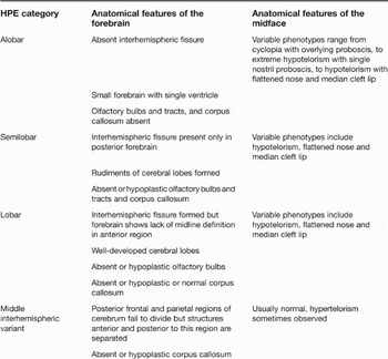
The incidence of HPE is ∼1 in 250 first-trimester conceptuses but, because of intrauterine lethality, live-born prevalence is ∼1 in 16 000 (Refs Reference Muenke, Beachy and Scriver1, Reference Yamada2). The primary brain malformation is lack of, or incomplete, separation of the forebrain into right and left hemispheres (for detailed reviews on clinical features of HPE, see Refs Reference Muenke, Beachy and Scriver1, Reference Cohen3, Reference Dubourg4). Three levels of severity are commonly distinguished (Table 1). Alobar HPE, the most severe type, is characterised by complete failure to form an interhemispheric fissure in the prosencephalon, resulting in a single, small brain ventricle. Semilobar HPE cases display rudimentary cerebral lobes and partial formation of a fissure in the posterior region of the forebrain, but subdivision is incomplete. In lobar HPE, the cerebral hemispheres are well developed, with a defined interhemispheric fissure, but the most anterior midline region is continuous, and some anterior structures, such as the corpus callosum and olfactory bulbs, may be missing or hypoplastic. A somewhat distinct defect, syntelencephaly, is widely considered to be a subtype of HPE, called the middle interhemispheric variant (MIH) (Ref. Reference Simon5). MIH is characterised by a failure of the cerebrum to divide in the posterior frontal and parietal regions, whereas forebrain structures anterior and posterior to this segment are separated (Ref. Reference Simon5). Although most holoprosencephalic brains can fit into these categories, it is important to note that each case is distinctive, and the range of midline defects extends from most to least severe in an uninterrupted continuum referred to as the HPE spectrum (Refs Reference Muenke, Beachy and Scriver1, Reference Ming and Muenke6).
Categorisation of holoprosencephaly phenotypes by severity

Abbreviation: HPE, holoprosencephaly.
Among the most important and informative features of HPE are craniofacial anomalies, which occur in 80–90% of patients and can lead to the correct diagnosis (Refs Reference Muenke, Beachy and Scriver1, Reference Cohen3, Reference Dubourg4). In a classic study, DeMyer coined the aphorism “The face predicts the brain” (Ref. Reference DeMyer7); accordingly, facial defects associated with HPE are extremely variable in severity and range from cyclopia with an overlying proboscis in the most severe cases, to slight hypotelorism (i.e. close-set eyes) and a solitary median maxillary central incisor in the mildest cases (Table 1). Such mild facial abnormalities can occur in the absence of brain malformations and are sometimes referred to as ‘microforms’ of HPE. It should be noted that, although the face predicts the brain most of the time, in approximately 20% of HPE cases facial features do not correlate with forebrain phenotypes (Ref. Reference Cohen3).
Development of structures that are defective in HPE
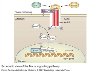
Development of the prospective forebrain is a multi-step process that includes: acquisition of a neural fate by ectodermal cells; the assumption of anterior characteristics by neural tissue at the rostral end of the neuraxis; morphogenetic transformation of the anterior neural plate; and dorsoventral patterning of the resulting prosencephalon (for detailed reviews on forebrain development, see Refs Reference Fuccillo, Joyner and Fishell8, Reference Hebert9, Reference Lupo, Harris and Lewis10, Reference Monuki and Walsh11, Reference Rallu, Corbin and Fishell12, Reference Wilson and Houart13). Embryologically, HPE is most often associated with a disruption in development of the ventral forebrain (Ref. Reference Muenke, Beachy and Scriver1), which is derived from the medial portion of the anterior neural plate. The underlying prechordal mesendoderm functions as a source of signals required for specification of ventral forebrain structures and subsequent formation of the midline, including septation of the forebrain into hemispheres and bilateral subdivision of the eye field (Refs Reference Wilson and Houart13, Reference Rubenstein and Beachy14); disruption of either its formation or function results in HPE. Surgical removal of the prechordal mesendoderm from avian and amphibian embryos results in severe HPE, including cyclopia (Refs Reference Wilson and Houart13, Reference Rubenstein and Beachy14, Reference Lowe, Yamada and Kuehn15, Reference Shen16, Reference Weng and Stemple17, Reference Chen18 and references therein). Formation of the prechordal mesendoderm is dependent on a signalling pathway initiated by the secreted factor Nodal, a member of the transforming growth factor (TGF) β superfamily (Refs Reference Lowe, Yamada and Kuehn15, Reference Shen16, Reference Weng and Stemple17). Nodal initiates signalling by binding to specific members of the TGFβ receptor family (ActRIB and ActRIIA/B), along with the co-ligand/co-receptor Cripto. Consequently, the cytoplasmically localised transcription factors Smad2 and Smad3 are phosphorylated by ActRIB and, with their dimeric partner Smad4, accumulate in the nucleus (Refs Reference Shen16, Reference Weng and Stemple17). Smad complexes, in turn, interact with additional transcription factors, including those of the FoxH subfamily, to drive expression of pathway-specific genes (Refs Reference Chen18, Reference Yamamoto19). Consistent with an important role for Nodal signalling in development of prechordal mesendoderm, is the finding that mutations in specific components of this pathway in zebrafish, mice and humans result in HPE (Refs Reference Chu20, Reference de la Cruz21, Reference Heyer22, Reference Nomura and Li23, Reference Pogoda24, Reference Sampath25, Reference Sirotkin26, Reference Song27, Reference Zhang, Talbot and Schier28) (Fig. 1).
Schematic view of the Nodal signalling pathway. Homodimeric Nodal family ligands, including Gdf1, bind to the co-receptor Cripto/TDGF1 in a complex with ActRlB and ActRIIA/B, which are dimerised members of the TGFβ receptor family. Cripto can also be produced in a soluble form and function as a co-ligand with Nodal (not shown). Receptor activation by ligand binding leads to phosphorylation of ActRlB by ActRIIA/B, and ActRlB in turn phosphorylates Smad2 and Smad3 (Smad2/3). Phosphorylated Smad2/3 interact with Smad4 and accumulate in the nucleus where they interact with additional transcription factors such as FoxH1 to stimulate target gene expression. Factors whose mutation has been implicated in holoprosencephaly in mice or humans are shown in colour. Note that Cripto and TDGF1 are the mouse and human names, respectively, for the same protein. Simplified and adapted from Ref. 20.

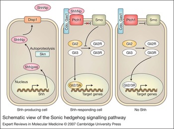
A major signal produced by the prechordal mesendoderm that is involved in patterning the ventral forebrain and anterior midline is the secreted factor Sonic hedgehog (Shh). In fact, Shh is produced throughout the axial mesoderm, including the prechordal mesendoderm and notochord, and plays a key role in ventral patterning throughout the neuraxis (Refs Reference Fuccillo, Joyner and Fishell8, Reference Lupo, Harris and Lewis10). Shh activates a complex signal transduction cascade in which the transmembrane protein Patched1 (Ptch1) functions to inhibit the activity of a second transmembrane protein, Smoothened (Smo). Binding of Shh to Ptch1 relieves this inhibition, stimulating a pathway that results in induction of target genes via the Gli family of transcription factors (Refs Reference Huangfu and Anderson29, Reference Wang, McMahon and Allen30) (Fig. 2). Mutations in components of this pathway result in HPE in mice and humans (Refs Reference Chen31, Reference Chiang32, Reference Dubourg33, Reference Ma34, Reference Ming35, Reference Nanni36, Reference Rahimov37, Reference Ribeiro, Murray and Richieri-Costa38, Reference Roessler39, Reference Roessler40, Reference Zhang, Ramalho-Santos and McMahon41). Gli2 is the major, immediate, pathway-responsive transcriptional activator, with Gli3 playing a more limited role in gene activation; in the absence of Shh, however, Gli3 is proteolytically processed into a transcriptional repressor that is a major antagonist of the Hedgehog pathway (Refs Reference Koebernick and Pieler42, Reference Sasaki43, Reference Wang, Fallon and Beachy44). Shh −/− mice lack ventral structures of the forebrain and spinal cord, and display alobar HPE and cyclopia (Ref. Reference Chiang32). By contrast, Gli3 −/− mice display an opposite phenotype: ventralisation of the forebrain and exencephaly (Refs Reference Grove45, Reference Tole, Ragsdale and Grove46). Reduction of Gli3 dosage in Shh mutant mice largely restores dorsoventral patterning of the telencephalon; unlike Shh −/− mutants, Shh −/−;Gli3 +/− embryos cleaved the forebrain and had two, albeit fused, eyes (Ref. Reference Rallu47). These results led to several important conclusions: (1) dorsoventral patterning of the forebrain is regulated by a balance between Shh and Gli3 function; (2) a major effect of Shh in forebrain development is to inhibit Gli3 repressor activity; and (3) Shh is not strictly required for induction of the ventral forebrain, so long as Gli3 function is also reduced. This last point indicates that additional factors are capable of determining the fate of ventral cells in the forebrain. These factors are very likely to be fibroblast growth factors (FGFs), because mice deficient for both FGF receptors 1 and 2 display Shh-independent HPE, as do mice lacking FGF8 (Refs Reference Gutin48, Reference Storm49). Significantly, maintenance of Fgf8 expression in the prosencephalon requires Shh, and Gli3 −/− mice have an expanded Fgf8 expression domain (Ref. Reference Aoto50). These results place FGF signalling downstream of Shh, but mutations in components of the FGF signalling pathway have not yet been reported in human HPE.
Schematic view of the Sonic hedgehog signalling pathway. Sonic hedgehog (Shh) is initially produced (left cell) as a precursor (pre) that undergoes autocatalytic proteolysis and cholesterol adduction, and palmitoylation catalysed by Skinny hedgehog (Skn) to form the mature signalling molecule ShhNp. ShhNp is released from producing cells by a mechanism that involves Dispatched1 (Disp1). In cells not exposed to ShhNp (right cell), Patched1 (Ptch1) inhibits the activity of Smoothened (Smo), blocking signalling. The transcription factors Gli2 and Gli3 are proteolytically processed to repressor forms (Gli2R and Gli3R) that can translocate to the nucleus and repress pathway target genes (note that Gli3R is the major repressor). Binding of ShhNp to Ptch1, possibly in conjunction with Gas1 or Cdo, relieves inhibition of Smo; Smo-mediated signalling prevents processing of Gli factors, which, either directly or via additional steps to produce activator forms (Gli2A and Gli3A), can translocate to the nucleus and activate pathway target genes (note that Gli2 is the major activator). Factors whose mutation has been implicated in holoprosencephaly (HPE) in mice or humans are shown in colour. Mutation of Gli2 but not Gli3 is implicated in HPE, but it is presumably loss of the activator form of Gli2 that is involved. Note that Cdo and Gas1 are structurally unrelated proteins that each bind Shh to promote signalling; they are drawn together for simplicity, not because they are known to physically interact. For more detail, see Refs Reference Dubourg33 and Reference Ma34.

Factors other than Gli3 are also involved in providing dorsal character to the forebrain. In particular, members of the bone morphogenetic protein (BMP) family and their receptors appear to be involved in formation or maintenance of the dorsal midline (Refs Reference Hebert9, Reference Wilson and Houart13). Strikingly, implantation of BMP4- or BMP5-soaked beads into the rostral neural tube of the chick embryo resulted in HPE, including cyclopia and loss of ventral midline structures; this appeared to be due to excessive cell death in the basal telencephalon rather than altered specification of ventral cell fates (Ref. Reference Golden51). Mice genetically deficient for antagonists of BMP signalling also show HPE phenotypes (see below), although mutations in the genes encoding these factors have not yet been reported in human HPE cases.
The association of cyclopia with severe (alobar) HPE is consistent with a requirement for the prechordal mesendoderm in bilateral subdivision of the eye field (Refs Reference Rubenstein and Beachy14, Reference Dale52, Reference Li53, Reference Pera and Kessel54, Reference Varga, Wegner and Westerfield55). This may occur via a need for the mesendoderm to orchestrate morphogenetic movements that physically split the developing eye field and/or direct signalling from the mesoendoderm to induce medial neural plate cells to adopt a nonretinal cell fate (Ref. Reference Wilson and Houart13). However, as mentioned above, non-neural facial structures are also affected in HPE. Shh is expressed in the ectoderm of the frontonasal and maxillary processes of the developing face and patterns neural crest-derived facial primordia to produce specific cartilaginous structures and bones of the facial midline (Refs Reference Helms, Cordero and Tapadia56, Reference Hu and Helms57). Disruption of Shh signalling, specifically in the face, via surgical removal of ectoderm, application of anti-Shh antibodies or treatment with the Shh pathway inhibitor cyclopamine, results in microforms of HPE (Refs Reference Hu and Helms57, Reference Cordero58). These results strongly suggest that mutations that compromise Shh signalling contribute to HPE phenotypes through loss of signalling function both from prechordal mesendoderm and directly in the developing face.
Aetiology of human HPE
Both genetic and environmental aetiologies for HPE have been described (Refs Reference Muenke, Beachy and Scriver1, Reference Cohen3, Reference Dubourg4, Reference Cohen and Shiota59). HPE exists in inherited, autosomal dominant (familial) forms and mutation-associated sporadic forms, and it is also observed as a component of several different multiple-malformation syndromes. Chromosomal anomalies are observed in many HPE live-birth cases [estimates range from 24% to 45% (Ref. Reference Cohen3)], and mapping of recurrent chromosomal rearrangements has implicated up to 12 loci in karyotypically abnormal human HPE (Ref. Reference Roessler and Muenke60). These studies led to the identification of four genes that are mutated in isolated, non-syndromic human HPE, including several of the familial forms: SHH, ZIC2, SIX3 and TGIF. Mutations in four additional genes have also been reported: PTCH1, GLI2, TDGF1 (also known as CRIPTO) and FOXH1 (also known as FAST1) (Refs Reference Cohen3, Reference Dubourg4, Reference Ming and Muenke6).
Types of mutations observed include missense and nonsense mutations, and small deletions that produce frameshifts or loss of several amino acids (for a review of the numbers and types of mutations in human HPE genes and the phenotypes of the associated cases, see Ref. Reference Cohen3). Recently, submicroscopic deletions in SHH, ZIC2, SIX3 and TGIF have been observed in cytogenetically normal HPE cases, and these may correlate with a more severe outcome than point mutations (Refs Reference Bendavid61, Reference Bendavid62). Taken together, mutations in these eight genes account for only ∼25% of the total cases examined, suggesting that additional HPE genes remain to be discovered. All mutations identified to date are found in the heterozygous state and, where examined, are loss-of-function alleles, resulting in haploinsufficiency (Refs Reference Roessler40, Reference El-Jaick63, Reference Maity, Fuse and Beachy64, Reference Schell-Apacik65, Reference Traiffort66) (a predicted exception are mutations in PTCH1, which, based on its role as a negative regulator of the SHH pathway, would be predicted to be genetic gain-of-function alleles).
Strikingly, a complete spectrum of HPE phenotypes is observed even in familial forms, in which it is estimated that among obligate mutation carriers, 37% have HPE, 27% display microforms of HPE and 36% have no clinical manifestation (Refs Reference Ming and Muenke6, Reference Cohen67). As discussed in detail below, several potential explanations for this variability exist, but there is little evidence to support one over another in the human condition. Nevertheless, the lack of a genotype–phenotype correlation in familial HPE, as well as in sporadic cases with mutations in the same gene, suggests that heterozygous mutation of the known HPE genes may be a contributing but insufficient factor (Ref. Reference Ming and Muenke6).
Environmental or teratogenic exposure is also implicated in HPE, and epidemiological studies of cytogenetically normal, non-syndromic HPE suggest that maternal diabetes mellitus and maternal alcohol consumption during early pregnancy may be important factors (Refs Reference Barr68, Reference Croen, Shaw and Lammer69). The incidence of HPE in infants of diabetic mothers is very high (∼1%), but the mechanism(s) by which maternal diabetes influences fetal forebrain development is unknown. By contrast, the magnitude of the contribution of fetal alcohol exposure to HPE incidence is not clear, but studies in animals suggest that alcohol may interfere with Shh signalling (Refs Reference Ahlgren, Thakur and Bronner-Fraser70, Reference Li71). Case reports and controlled animal studies are consistent with the possibility that fetal exposure to numerous different teratogens, including retinoids, plant alkaloids, and specific pharmaceutical drugs and infectious agents, could increase the risk of HPE (Ref. Reference Cohen and Shiota59). Given the relatively low percentage of cases with defined mutations and the possibility of gene–environment interactions in HPE (see below), additional epidemiological studies are warranted.
An aetiological factor that may encompass both genetic and environmental variables is cholesterol biosynthesis and utilisation. A small percentage (∼5%) of patients with Smith-Lemli-Opitz (SLO) syndrome display HPE; this syndrome arises from recessive mutations in 3β-hydroxysterol-Δ7-reductase, which catalyses the final step in cholesterol synthesis (Ref. Reference Kelley72). Furthermore, cholesterol-lowering drugs can produce HPE in experimental animals (Ref. Reference Cohen and Shiota59) and, perhaps, humans (Ref. Reference Edison and Muenke73). Cholesterol is required for transduction of the Shh signal (Ref. Reference Cooper74), providing a probable mechanistic explanation for these observations. Interestingly, ∼10% of cell lines from non-syndromic HPE cases displayed abnormally low cholesterol biosynthesis (Ref. Reference Haas75), but the underlying basis of the defect (or defects) is unknown. Approximately 20% of the patients with sterol abnormalities also had mutations in known human HPE genes (Ref. Reference Haas75).
Phenotypic variability in HPE and animal models
The incomplete penetrance and highly variable expressivity observed in familial HPE, as well as the wide phenotypic spectrum of sporadic HPE generally, suggests that factors in addition to heterozygous mutations in HPE genes may be required to produce this defect. Although limited information is available for human HPE, animal models offer experimental evidence for multiple potential mechanisms, including both genetic and environmental factors.
Digenic mutation
One mechanism observed in human HPE cases and formally proposed by Ming and Muenke as a likely explanation for variable penetrance and expressivity (Ref. Reference Ming and Muenke6) is that heterozygous mutation of two HPE genes, and the consequent double haploinsufficiency, is required to produce a severe phenotype. HPE patients with mutations in both SHH and TGIF (two cases); SHH and ZIC2; and PTCH1 and GLI2 have been identified (Refs Reference Nanni36, Reference Rahimov37). Thus, although the number of instances is small, it appears that digenic mutation might result in HPE whereas single haploinsufficiency may be insufficient. Interestingly, in these cases the mutant genes may encode factors in the same signalling pathway (e.g. PTCH1 and GLI2) or factors whose mechanistic relationship is unclear and that may exert their effects through parallel or unrelated pathways (e.g. SHH and TGIF).
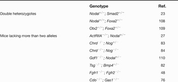
Mice heterozygous for known HPE genes generally do not display any phenotype, which is consistent with the notion that the heterozygous mutations seen in human HPE patients may not be sufficient to produce significant structural defects. However, double heterozygosity produces HPE phenotypes in several different mouse models (Table 2). Like the human situation, digenic inheritance of HPE in mice can occur when the mutant genes encode factors in the same signalling pathway (e.g. Smad2 and Nodal) or when they encode factors that may act in a parallel or complementary fashion (e.g. Otx2 and Foxa2/Hnf3b). Genetic interactions that lead to HPE have also been observed in mice carrying homozygous mutations in one gene and heterozygous or homozygous mutations in a second gene (Table 2). In certain cases, these interactions probably reveal redundant or overlapping function between the mutant factors (e.g. the BMP antagonists Noggin and Chordin; the TGFβ superfamily members GDF1 and Nodal). Interestingly, mice that lack the Shh-binding proteins Cdo and Gas1 individually each display microforms of HPE, whereas mice lacking both factors have severe HPE (Refs Reference Allen, Tenzen and McMahon76, Reference Martinelli and Fan77, Reference Seppala78, Reference Cole and Krauss79).
Digenic involvement in murine holoprosencephaly

Modifier genes
A second genetic, two- (or multiple-) hit model for producing the HPE spectrum in mice involves placement of a mutation predisposing to HPE onto different genetic backgrounds (i.e. different inbred mouse strains that are homozygous for variant alleles, some of which may affect the phenotypic manifestation of unlinked mutant genes; such loci are called modifier genes). In this case, one hit is supplied by a loss-of-function mutation at a single locus and an additional hit (or hits) provided by uncharacterised modifier loci that differ between distinct inbred strains. No such modifier loci have been identified, but they could be variants of bona fide HPE genes or, alternatively, genes whose complete loss of function is insufficient to produce HPE, but is sufficient to influence (directly or indirectly) the intensity or timing of the signal output of pathways associated with HPE. A signalling pathway that is partially compromised by mutation could be very sensitive to alleles that further modify the efficiency of signal flow through that pathway. Although no modifier gene effects have been identified in human HPE, several examples now exist in mice.
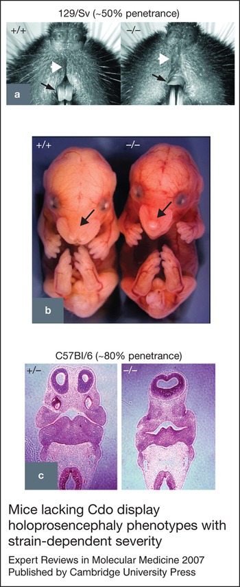
Mice lacking the Shh pathway regulator Cdo on a 129/Sv background display normal midline patterning of the forebrain but have facial microforms of HPE with ∼50% penetrance (Refs Reference Cole and Krauss79, Reference Zhang80). By contrast, ∼80% of Cdo −/− mice on a C57Bl/6 background display a severe form of HPE, typically characterised by a semilobar forebrain, single nostril and absence of, or rudimentary, olfactory bulbs (Ref. Reference Zhang80) (Fig. 3). However, even on the C57Bl/6 background, Cdo mutant mice are not completely alobar or cyclopic, suggesting that residual Shh activity remains. The C57Bl/6 background similarly sensitises mice lacking the BMP pathway regulator Tsg to HPE and additional non-HPE-related phenotypes (Refs Reference Petryk81, Reference Zakin and De Robertis82). Interestingly, removal of one copy of Bmp4 from Tsg −/− animals of a resistant, hybrid genetic background also resulted in HPE, suggesting that loss of Bmp function in these mice was responsible for the HPE phenotype (Ref. Reference Zakin and De Robertis82). As gain of Bmp function can also cause HPE and cyclopia (Refs Reference Golden51, Reference Anderson83, Reference Bachiller84), these results and additional reports (Ref. Reference Ohkubo, Chiang and Rubenstein85) highlight the notion that Bmp signalling plays multiple and complex roles in forebrain patterning and that both the timing and nature of a defect may dictate the outcome.
Mice lacking Cdo display holoprosencephaly phenotypes with strain-dependent severity.Figure 3. Mice lacking Cdo display holoprosencephaly phenotypes with strain-dependent severity. (a) Frontal view of the midfacial region of 4-week-old adult mice with the indicated Cdo genotypes. The Cdo −/− animal shows microforms of holoprosencephaly (HPE), including a single, central maxillary incisor (arrows) and a dysgenic philtrum (arrowheads). Cdo −/− mice on the 129/Sv background show such phenotypes with ∼50% penetrance. (b) Whole embryos of the indicated Cdo genotypes at embryonic day (E) 15.5. The arrows indicate that the Cdo+/+ embryo has two nostrils and a normal midface, whereas the Cdo −/− embryo displays a single nostril and hypoplastic midface. (c) Sections of E11.5 embryos with the indicated Cdo genotypes. Note the paired lateral ventricles in the Cdo +/− (control) embryo and the single ventricle in the Cdo −/− embryo. Cdo −/− mice on the C57Bl/6 background show the more severe HPE phenotypes shown in (b) and (c) with ∼80% penetrance. (a) is reproduced, with permission, from Ref. Reference Cole and Krauss79 (© 2003 Elsevier). (b) and (c) are reproduced, with permission, from Ref. Reference Zhang80 (© 2006 Elsevier).

A third factor that shows mouse-strain sensitivity in development of HPE is Tgif, a homeodomain-containing protein that functions as a repressor of TGFβ and retinoic-acid-mediated transcriptional responses; however, this effect appears to depend on the allele studied. Heterozygous missense and nonsense mutations in TGIF are found in human HPE, but four independent lines of mice carrying homozygous null mutations in Tgif did not reveal HPE phenotypes (Refs Reference El-Jaick63, Reference Bartholin86, Reference Jin87, Reference Shen and Walsh88, Reference Mar and Hoodless89). These lines were generally studied on hybrid backgrounds. An additional, distinct, Tgif mutation was reported, in which the third exon was deleted and replaced with a selectable marker gene cassette, theoretically allowing production of a truncated protein product that would be of a size similar to some of the predicted protein products of human TGIF nonsense mutations (Ref. Reference Kuang90). When crossed onto a C57Bl/6 background, mice carrying this mutation in either the heterozygous or homozygous state developed a variety of neural patterning defects, most often exencephaly, but more rarely, HPE (Ref. Reference Kuang90). It was suggested that the formation of a truncated protein product with dominant negative properties accounted for the phenotypes observed in heterozygous mutants. Although this is a reasonable conjecture, stable production of a truncated protein by this allele has not been demonstrated nor has any dominant interfering function. However, it seems unlikely that the effects seen in these Tgif +/Δexon3 mutants are due to true haploinsufficiency, because two of the null mutations did not produce HPE phenotypes on a C57Bl/6 background (Ref. Reference El-Jaick63, Reference Mar and Hoodless89). These results suggest that the combination of a specific allele plus C57Bl/6 modifier effects were required to observe HPE, even infrequently, in mouse Tgif mutants.
It is intriguing that the C57Bl/6 background sensitises disparate mutant mouse lines to HPE, but it is not certain that the same modifier gene (or genes) is at play in each case. Because most targeted mutations in the mouse begin on a mixed 129/Sv × C57Bl/6 background, it is logical that the effects of these strains have been observed first, but it will be important to determine the effects of additional backgrounds on these animals. Although no modifier effects have been described for human HPE, the relatively low mutation frequency of each HPE gene in human cohorts complicates such analyses, although study of HPE families could potentially shed light on this issue. Nevertheless, it is highly likely that genetic variation analogous to that found in different inbred strains of mice is widespread in the human population. It therefore seems eminently reasonable to predict that modifier genes, present at much higher frequency than bone fide HPE mutations, play a role in the variable penetrance and expressivity of phenotypes seen in mutation carriers. The types of genetic variation that confer HPE-modifier properties on mouse or human alleles are not known but could include changes in coding or regulatory regions, or copy-number variation.
Stochastic events
Although much evidence suggests that heterozygous mutation of HPE genes is not sufficient to produce a severe phenotype, it is possible that, at least in some cases, haploinsufficiency may indeed be enough, because early forebrain patterning may be a particularly sensitive developmental window. For example, induction of the ventral forebrain and midface structures by Shh may require very high pathway activity during a relatively short period of time. Loss- and gain-of-function experiments with Shh in the chick embryo indicate that, at least for the face, this process and the consequent developmental outcomes are highly dosage sensitive (Ref. Reference Hu and Helms57).
Genetic experiments in the mouse provide some evidence for this. Cripto encodes a co-ligand/co-receptor for Nodal in the mouse, and heterozygous mutations in the human orthologue TDGF1 have been identified in human HPE (Ref. Reference de la Cruz21). An allelic series of mutations in Cripto indicates dosage sensitivity in the production of HPE phenotypes. Cripto +/− mice are phenotypically normal but Cripto-null animals die early during embryogenesis because of failure in anterior–posterior axis positioning before gastrulation (Ref. Reference Ding91). By contrast, although mice homozygous for a hypomorphic Cripto allele, Cripto3loxP, are viable and recovered at predicted mendelian ratios, compound heterozygous mice (Cripto3loxP/null) show extremely variable phenotypes, ranging from early embryonic lethality (similar to fully null animals) to viable and fertile adults that lack overt defects (Ref. Reference Chu20). Importantly, some Cripto3loxP/null mice show a spectrum of HPE phenotypes, including cyclopia (Ref. Reference Chu20). As these mice were on a hybrid genetic background, it seems that this low level of Cripto may result in anything from normal development to severe defects, depending on the stochastic nature of specific, but unidentified, early developmental processes. Cripto3loxP/null mice must have <50% Cripto function (or presumably they would behave like Cripto +/− mice), and are therefore not haploinsufficient. However, it is possible that human development is different from mouse development, and haploinsufficiency for specific genes may in fact be sufficient to produce variable HPE phenotypes when the gene products are involved in highly dosage-sensitive developmental events that may display a stochastic nature when partially compromised.
Environmental factors: production of the HPE spectrum in the absence of mutation
Environmental and/or teratogenic factors are implicated in human HPE; could they play a role in the variability that characterises this anomaly? One logical possibility is a dose–response relationship between the exposure level of an HPE-producing teratogen and the severity of the response, but there is no clear evidence that this is true either in humans or experimental animals. An alternative possibility is that the time of exposure during development to an effective dose of a teratogen could produce distinct outcomes. In a striking study, Helms and colleagues reproduced the entire HPE spectrum by exposing chick embryos to the Shh pathway inhibitor cyclopamine at different times during gestation (Ref. Reference Cordero58). These results clearly indicate that the full range of HPE expressivity can occur in the absence of mutation.
Expression of Shh in the prechordal mesendoderm induces Shh expression in the developing ventral forebrain; forebrain expression is, in turn, required for Shh expression in the ectoderm of the frontonasal and maxillary processes of the developing face, probably via a neural crest intermediate (Refs Reference Rubenstein and Beachy14, Reference Cordero58, Reference Marcucio92). Thus, a relay system exists, in which Shh expression and signalling progressively pattern the midline of the forebrain and midface. Blocking Shh signalling in the chick with cyclopamine during the early stages of neural plate patterning resulted in alobar HPE and cyclopia, whereas inhibiting the pathway during ventral forebrain patterning produced forebrain fusion and pronounced midface hypoplasia, but not cyclopia (Ref. Reference Cordero58). Finally, if cyclopamine exposure occurred after ventral forebrain patterning, but before Shh is expressed in the facial ectoderm, defects were restricted to hypotelorism and milder midface hypoplasia – that is, HPE microforms (Ref. Reference Cordero58). It was suggested that inhibition of Shh signalling from the prechordal mesendoderm, ventral forebrain and ectoderm of the facial processes, respectively, resulted in these graded responses. One can therefore envisage that – even in the absence of any predisposing genetic deficit – a full spectrum of HPE phenotypes might occur in human cohorts exposed to teratogens at defined times during fetal development.
The development of only HPE microforms when chick embryos are treated with cyclopamine just before Shh expression in the facial ectoderm is reminiscent of the phenotype of Cdo −/− mice of the 129/Sv background (Refs Reference Cordero58, Reference Cole and Krauss79, Reference Zhang80) and suggests that the latter animals have defects in Shh signalling specifically in the midface. Furthermore, the phenotype of chick embryos treated with cyclopamine just before Shh expression in the ventral forebrain resembles that of Cdo −/− mice on the C57Bl/6 background (Refs Reference Cordero58, Reference Zhang80). The notion that the HPE spectrum can be produced by either genetic or teratogenic means raises the interesting possibility of gene–environment interactions in human HPE. It is important to note that genetic and environmental causes are not mutually exclusive and that gene–environment interactions are very likely to be involved in aetiologically complex maladies (such as HPE) that occur in a genetically heterogeneous population with uncharacterised exposures to naturally occurring and synthetic chemical compounds. A particularly important issue, therefore, is whether genetic and environmental causes of HPE interact in the production of this common birth defect. More specifically, it can be asked, do teratogenic exposures worsen the outcomes of individuals with genetic predisposition to HPE phenotypes? Furthermore, does genetic predisposition to HPE sensitise individuals to lower doses of teratogen than might ordinarily be required to induce HPE in a ‘normal’ individual? Very little information on this issue exists, either in human cohorts or experimental animals. Exposure of mice that are genetically predisposed to HPE but not overtly holoprosencephalic (e.g. Cdo −/− mice on a 129/Sv background; possibly Tgif −/− mice) to teratogens, diabetes-inducing diets or other environmental factors might be a way to identify which combinations of gene–environment interaction are most likely to be synergistic. Along these lines, Sulik and colleagues treated mice lacking apolipoprotein B (which have low plasma cholesterol levels) to the cholesterol-lowering drug BM15.766; the mutant mice were sensitised to the drug and displayed, among other malformations, HPE phenotypes (Ref. Reference Lanoue93).
Some additional unexplained aspects of HPE
Although variable penetrance and expressivity is the most obvious conundrum of HPE, additional unexplained features of this anomaly exist. First, Shh signalling regulates growth, patterning and morphogenesis of a wide variety of vertebrate body structures outside the central nervous system (CNS) and midface, including the limbs, axial skeleton and several internal organs (Refs Reference Ingham and Placzek94, Reference McMahon, Ingham and Tabin95). However, even severely affected HPE patients, including those with mutations in SHH pathway genes, rarely display defects in the development of limbs and internal organs (Ref. Reference Muenke, Beachy and Scriver1). How might this occur?
An obvious possibility, discussed above in relation to the potential stochastic nature of specific events in forebrain patterning, is that the development of the forebrain is simply more sensitive to partial losses of function than is the development of limbs. A second possibility is that mutations occur in genes that regulate the SHH pathway (or other relevant pathways) specifically in the affected regions. Cdo appears to fall into this category. Cdo and its paralogue, Boc, both positively regulate Shh signalling, but expression of the Cdo and Boc genes are each negatively regulated by high Shh-pathway activity (Ref. Reference Tenzen96). Although Cdo and Boc are negative targets of Shh signalling in the CNS, somites and limb buds, exceptions to this general pattern are observed, including expression of Cdo, but not Boc, in the prechordal mesendoderm (Refs Reference Zhang80, Reference Tenzen96). Accordingly, Cdo −/− mice show strain-dependent HPE phenotypes, probably because of loss of function in prechordal mesendoderm, but Boc −/− mice are not holoprosencephalic (Ref. Reference Okada97). Conversely, Boc, but not Cdo, is expressed in commissural neurons of the developing spinal cord and, Boc −/− mice (but not Cdo −/− mice) show defects in Shh-mediated guidance of commissural axons (Ref. Reference Okada97). Cdo and Boc mutant mice presumably show these very selective loss-of-Shh-pathway phenotypes because in other Shh-dependent structures they are either not expressed because of high-level pathway activity (and are dispensible at these sites) or, where pathway activity is lower, they are co-expressed and display some functional redundancy. The HPE phenotype of Chrd−/−;Nog +/− mice (Table 2) is also very specific and may similarly arise as a result of expression specificity and redundancy between antagonists of Bmp signalling (Ref. Reference Anderson83).
A third possible mechanism for the spatial restriction of HPE phenotypes is mutation of transcriptional regulatory regions, rather than coding regions. Six distinct enhancer elements distributed over 400 kb control Shh expression in the developing murine CNS (Ref. Reference Jeong98). Interestingly, three ventral forebrain enhancers are located distal to chromosomal translocation breakpoints that occurred upstream of the SHH transcriptional unit in several reported cases of human HPE (Ref. Reference Jeong98). It is not clear that the spatial restriction in phenotype in these individuals was specifically due to loss of region-specific enhancers, because these translocations probably also removed a limb-specific enhancer (Ref. Reference Lettice99); nevertheless, the disruption of SHH expression, rather than its coding sequence, was presumably the cause of HPE in these cases. Interestingly, point mutations in the limb-specific enhancer sequence result in a form of polydactyly (Ref. Reference Lettice99), indicating that regionally selective phenotypes can be produced by mutations in regulatory sequences. It therefore seems likely that mutations or small deletions that interrupt the function of region-specific enhancers could be involved in the spatial restriction of HPE phenotypes in some cases.
Obviously, the latter two potential mechanisms described above do not explain how heterozygous null mutations in SHH produce forebrain and midface phenotypes without affecting, for example, limb outgrowth and digit patterning. It is possible that additional genetic events – either digenic mutation or modifier genes – could act to regulate not only the severity of the phenotype, but also its spatial restriction.
Another unanswered question on HPE is how disruption of ventral forebrain patterning by ventrally located signalling molecules, such as Shh, can result in a failure of the dorsal midline invagination required to produce cerebral hemispheres and midline structures, such as the choroid plexus (Ref. Reference Hayhurst and McConnell100). Partitioning of the forebrain initiates dorsally, and selective cell proliferation and cell death in the dorsomedial forebrain are thought to be involved in creating the midline recess (Refs Reference Hebert9, Reference Monuki and Walsh11, Reference Furuta, Piston and Hogan101). The roof plate, which is a dorsal structure, is thought to be important in the development of HPE. Genetic ablation of the forebrain roof plate in mice led to a phenotype similar to MIH/syntelencephaly (Ref. Reference Cheng102). MIH, as well as the classical categories of HPE, is sometimes associated in people with mutations in ZIC2, although facial defects are mild in most such patients (Ref. Reference Brown103). Zic2 mutant mice also display HPE and, interestingly, Zic2 expression is highly enriched in the dorsal CNS, rather than the ventral CNS (Refs Reference Nagai104, Reference Nagai105).
A recent study reported that mice heterozygous for a Shh allele, encoding a protein that lacks its cholesterol adduct (ShhN), displayed several HPE-related phenotypes, including some similar to those seen in MIH (Ref. Reference Huang, Litingtung and Chiang106). A patient with a SHH truncation mutant that would be predicted to lack the cholesterol moiety, had a similar phenotype (Ref. Reference Roessler40). Analysis of Shh +/ShhN mouse embryos suggests that ShhN has an expanded range of signalling, resulting in a disruption of the normal signalling network that regulates dorsal cell fates (Ref. Reference Huang, Litingtung and Chiang106). Specific defects in dorsal patterning may therefore arise in a number of ways to produce HPE phenotypes, including MIH, and this area deserves further study.
HPE may also arise by mechanisms not obviously accounted for by the current models. For example, conditional mutation of the small GTPase Cdc42 in the developing mouse telencephalon resulted in HPE, in the apparent absence of dorsoventral patterning defects (Ref. Reference Chen107). Cdc42 regulates cell–cell adhesive contacts and the actin cytoskeleton, and it was proposed that disruption of apical-basal polarity in Cdc42-null cells prevented formation of an appropriate telencephalic architecture, leading to a failure in midline partitioning.
Concluding remarks
HPE presents a number of features that are poorly understood. Among these are the broad range of phenotypes encompassed by the HPE spectrum and the lack of involvement of body structures that are also dependent on the pathways known to be defective in many cases of HPE (e.g. the Shh pathway). Animal models have provided some insight into these puzzling features, but it will be challenging to prove which potential mechanisms are at play in the human condition. However, it seems likely that just as multiple genes and distinct environmental influences are implicated in the development of HPE, multiple mechanisms will underlie the associated variability and spatial restriction. Furthermore, the effects of any interaction between genetic variation (both true mutations and modifiers) and exposure to environmental agents on signalling pathways during key developmental windows could be enormously complex; this may be one reason why the HPE spectrum is so broad and each case distinctive. Finally, it is possible that the concepts outlined here for HPE may also apply to other human disorders that arise from defects in key dosage-dependent and time-dependent developmental events.
Acknowledgements and funding
Work in the author's laboratory on this topic is funded by the National Institutes of Health, the March of Dimes and the T.J. Martell Foundation. I thank Wei Zhang, Karen Schachter, James Briscoe and the anonymous referees for critical comments on the manuscript.