Introduction
Micro- and nanostructures are widely found in nature, although by virtue of their size, they are individually invisible to the naked eye. The utility of these structures, however, are often manifested in their cumulative effect, which may be optical (butterfly wing),Reference Vukusic, Sambles, Lawrence and Wootton1 adhesive (gecko feet),Reference Autumn, Liang, Hsieh, Zesch, Chan, Kenny, Fearing and Full2 or mechanical (muscle)Reference Sharafi and Blemker3 in nature. Another class of natural micro-/nanostructures with profound influence in the development and maintenance of organisms, but which is even less apparent, is structures that affect cell behavior and function. This class of “biological structures” is of great relevance with regard to application in areas of biology and medicine and will thus receive greater emphasis in the present review. By understanding how fabricated structures interact with their biological counterparts of the same length scale, one can gain an understanding of how laser-based fabrication may be effectively applied in biomedicine.
Importance of nanostructured features in biology and medicine
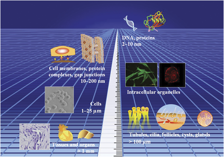
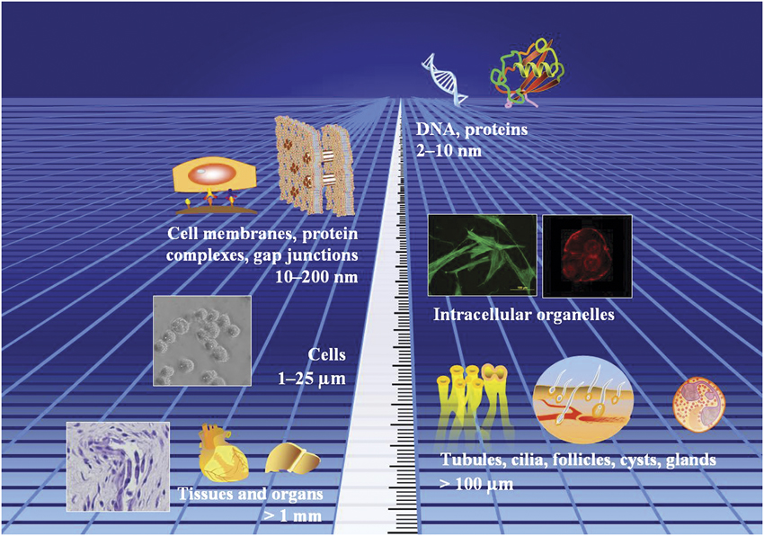
In examining the importance of micro- and nanostructures in nature in relation to those that are fabricated, it is instructive to first look at the different length scales of biological objects (Figure 1). At the lower end of the spectrum (2–10 nm), one finds the basic unit of heredity, DNA, as well as other biological molecules such as proteins, polysaccharides, and glycoproteins. Moving one notch higher on the length scale (10–200 nm), we find the subcellular structures, such as the cell membrane, protein complexes (gap junctions, focal adhesion complexes), and intracellular organelles, the latter of which may span several microns. At a length scale of 1–25 μm, the major biological structure is the cell. Micro-tissue structures, such as tubules, follicles, cysts, and glands, are seen at length scales of several hundred microns, while other tissues and organs are formed at mm length scales and larger.
The length scales of biological objects.
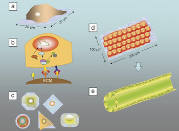
Fabricated structures of a particular length scale correlate with natural structures of the corresponding dimensions and interact accordingly to produce the designed response. As an illustration, consider a culture of cells on a two-dimensional (2D) extracellular matrix (ECM)-micropatterned substrate, where the ECM-free areas are non-cell adhesive (Figure 2). Such micropatterns can be formed using focused lasers at dimensions that allow only a single cell to occupy and adhere to each micropattern (Figure 2a). The microstructure consists of components at two length scales—the ECM molecules (50–300 nm) and the dimensions of the pattern itself (20 μm). The ECM molecules possess ligands that may bind to appropriate receptors on the cell membrane (molecular interaction), activating a signaling cascade and ultimately producing a cellular response (Figure 2b). The geometry of the pattern, on the other hand, determines the area of cell membrane that may interact with the mentioned receptors via focal adhesion complexes (subcellular interaction), and thus controls the shape of the cell when attached to the substrate (cellular interaction) (Figure 2c). Cell shape, in turn, modulates the signaling events via modification of the cell cytoskeleton.
(a) A single cell occupies an extracellular matrix (ECM) micropattern. (b) Binding of ECM ligands to cell receptors activates a signaling cascade that produces a cellular response. (c) The geometry of the micropattern controls cell shape. (d) A larger ECM micropattern can accommodate more than one cell, allowing intercellular interactions that lead to the formation of (e) a tubular tissue structure.

Taking a second example of an ECM strip of larger dimensions, for example, 100 μm × 200 μm, more than one cell can occupy such a pattern, leading to the potential of intercellular interaction (Figure 2d). If interaction occurs, it introduces the additional possibility that the cells may self-assemble to form tissue structures such as cysts and tubules (for epithelial cells, which make up blood capillaries) and attain dimensions that correspond to the dimensions of the microfabricated structure (Figure 2e).
These examples thus serve to illustrate that fabricated micro- and nanostructures are able to control cell area, shape, and organization into higher order structures, and how a good size correlation exists between the cellular and fabricated structures. Considering the importance of length scale in the application of microfabrication in biomedicine, this review will be structured such that the various synthetic micro- and nanostructures are discussed in relation to their level of interaction with the corresponding natural objects.
Molecular interactions
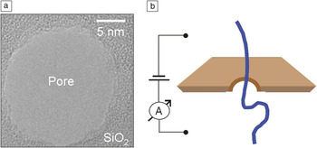
The fabricated nanostructure that has gained the most appeal where biomolecules are concerned is not an ECM coating or a built-up feature, but rather a pore. A paper by Kasianowicz et al. in 1996 sparked keen interest in using nanopores as a method for analyzing DNA molecules.Reference Kasianowicz, Brandin, Branton and Deamer4 An electric field could be employed to drive single-stranded DNA and RNA molecules through the ion channel of a protein, resulting in an ion current that was proportional to the dimensions of the molecule. Blockage of the current occurs when DNA or other analyte molecules enter the pore. The duration and amplitude of blockage provide a readout of the analyte’s properties, such as molecular mass, composition, or even sequence.Reference Griffiths5 The attractiveness of using nanopores to elucidate molecular information lies partly in the ease of its setup—two chambers of buffer separated by a membrane that contains the pore, electrodes to apply a voltage difference across the membrane, and a method to measure the resulting ion current (Figure 3).
(a) Electron micrograph of a nanopore on a SiO2 membrane. (Reprinted with permission from Reference Reference Martin and Siwy7. ©2007, AAAS.) (b) Schematic of a basic setup used to elucidate molecular information using nanopores.

The earliest and still widely used nanopore is constructed of the ion channel protein, α-hemolysin, which is inserted into a lipid membrane.Reference Kasianowicz, Brandin, Branton and Deamer4 α-hemolysin is particularly stable, possesses the correct size pore (1.5 nm) for single-stranded DNA molecules to pass through, and is amenable to modification. More recently, efforts to create nanopores on a synthetic, solid platform have yielded advantages such as improved stability and flexibility of the pore dimensions.Reference Nilsson, Lee, Ratto and Létant6, Reference Martin and Siwy7 Unlike α-hemolysin, synthetic nanopore sizes are not limited by the inherent dimensions of the protein channel, allowing them to accommodate longer DNA strands and even microbes. Materials such as silicon dioxide, silicon nitride, and gold have been used to fabricate nanopores for a variety of applications, such as DNA sequencing and detection, and measurement of biophysical properties such as association rate constants and DNA binding kinetics.Reference Kasianowicz, Henrickson, Weetall and Robertson8–Reference Hornblower, Coombs, Whitaker, Kolomeisky, Picone, Meller and Akeson10 Earlier methods employed electron or ion beam lithography and track etching to synthesize nanopores, but recently, lasers have also been used.
Methods for the chemical functionalization of nanopores have also been developed to widen their capabilities and application set by combining size control with the presence of appropriate functional groups in the pore region. For example, lining the pore channel with a protein called FG-nucleoporin admitted molecules that bound the protein, but excluded those that did not. In this way, the functionalized channel mimicked the selectivity of the pore complex found in the cell nucleus.Reference Jovanovic-Talisman, Tetenbaum-Novatt, McKenney, Zilman, Peters, Rout and Chait11 Similarly, other workers have immobilized DNA probesReference Nilsson, Lee, Ratto and Létant6, Reference Bayley and Cremer12 to achieve specificity in the counting and detection of molecules. Another application of fabricated pores exploits their size selectivity for molecular filtration.Reference Han, Fu and Schoch13 As pores of dimensions smaller than 0.1 μm exclude bacteria, these could also be used for low-temperature sterilization of fluids.Reference Hassel, Bello-Rodriguez, Milenkovic and Schneider14
Subcellular interactions
While there are various organelles that reside within the cell, such as the endoplasmic reticulum and nucleus, the subcellular feature most directly relevant to microfabricated structures are the focal adhesion complexes (FACs). The location of FACs on the cell membrane allows them to interact directly with ECM-coated structures and function like a switchboard operator. Cell regulatory signals from the ECM can be relayed to the cell nucleus via these FACs, which assemble and disassemble each time they come into contact with the appropriate ECM molecules. These signals tell the cell whether to migrate, proliferate, or undergo cell death. Here, both the size and distribution of the ECM microstructures would matter.
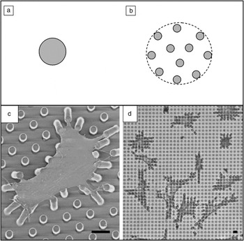
For example, ECM islands of different sizes (3–50 μm) and geometries (e.g., squares) have been created on planar glass substrates by printing self-assembled monolayers of protein-adhesive molecules in square islands and filling the areas in between with non-adhesive molecules. Thus, incubation of the substrates in a solution of fibronectin, an ECM protein, localizes it to the surface of the square islands and not the surrounding regions. By changing the size and spacing of these cell-adhesive fibronectin islands, the area of cell spreading and total area of cell-fibronectin contact could be independently varied, providing important basic information on cell behavior.Reference Chen, Mrksich, Huang, Whitesides and Ingber15 To illustrate, a cell could be confined in a 20-μm circular adhesive island, as depicted in Figure 4a. By creating an array of smaller, closely spaced adhesive islands (Figure 4b), cells could be made to spread on a larger area (dotted line), while maintaining the same area of adhesive contact (shaded regions). In the earliest work of this sort, it was found that the total area of cell coverage, and not area of adhesive contact of cells, controls cell death in endothelial cells via FACs.Reference Chen, Mrksich, Huang, Whitesides and Ingber15 Using similar micropatterns, further work has shown that the amount of focal adhesions increases in proportion to cell spreading.Reference Chen, Alonso, Ostuni, Whitesides and Ingber16
(a–b) The size and spacing of cell-adhesive islands can be used to study cell behavior. (a) A cell can be confined in a circular adhesive island, whereby it assumes the shape of the island. (b) Alternatively, the same cell can be made to spread on a larger area (dotted line), while keeping the area of adhesive contact (shaded area) constant. Scanning electron micrograph (c) and phase contrast micrograph (d) of smooth muscle cells attached to posts where the tips of the posts have been printed with fibronectin (scale bar = 10 μm). (Reprinted with permission from Reference Reference Galbraith and Sheetz19. ©2003, National Academy of Sciences.)

Microcontact printed arrays of fibronectin strips of dimensions similar to FACs have also been achieved by poly(dimethylsiloxane) (PDMS) stamping.Reference Goffin, Pittet, Csucs, Lussi, Meister and Hinz17 When cells were grown on ECM islets that were 1.25 μm wide and 2–20 μm long, two types of FACs (termed classical FACs and super FACs) were generated. In the same experiment, the threshold tension required to recruit a mechanosensitive protein, smooth muscle actin, to the stress fibers could also be identified.
Considering the usefulness of ECM microstructures in controlling cell behavior, as presented previously for the case of a single ECM protein (fibronectin), there would be value in developing methods to simulate the chemical complexity of native ECM. Toward that end, Mei et al. have developed automated printing techniques based on atomic force microscopy to generate two-component ECM arrays of varying spacing and composition.Reference Mei, Cannizzaro, Park, Xu, Bogatyrev, Yi, Goldman, Langer and Anderson18 Precise tuning of the 6–9 μm features led to an increase in the alignment of mouse muscle fibroblasts.
Subcellular microstructures can also be employed as devices to measure cell forces.Reference Galbraith and Sheetz19, Reference Tan, Tien, Pirone, Gray, Bhadriraju and Chen20 Understanding cell forces and contractility is critical in understanding wound healing and organ regeneration and, thus, the practice of tissue engineering. For example, the level of forces generated by cells will lead to either the formation of scar tissue or the regeneration of healthy tissue. One particular device comprises an array of vertical ECM-coated posts upon which cells are cultured, attached, and spread out across multiple posts, akin to having them lie on “a bed of needles” (Figure 4c–d).Reference Tan, Tien, Pirone, Gray, Bhadriraju and Chen20 From the deflection of the posts, the traction force generated by the attached part of the cell can be calculated. By changing the geometry of the posts, such as their heights and shapes, spatial changes in the local substrate stiffness could be affected—a useful feature since substrate stiffness has been shown to affect cell behavior and the differentiation of stem cells to other cell types. For example, by decreasing the height of the posts by half, the stiffness could be increased eight-fold. It has been shown that cell shape affected the ability of the cells to contract. When the cells were exposed to lysophosphatidic acid, a chemical found in serum that stimulates cells to contract, contraction was only observed when the cells were spread out. The size of the focal adhesions on the cells could also be correlated with stresses experienced at the attachment sites.
Interesting results have been obtained from other experiments, where a higher density of microfabricated pillars was used to better resolve the cell forces generated by different parts of the cell.Reference du Roure, Saez, Buguin, Austin, Chavrier, Silberzan and Ladoux21 It was found for the case of kidney epithelial cells that the highest traction stress was localized at the cell edges. The traction stress for a single cell was, in turn, smaller than that of a cell layer; the cells in a layer could work together as a cell sheet, accounting for the greater traction. The mechanical activity of these epithelial cells was also found to be initially higher and slowed down as the layer of cells began to completely cover the substrate surface (i.e., become confluent).
As most cells within the body reside within a three-dimensional (3D) microenvironment, it would be desirable to develop micro- and nanostructures that are able to capture the complexity of 3D scaffolds for tissue engineering. As a step in that direction, laser direct writing (LDW) has been used to fabricate 3D structures for cell culture in the form of submicron elastic beams made from an inorganic (Si–O–Si)–organic hybrid polymer system.Reference Klein, Striebel, Fischer, Jiang, Franz, von Freymann, Wegener and Bastmeyer22 Cardiomyocytes (heart muscle cells) cultured on these structures spread out and attached to different beams. When the cells contracted, the elastic beams bent and stretched accordingly, allowing the cellular force to be characterized.
In another example of a 3D LDW scaffold from the same group, selected parts of the scaffold were functionalized by embedding 4 μm × 4 μm squares of a protein-adsorbing material within a 3D protein-repelling matrix. The entire system was then incubated in the cell-adhesive ECM protein, fibronectin.Reference Klein, Richter, Striebel, Franz, von Freymann, Wegener and Bastmeyer23 Cells cultured on these scaffolds were observed to adhere selectively to the ECM-modified parts, providing a method to control cell shape in 3D. Ochsner et al.Reference Ochsner, Dusseiller, Grandin, Luna-Morris, Textor, Vogel and Smith24 introduced another method to control cell shape in 3D, which also involved a flexible substrate. Endothelial cells were cultured in arrays of PDMS microwells coated with fibronectin; fluorescent confocal microscopy showed that the actin cytoskeleton of the cell was 3D in nature and not just limited to the cell-substrate interface.
Cellular interactions
ECM micropatterns of dimensions comparable to those of a typical cell are able to control the cell size and shape by virtue of providing sites for the cell to attach relative to the surrounding non-adhesive surface. Thery et al. have shown that the geometry of micropatterned ECM affects the polarity of the cell surface and the organization of intracellular organelles, such as microtubules, nucleus, centrosome, and Golgi Apparatus.Reference Théry, Racine, Piel, Pépin, Dimitrov, Chen, Sibarita and Bornens25 Here, the ability to generate micropatterns of defined shape and size is of great value, as this guarantees that the shape and position of the organelles do not vary too much from one cell to another. This, in turn, allows for fine and automated measurements of the cell phenotype. Having mapped out the relative positions of intracellular organelles of a normal cell as the standard reference, significant differences can then be found for abnormal cells of the same type.
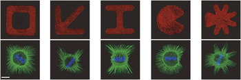
The geometry of ECM micropatterns can also control the way cells divide. During cell division, a mitotic spindle is formed, which helps to separate the chromosomes into the daughter cells. The axis of cell division is always perpendicular to the mitotic spindle. By culturing cells on micropatterned ECM, it was established that spindle orientation of the cells was dependent of the spatial distribution of the ECM (Figure 5).Reference Théry, Jiménez-Dalmaroni, Racine, Bornens and Jülicher26, Reference Théry, Racine, Pépin, Piel, Chen, Sibarita and Bornens27 In simplified terms, fibers form in the regions where the cell adheres to the ECM, and these fibers help to stabilize and pull on proteins associated with the mitotic spindle, thus aligning it.
Mitotic cells on different fibronectin micropatterns (red, top row). Retraction fibers (green, bottom row), spindle poles (red) and chromosomes (blue) were labeled. Scale bar, 10 μm. (Reprinted with permission from Reference Reference Théry, Jiménez-Dalmaroni, Racine, Bornens and Jülicher26. ©2007, Nature Publishing Group.)

In high-content screening, image analysis is used to measure changes in cells brought about by a biochemical treatment, such as changes in the orientation of cell division or transport of a protein into the cell. In conventional culture, many of the changes go unnoticed due to the high variability of cells cultured on conventional substrates, such as tissue culture flasks and petri dishes. Regular arrays of highly reproducible micropatterns and identically polarized cells ensure that the cellular response to any external challenge would be homogeneous. This narrower range of response would clarify the effect of biochemical drugs on individual cells in high-throughput screens.
Confinement of groups of cells can be equally revealing. Ruiz et al. used arrays of cell-adhesive patterns in the form of interconnected squares, observing migration and outgrowth of neural stem cells between the squares, with the outgrowth resembling the thin projections (axons) of neurons.Reference Ruiz, Buzanska, Gilliland, Rauscher, Sirghi, Sobanski, Zychowicz, Ceriotti, Bretagnol, Coecke, Colpo and Rossi28 Within the squares themselves, cell density was found to affect the morphology of the cells. Cells plated at low density spread out according to the shape of the pattern, while those at high density remained round.
Intercellular interactions
As suggested previously, most of the micro- and nanostructures that have found biomedical applications at the molecular, subcellular, and cellular levels have been fabricated on 2D substrates. However, the main value of studying intercellular interactions in medical sciences is the knowledge that may contribute toward tissue healing and regeneration. As most tissue is 3D in nature, translation of this basic knowledge to therapeutic applications requires that microstructures be designed to provide a 3D microenvironment, in conjunction with 3D culture and growth of cells.
As described in the introduction to this review, a 2D ECM strip pattern of dimensions several times the cell diameter can guide the localization of cells within a defined area, thus facilitating their self-assembly into tissue structures. 3D microstructures function in a similar way to confine cells, but their “sphere of influence,” comprising soluble and insoluble ECM components, literally surrounds the cell. One type of 3D structure that is relevant to the control of the cellular microenvironment is the hydrogel. Being a macromolecular network that incorporates a high volume fraction of water, hydrogels are suitable for cell encapsulation due to their favorable mechanical and mass transfer properties. The range of stiffness exhibited by hydrogels coincides well with that of tissue (which is itself a type of hydrogel). Oxygen and nutrients are available to cells via diffusion through the hydrogel structure, and the permissive nature of the matrix allows cells to migrate and self-assemble.
Microgels are a subset of hydrogels, in which the gel polymer takes the form of microparticles.Reference Tumarkin and Kumacheva29 The earliest type of microgels was produced by introducing a stream of polymer solution into a second solution with a cross-linker, which led to its coagulation into microcapsules.Reference Lim and Sun30 A widely used system involved the use of alginate as the polymer and calcium as the cross-linker. It was proposed that the encapsulation of cells in this microgel could be a viable method for reducing the effects of immune rejection when these cells were transplanted. Since then, cell encapsulation in microgels has been applied in therapies for diseases such as cancer,Reference Cirone, Bourgeois and Chang31 diabetes,Reference Lim and Sun30 liver failure,Reference Liu and Chang32 and enzyme deficiency.Reference Ross, Bastedo, Maier, Sands and Chang33
Due to the more recent paradigm of looking at cell behavior in the context of its microenvironment,Reference Lutton and Goss34 another important advantage of microgels has become apparent—that of being able to tailor the local cell microenvironment. Cells that are encapsulated within microgels can be exposed to defined ECMs with specific biochemical and physical characteristics to achieve the desired expression of cell type.Reference Ng, Wu, Zhou, Toh, Ho, Chia, Zhu, Mao and Yu35, Reference Albrecht, Underhill, Mendelson and Bhatia36 At the same time, different modular microenvironments containing different cell types could be combined, allowing tissue architecture to be engineered by a bottom-up approach.Reference Rivron, Rouwkema, Truckenmuller, Karperien, De Boer and Van Blitterswijk37, Reference Khademhosseini and Langer38 Microgels in the form of pre-molded structures,Reference Du, Lo, Ali and Khademhosseini39 microfibers,Reference Fedorovich, De Wijn, Verbout, Alblas and Dhert40, Reference Wan, Yim, Liao, Le Visage and Leong41 microdroplets,Reference Moon, Hasan, Song, Xu, Keles, Manzur, Mikkilineni, Hong, Nagatomi, Haeggstrom, Khademhosseini and Demirci42 and microspheresReference Albrecht, Underhill, Mendelson and Bhatia36 could be assembled, leading to larger tissue constructs useful for drug screening or as implantable materials.
An important application of micro- and nanostructures relates to their ability to control interactions between cells, by spatially defining the location of either the cells themselves or biological cues that attract or engage cells to arrange themselves accordingly. Such interactions are important in embryonic development, and recapitulation of these interactions would be important in efforts to engineer tissue de novo. For example, interactions of endothelial cells (which make up blood capillaries) and parenchymal cells (the major cell type in the tissue or organ of interest) have been shown to be important for the function of the tissues concerned.Reference Red-Horse, Crawford, Shojaei and Ferrar43 A combination of cell-laden microgels provides a means of interacting two or more cell types. Such interactions, as found in the so-called Janus-type microgels comprising two distinct physical and chemical hemispheres,Reference Shepherd, Conrad, Rhodes, Link, Marquez, Weitz and Lewis44, Reference Nie, Li, Seo, Xu and Kumacheva45 would help elucidate the biology of cell-cell interactions.
ECMs are rich in chemical factors and signaling molecules that regulate the behavior of cells. However, microstructures can endow further functionality to ECMs, as described in the review by Nelson and Tian.Reference Nelson and Tien46 By spatially confining the growth of cells, microstructures are able to influence the distribution of mechanical stresses and biochemical stimuli on cell populations over length scales larger than the individual cell dimensions. Spatial confinement in microstructures can also regulate other factors behind tissue formation, such as cell-cell interactions and cell-ECM interactions.
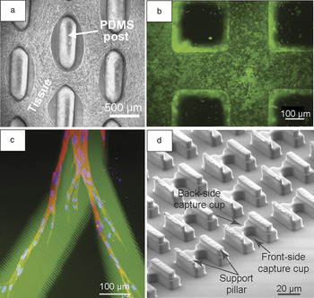
At its simplest, the culture of cells in microstructures serves to confine their growth and proliferation to obtain multicellular tissue structures, or organoids. For example, by casting hydrogels containing skeletal muscle cells in microfabricated molds consisting of staggered, elongated posts, porous and aligned muscle tissue networks have been achieved, which exhibited better viability as compared to similarly fabricated non-porous muscle sheets (Figure 6a).Reference Bian and Bursac47 Other researchers have cultured heart muscle cells (cardiomyocytes) with non-muscle cell types in matrigel-coated microchannels.Reference Iyer, Chui and Radisic48 Compared to the simultaneous seeding of different cell types, it was found that seeding non-muscle cells to form organoids, followed by seeding of muscle cells, yielded more elongated and densely arranged cells. This led to cardiac tissues with better functionality. Such a result may not be surprising, considering the fact that in tissue development, cells of the same type often interact with each other within the cell layers (homotypic interaction), while different cell types interact at the interface between the cell layers (heterotypic interaction). Toward the same end, spatially defined microscale co-cultures of three vascular cell types in different layers have been designed,Reference Tan and Desai49 resulting in a biomimetic system whereby the degree and nature of cell-cell interactions were more similar to those observed in vivo.
Various fabricated microstructures and their biomedical applications. (a) An array of posts enables porous and aligned muscle tissue structures to be achieved. (Reprinted with permission from Reference Reference Bian and Bursac47. ©2009, Elsevier.) (b) Liver cells cultured within a porous 3D structure fabricated by two-photon laser scanning photolithography. (Reprinted with permission from Reference Reference Hsieh, Ng, Narayanan, Wan and Ying52. ©2010, Elsevier.) (c) Cell-adhesive channels (green) within a photo-cross-linked hydrogel guide the growth and migration of fibroblasts (nuclei labeled blue, actin labeled red). (Reprinted with permission from Reference Reference Lee, Moon and West54. ©2008, Elsevier.) (d) Details of the trap structure used in a microfluidic device for cell capture and pairing. (Reprinted with permission from Reference Reference Skelley, Kirak, Suh, Jaenisch and Voldman56. ©2009, Nature Publishing Group.)

Our understanding of the mechanism of endothelial tube branching has been advanced using microfabricated tubules, formed by embedding endothelial cells within collagen channels and allowing the cells to self-assemble.Reference Nelson, Van Duijn, Inman, Fletcher and Bissell50 Through an elegant set of experiments involving different tubule geometries coupled with modeling, it was shown that branching of tubules generally occurred at their ends, due to low concentrations of an inhibitory molecule. Other researchers have functionalized microfabricated scaffolds with ECM components, such as RGD (a cell-adhesive sequence) and heparan sulfate (a proteoglycan), to ensure efficient cell attachment and binding of growth factors, once again, imbuing ECM with a microstructural aspect for further functionality.Reference Mapili, Lu, Chen and Roy51 These structures could be used to study the behavior of stem or progenitor cell populations in a complex, patterned microenvironment, with the ultimate aim being tissue engineering. Similarly, we have microfabricated porous, 3D structures using two-photon laser scanning photolithography (TPLSP), and coated these with ECM. Liver cells cultured within this 3D environment exhibited higher liver-specific cell functions as compared to cells cultured on the ECM-coated 2D controls. This could be attributed to a localized concentration of growth factors (Figure 6b).Reference Hsieh, Ng, Narayanan, Wan and Ying52
Microstructures have proven useful to guide the migration of cells or their projections. One paradigm involves the use of biochemical channels in cell-invasive hydrogel matrices to guide the growth of nerve axons.Reference Luo and Shoichet53 In an example employing an agarose hydrogel, the general strategy involved creating channels within the agarose (using a focused laser source), which were modified with light-sensitive groups that were later coupled to the desired biomolecules. In this way, channels of GRGDS, a cell-adhesive sequence from the protein fibronectin, were shown to guide the axonal projections of spinal nerve cells. Poly(ethylene glycol)-acrylate based hydrogel microstructures were employed in essentially the same way to guide the migration and growth of human dermal fibroblasts. However, in this case, the use of a two-photon laser allowed the channels to be placed in the center of the hydrogel matrix (Figure 6c).Reference Lee, Moon and West54
Microstructures in microsystems
One major advantage of using microstructures for cell culture is that their length scale allows them to be integrated within other microsystems. These could be analytical devices to probe biochemical processes that regulate cell behavior, microfluidic systems that provide flow regimes beneficial to cell function, or microdevices for lysing cells for further genetic analysis. The interfacing of microscaled cell culture with such microsystems simplifies cell handling and leads to reductions in cost and the biological sample required.Reference El-Ali, Sorger and Jensen55 As an illustration, microsystems have been used to pair cells to achieve cell fusion,Reference Skelley, Kirak, Suh, Jaenisch and Voldman56 which is an important tool for gene expression studies and antibody production. The device comprised a dense array of cell traps containing both front-side and back-side capture cups (Figure 6d), and essentially worked in the following way: the first cell type was loaded into the capture cups, followed by the second cell type, which was trapped immediately in front of the previously trapped cells. Microfluidic flow through the device enabled thousands of cells to be immobilized and paired at once.
An integral part of all the microsystems described are microfluidic channels, which are essentially precise, microfabricated channels through which liquid is made to flow. Microfluidic devices themselves allow important insights into cellular processes, such as the migration of neutrophils in response to gradients of chemical signals,Reference Lin, Nguyen, Wang, Saadi, Gross and Jeon57 the effect of shear stress on cell adhesion,Reference Lu, Koo, Wang, Lauffenburger, Griffith and Jensen58 cell alignment,Reference Song, Gu, Futai, Warner, Nor and Takayama59 stem cell differentiation,Reference Toh, Blagovic and Voldman60 and cell-cell interactions in patterned co-cultures.Reference Torisawa, Mosadegh, Luker, Morell, O’Shea and Takayama61 One major advantage of culturing cells within microfluidic channels is the ability to introduce microscaled flows in the system.Reference Griffith and Swartz62 Cells in the body are subject to fluid shear stresses, which affect the way they behave and the way they assemble to form tissues. Fluid flow also influences the mass transfer of nutrients and oxygen to cells and their exposure to growth factors and cell signaling molecules such as chemokines. Microscale flows were incorporated in a 3D liver culture model, which, together with the appropriate cell-cell interactions, was designed to optimize liver functionality in vitro. Reference Powers, Janigian, Wack, Baker, Stolz and Griffith63 The bioreactor comprised of a silicon scaffold containing an array of 100 channels, each with a cross-sectional area (300 mm × 300 mm) that was large enough to facilitate 3D liver cell aggregation. Perfusion flow through the cell mass simulated flow rates and fluid shear stresses at or below the physiological shear range in vivo.
Another application of microfluidic channels in tissue engineering is to create vascular networks. Shin et al. fabricated closed channels of vascular-like geometries from PDMS and used them to culture human microvascular endothelial cells for two weeks under dynamic flow, upon which the cells lined the channels as a monolayer and expressed endothelial cell genes.Reference Shin, Matsuda, Ishii, Terai, Kaazempur-Mofrad, Borenstein, Detmar and Vacanti64 Microvasculature could also be created by seeding endothelial cells on the walls of pre-formed collagen channels, allowing them to attach and grow.Reference Chrobak, Potter and Tien65 Flow of nutrient media through these channels exposed the cells to shear stress and chemical factors, leading to stable endothelial tubes exhibiting functional behaviors such as leukocyte (white blood cell) adhesion and barrier function. Other workers have shown how different types of ECM gels could be perfused into the channels and induced to gel with the appropriate external stimuli (e.g., chemicals, temperature changes). The different gel phases could be anchored together within the microfluidic channels by using the nucleating effect of collagen fibers.Reference Gillette, Jensen, Tang, Yang, Bazargan-Lari, Zhong and Sia66 The collagen fiber-mediated interfacing stabilized the interface between the different gels and prevented them from shrinking due to cellular contraction.
Cells can also be cultured in separate microscaled chambers representing different tissue compartments, such as lung and liver, connected by a recirculating tissue culture medium that allows metabolic interaction between the organs.Reference Sin, Chin, Jamil, Kostov, Rao and Shuler67 Integrated with sensors for oxygen and pH, such models may be useful for the evaluation of drug toxicity and pharmacokinetic profiles to supplement/complement animal studies and predict human response in clinical trials.
Conclusions
From the present review, it is evident that fabricated micro- and nanostructures play an important role in biology and medicine. This is because the length scale of biological objects must match the length scale of any structure intended to probe, analyze, perturb, or influence the behavior of the biological object in question. This basic concept can be seen in nanopores that probe the molecules filtering through it, extracellular matrix-coated flexible posts that measure the traction force of cells, and microchannels that confine groups of cells and thus their growth into tissue. Likewise, further advances in the application of micro- and nanofabricated structures in biomedicine would rely on progress in fine-tuning the interactions or discovery of new modes of interaction between these structures and biological objects.
New technologies such as two-photon laser scanning photolithography can enable the fabrication of finer and more complex structures to better match the resolution and complexity of biological materials. Development of better microsystems to interface with these microstructures would also benefit from increased capabilities in the areas of microfluidics and biosensing. In the area of microgels for tissue engineering, better methods to pattern these modular microenvironments in 3D are foreseen to gain importance, especially as they begin to demonstrate increased functionality of the resulting tissues. To achieve the latter goal, use of the appropriate biomaterials and materials chemistry compatible to cells and in vivo conditions would be essential. New strategies aimed at establishing interactions between the biological objects and the microstructures would also lead to more advanced applications. For example, now that 3D cell culture is gaining wider acceptance and practice in the life sciences community, recent research efforts have been directed toward developing time as the fourth dimension. Here, light-reactive chemistries have been developed to avail ligands in a hydrogel, and thus controlling the interactions between the ligands and cells as a function of time.Reference Kloxin, Kasko, Salinas and Anseth68
Our ability to harness the length scale and complexity of micro- and nanostructures will depend on our current understanding of biology at the same length scales. As this understanding can be advanced using the fabricated structures themselves, a feedback loop should result. This will hopefully spur further developments in the field and uncover solutions to various diseases that afflict humankind today.
Acknowledgment
This work is funded by the Institute of Bioengineering and Nanotechnology (Biomedical Research Council, Agency for Science, Technology and Research, Singapore).