Introduction
The cupula consists mainly of mucopolysaccharide and is very adhesive in nature. This adhesiveness facilitates easy attachment of the otoconia, resulting in cupulolithiasis. SchuknechtReference Schuknecht1 reported cupulolithiasis in which the otoconia attach to the cupula, making it gravity-sensitive. In our experience, cases of intractable cupulolithiasis need careful treatment. Therefore, physical therapy with maximal efficacy should be developed.
This study aimed to investigate what kinds of stimuli are effective in detaching the otoconial mass from the cupula in experimental models of cupulolithiasis.
Materials and methods
The experimental models of cupulolithiasis were prepared using bullfrogs (Rana catesbeiana) weighing 110–220 g. After induction of deep anaesthesia with ether, the bullfrogs were decapitated and the bony labyrinth was removed and placed in Ringer solution according to the method of Suzuki et al. Reference Suzuki, Harada, Hirakawa, Hirakawa and Omura2 The caudal otic capsule was chiselled so that the entire posterior semicircular canal was exposed. The remaining membranous labyrinth was left encapsulated in the bony capsule. The membranous labyrinth was carefully cut at the crus commune to create a tiny, 0.5 mm opening. A small piece of otoconia, removed from the sacculus of the other ear, was introduced through this opening into the canal lumen. The position of the labyrinth preparation was adjusted so that the otoconial mass was fixed onto the cupula.
Three different kinds of stimulus were then used in this experimental model of cupulolithiasis, in order to determine the most effective stimulus for detaching the otoconial mass: gravity, sinusoidal oscillation and vibration.
A dissection microscope (SZX12; Olympus, Tokyo, Japan) was used for observation of the otoconial mass.
All experiments were conducted according to the ethical rules for animal experiments at Tokyo Medical University.
Experiment one: gravity
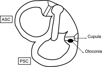
The first experiment tested the effect of gravity, using 10 membranous labyrinth preparations. The labyrinth preparation was placed in a glass dish filled with Ringer solution (100 ml) so that the cupula-to-crista axis was in the horizontal plane with the canal side in the downward position. The labyrinth preparation was maintained in this position for 30 minutes. The otoconial mass attached to the cupula was checked every 10 minutes (Figure 1).
Diagram representing experiment one (gravity). The labyrinth preparation was placed so that the cupula-to-crista axis was in the horizontal plane with the canal side in the downward position. The otoconial mass was checked every 10 minutes for 30 minutes. ASC = anterior semicircular canal; PSC = posterior semicircular canal.

Experiment two: sinusoidal oscillation
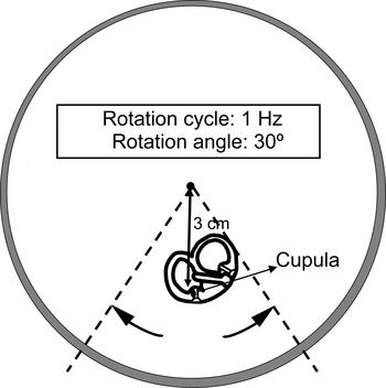
The second experimental model assessed the effect of sinusoidal oscillation, using 10 membranous labyrinth preparations. The labyrinth preparation was placed in a glass dish filled with Ringer solution (100 ml), which was magnetically fixed to a turntable (FRA-02; First, Tokyo, Japan). The labyrinth preparation was placed 3 cm from the rotational centre. The cupula-to-crista axis was placed along a line pointing towards the rotational centre. The turntable was sinusoidally rotated with a rotational cycle of 1 Hz and a rotational angle of 30°. The sinusoidal rotation was maintained for 30 minutes. The otoconial mass attached to the cupula was checked every 10 minutes (Figure 2).
Diagram representing experiment two (sinusoidal oscillation). The labyrinth preparation was placed 3 cm from the centre of a turntable. The cupula-to-crista axis was placed along a line pointing towards the centre. Oscillation was applied for 30 minutes. The turntable was rotated with a rotational cycle of 1 Hz and a rotational angle of 30°. The otoconial mass was checked every 10 minutes.

Experiment three: vibration
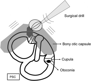
The third experimental model tested the effect of vibration, using 14 membranous labyrinth preparations. The labyrinth preparation was placed in a glass dish (100 ml) in the same position as in experiment one. Mechanical vibration was applied to the top surface of the bony otic capsule of the labyrinth preparation using a surgical drill (BL-F5A; Osada Electric, Tokyo, Japan). A cutting burr with a 5 mm tip diameter was used. The frequency of the vibration, as measured by a vibration analyser (VA-12; Rion, Tokyo, Japan), was 340 Hz. The otoconial mass attached to the cupula was checked during the procedure. Care was taken not to rupture the membranous labyrinth (Figure 3).
Diagram representing experiment three (vibration). Mechanical vibration at 340 Hz was applied to the bony capsule near the labyrinth using a surgical drill. The otoconial mass was checked during the procedure.

Results
Experiment one: gravity
The otoconial mass was detached within 30 minutes in 2 of the 10 labyrinth preparations used in experiment one; in these cases, the detachment times were 10 and 30 minutes.
Experiment two: sinusoidal oscillation
The otoconial mass was not detached from the cupula within 30 minutes in any of the labyrinth preparations in experiment two.
Experiment three: vibration
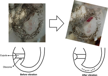
The otoconial mass was detached from the cupula in all labyrinth preparations in experiment three (Figure 4). The detachment times ranged from 10 seconds to 5 minutes 10 seconds. The mean detachment time was 2 minutes 20 seconds.
Magnified photographs and diagrams for experiment three (vibration). Before vibration, the otoconial mass was attached to the cupula. After vibration, the otoconial mass was detached from the cupula and was separated into pieces.

Discussion
Benign paroxysmal positional vertigo (BPPV) is mainly caused by movement of detached otoconia within the semicircular canal (canalolithiasis), or by movement of otoconia attached to the cupula (cupulolithiasis). In our group's previous experiments,Reference Suzuki, Kadir, Hayashi and Takamoto3, Reference Otsuka, Suzuki and Furuya4 both cupulolithiasis and canalolithiasis effectively stimulated the cupula and were thus potentially valid mechanisms of BPPV. The clinical features of typical BPPV are characterised by brisk, rotatory, dominant nystagmus with a latency of several seconds and a short duration. This feature could be explained more favourably by the mechanism of canalolithiasis. However, there is another type of positional vertigo with negligible latency and a long duration of more than several minutes. This type of vertigo could be explained by cupulolithiasis. Based on our experience, cases of intractable cupulolithiasis occur and can even require canal plugging surgery.Reference Suzuki, Ichimura, Ueda and Suzuki5 Therefore, the development of physical therapy with maximal efficacy for cupulolithiasis is highly desirable.
Chiou et al. Reference Chiou, Lee, Tsai, Yu and Lee6 have reported the use of a forced prolonged position, wherein patients with cupulolithiasis were required to lie on the side of weaker nystagmus for 12 hours. Symptoms were relieved in all patients after four sessions. However, in our experimental study the otoconial mass was detached in only 2 out of 10 labyrinth preparations following 30 minutes' exposure to gravity. With longer exposure times, it is possible that more otoconia could be detached. The forced prolonged position technique is effective but requires patients to remain in a lying position for a long period, which is uncomfortable for some.
White et al. Reference White, Coale, Catalano and Oas7 used several types of rolling or brisk deceleration manoeuvres (developed by Lempert and Tiel-Wilck,Reference Lempert and Tiel-Wilck8 Gufoni et al.,Reference Gufoni, Mastrosimone and di Nasso9 Asprella Libonati et al.,Reference Asprella, Gagliardi, Cifarelli and Larotonda10 and Brandt and Steddin)Reference Brandt and Steddin11 as well as head-shaking. The Gufoni manoeuvre first places patients in the sitting position, then makes them quickly lie down towards the affected side, and finally makes them rotate their head 45° downward and maintain this position for 2 to 3 minutes.Reference Gufoni, Mastrosimone and di Nasso9 The Vannucchi–Asprella manoeuvre rapidly moves patients from the sitting to the supine position, then turns the head rapidly to the unaffected side, then returns the patient to the sitting position, and finally returns the head to the midline. This manoeuvre is repeated five to eight times.Reference Asprella, Gagliardi, Cifarelli and Larotonda10 After these manoeuvres, White et al. Reference White, Coale, Catalano and Oas7 found that the symptoms of all canalolithiasis patients were resolved, but the symptoms of only 50 per cent of cupulolithiasis patients were resolved.
In our experimental study, none of the otoconial masses were detached by sinusoidal oscillation in any of the labyrinth preparations. This result is compatible with the low efficacies noted when the above-mentioned manoeuvres were applied to patients with cupulolithiasis.
Kim et al. Reference Kim, Jo, Chung, Byeon and Lee12 have reported a new cupulolith repositioning manoeuvre. Beginning with the patient in the supine position, the patient's head is turned 135° toward the affected side. Mastoid oscillation is then applied to the affected side, using a 60-Hz, hand-held vibrator, for 30 seconds. The otoconia at the canal side of the cupula will be detached by this manoeuvre. The patient's head is returned to the supine position. Next, the patient's head is turned 90° towards the unaffected side and mastoid oscillation is repeated once. The otoconia at the utricular side of the cupula will be detached by this manoeuvre. Finally, the patient's head is rotated 90° in the prone position and the patient is slowly brought to a sitting position. Using this manoeuvre, symptoms were resolved in 62 per cent of patients in one session, and in 97 per cent of patients after six sessions.
Our experimental study found that the otoconial mass was detached by mechanical vibration in all labyrinth preparations. This result is compatible with the high efficacy rate of Kim and colleagues' cupulolith repositioning manoeuvre.
• Cupulolithiasis is intractable because of otoconial adhesion to the cupula
• This experimental study tested the effectiveness of various stimuli in resolving cupulolithiasis
• The effect of gravity, sinusoidal oscillation and mechanical vibration was tested
• Vibration was most effective in detaching an otoconial mass from the cupula
However, the question remains whether vibration could also dislodge the utricular otoconia. Amir et al. Reference Amir, Young and Belloso13 reported a case of BPPV which occurred after the use of a whole-body vibration training plate. Similarly, our previous experiments, using bullfrog membranous labyrinth preparations, indicated that otoconia were dislodged from the utricle and moved into the posterior semicircular canal in half of the preparations after 15 minutes of vibration using a surgical drill.Reference Otsuka, Suzuki and Furuya4 It takes longer for vibration to dislodge the otoconia from the utricle than from the cupula. Fine morphological studies show that the utricular otoconia are connected to each other with fine fibrils and are embedded in a gelatinous substance made of mucopolysaccharide.Reference Kachar, Parakkal and Frex14, Reference Lins, Farina, Kurc, Riordan, Thalmann and Thalmann15 The whole otoconial mass is covered by a supraotoconial layer.Reference Nakai, Masutani, Kato and Sugiyama16 This may be one reason why short-duration vibration can dislodge the otoconia from the cupula without dislodging the utricular otoconia.
Conclusion
In this experimental study of cupulolithiasis in a bullfrog membranous labyrinth model, over 30 minutes, the otoconial mass was detached by gravity in only 2 of 10 labyrinth preparations, and was not detached by sinusoidal oscillation in any preparation. However, the otoconial mass was detached by mechanical vibration in all labyrinth preparations, over the same time period; the mean detachment time was 2 minutes 20 seconds. Thus, mechanical vibration was the most effective stimulus for detaching the otoconia from the cupula in this experimental model.
Acknowledgements
The authors are indebted to Dr Clifford Kolba and Associate Professor Edward Barroga for their editorial review of the manuscript. The authors also thank Dr Hirotoshi Hishida, Department of Mechanical Engineering, Faculty of Engineering, Kogakuin University. This study received financial support from a Grant-in-Aid for Scientific Research (C) from the Ministry of Education, Culture, Sports, Science, and Technology, Japan, and a Health and Labor Science Research Grant for Research on Specific Disease (Vestibular Disorders) from the Ministry of Health, Labor and Welfare, Japan and a follow-up grant for a Tokyo Medical University research project.