Introduction
Salivary gland tumours represent less than 5 per cent of all head and neck tumours. Metastatic disease within the parotid accounts for 9–14 per cent of all parotid tumours.Reference Batsakis1 Most parotid metastases (80 per cent) come from primary sites in the head and neck. Parotid metastases from distant tumours are rare (constituting 0.16–4 per cent).Reference Seijas, Franco and Sastre2
The past 30 years have seen a large increase in reports of gastroesophageal junction tumours. All are associated with very high mortality, with a five-year survival rate of less than 15 per cent.3
Gastroesophageal tumours can be classified as having their centre either 5 cm proximal or distal to the gastric cardia.Reference Siewert, Stein and Feith4 Based on this classification, tumour types I, II and III can be designated, as follows. Type I tumours are adenocarcinomas of the distal oesophagus which infiltrate the gastroesophageal junction and generally develop within Barrett's oesophagus. Type II tumours are adenocarcinomas of the cardia which arise from the cardiac mucosa. Type III tumours involve the sub-cardial gastric mucosa and infiltrate the cardia and lower oesophagus, generally submucosally.
Gastroesophageal junction tumours spread by local invasion, lymphatic spread and haematogenous metastasis. They metastasise to the liver, lung, peritoneum, small intestine mesentery and omentum. Less frequently, distant spread has been reported to the patella, retina, orbit, brain, mandible, pineal region, oral cavity and maxilla.Reference Ashby and Dappen5–Reference Willard, Weber and Buche12
Case report
A 66-year-old man presented to our rapid-access ENT clinic with a swelling below his right ear lobe, within the right parotid tail. There were no upper aerodigestive tract symptoms such as hoarseness, sore throat, pain, cranial nerve palsies or cough. However, he did complain of worsening dysphagia for solids over the last 12 weeks. He had also lost 10 kg in weight and felt weak and unwell.
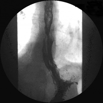
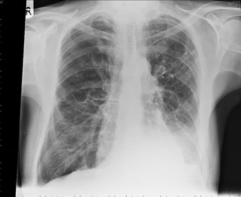
An urgent barium swallow showed irregular mucosa and narrowing at the gastroesophageal junction (Figure 1). A chest X-ray showed cannon-ball metastasis (Figure 2). Fine needle aspiration cytology of the parotid suggested malignant epithelial cells in a pattern indicating gland formation, suggesting adenocarcinoma.
Barium swallow study showing narrowing at the gastroesophageal junction.

AP chest X-ray showing canon-ball metastasis. R = right

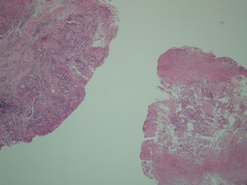
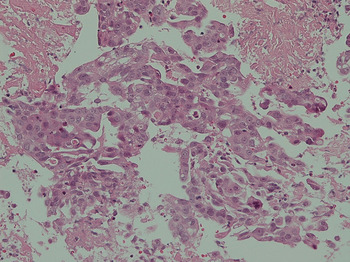
An urgent Oesophago-Gastro-Duodenoscopy (OGD) showed raised, ulcerated mucosa across the anterior aspect of the gastroesophageal junction; this region was biopsied. The endoscopy was otherwise normal. Histological examination of biopsy tissue confirmed moderately differentiated adenocarcinoma of the gastroesophageal junction (Figures 3 and 4) adjacent to an area with features of Barrett's oesophagus.
Photomicrograph of biopsy tissue from the gastroesophageal junction, showing neoplastic changes. (H&E; ×10)

Photomicrograph of biopsy tissue from the gastroesophageal junction, showing deeply stained nuclei and pleomorphism. (H&E; ×20)

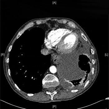
Computed tomography (CT) of the chest and abdomen indicated multiple hepatic and pulmonary metastases, along with a right adrenal metastasis. There were multiple enlarged lymph nodes visible below the carina (Figure 5) and along the lesser curve of the stomach.
Axial computed tomography scan showing enlarged mediastinal lymph nodes. A = anterior; R = right; L = left; P = posterior

An oesophageal stent was inserted in view of the patient's worsening dysphagia. He was then given palliative chemotherapy, and died one year following his initial presentation to our clinic.
Discussion
In 2007, Schoneveld et al. described a case of a 45-year-old man diagnosed with a metastasis in the left parotid gland, two months after trans-hiatal gastroesophagectomy for a primary gastric adenocarcinoma.Reference Schoneveld, Hesp and Teune13 They concluded that the tumour had probably spread via an alternate haematogenous route by way of Batson's plexus.Reference Batson14 Similarly, Öztürk et al. reported a case of a 73-year-old man with gastric adenocarcinoma which metastasised to the right parotid gland three years after the patient had undergone gastrectomy.Reference Öztürk, Benekli and Güler15 They too considered this metastasis to have occurred via haematogenous spread. We could find no previous report of gastroesophageal junction adenocarcinoma presenting with a parotid metastasis.
• Salivary gland tumours constitute less than 5 per cent of all head and neck tumours
• Parotid metastases from distant tumours are rare (representing only 0.16–4 per cent of parotid metastases) but well reported
• Oesophageal carcinoma metastasis to the salivary glands has been previously reported, but in cases with an established diagnosis of oesophageal carcinoma
• In the reported case, oesophageal carcinoma presented with parotid swelling alone
In our patient, a CT scan revealed multiple sub-carinal lymph nodes and lymph nodes along the lesser curvature of the stomach, along with pulmonary and hepatic metastases. The haematogenous route is the most common mode of spread to the liver and lung. In addition, tumour may spread via the paravertebral plexus (Batson's plexus), bypassing the portal vein and the filtering system of the lungs. Lymphatic spread is highly unlikely as the lymph from the gastroesophageal junction is drained to the perigastric region. Therefore, we believe that the rapid spread of our patient's gastroesophageal junction tumour could only have occurred via a haematogenous route, with or without involvement of Batson's plexus.
Conclusion
Whenever a parotid swelling is found, one should always consider the possibility of metastasis from distant organs, as rare as this may be. When faced with such a presentation, we would stress the importance of good history-taking and a broad approach to diagnosis and management.