Introduction
Actinobacteria are a large and diverse phyla of Gram-positive bacteria, with high guanine-cytosine content and a wide range of morphologies from coccoid or rod-coccoid to those with branched mycelia similar to fungi (Yoshida et al. Reference Yoshida, Seo, Suzuki, Nishino, Kobayashi, Hamada-Sato, Kogure and Imada2008). They play significant roles in several processes that contribute to soil fertility, such as nutrient cycling, decomposition of various compounds, e.g. complex biopolymers, and formation of beneficial soil humus (Kennedy Reference Kennedy1999).
Actinobacteria are also well-known for their ability to synthesize a wide variety of biologically active secondary metabolites, such as antibiotics, pigments, toxins, enzymes and other compounds of biotechnological interest with potential for commercial application. Several decades of effort focusing on the isolation and screening of thousands of Actinobacteria have shown them to be a most prolific source of nearly 10000 diverse bioactive compounds (Bérdy Reference Bérdy2005). Although Actinobacteria comprise several different genera (Stackebrandt et al. Reference Stackebrandt, Rainey and Ward-Rainey1997), the majority of the bioactive compounds (~76%) have been isolated from the Streptomyces genus (Bérdy Reference Bérdy2005). This genus has attracted great interest due to its well-known ability to synthesize many different bioactive secondary metabolites, including >80% of known antibiotics, antitumour compounds and growth effectors, among other compounds with medical importance exploited by the pharmaceutical industry (Bentley et al. Reference Bentley, Chater and Cerdeño-Tárraga2002, Bérdy Reference Bérdy2005).
Members of the Actinobacteria phyla are widely distributed in a multiplicity of natural environments: terrestrial, aquatic and extreme environments from tropical to polar regions (Subramani & Aalbersberg Reference Subramani and Aalbersberg2013). Actinobacteria are abundant in soil. Discovery of new antibiotics from Actinobacteria has been rare in recent years. As a result, scientists began to look for Actinobacteria from unusual environments with the hope of discovering novel antibiotics (Lazzarini et al. Reference Lazzarini, Cavaletti, Toppo and Marinelli2001, Srivibool & Sukchotiratana Reference Srivibool and Sukchotiratana2006, Thakur et al. Reference Thakur, Yadav, Gogoi and Bora2007, Okoro et al. Reference Okoro, Brown, Jones, Andrews, Asenjo, Goodfellow and Bull2009).
Antarctica is separated from the other continents by the Southern Ocean and the Antarctic Circumpolar Current (Smith Reference Smith1991). Besides being the coldest, windiest and driest place on Earth with the highest UV index, it is also one of the most hostile and pristine environments (Ross & Vincent Reference Ross and Vincent1998). Antarctic microorganisms, especially the psychrophiles and psychrotrophs, develop novel metabolic pathways as a result of the evolutionary selection to survive and grow in these extreme conditions (Van den Burg Reference Van den Burg2003). Antarctic bacteria are potential sources of novel enzymes and bioactive compounds with relatively few studies on them compared to their counterparts from other regions (Saadoun & Gharaibeh Reference Saadoun and Gharaibeh2003, Lam Reference Lam2006).
The aim of this study was to phylogenetically and physiologically characterize an Antarctic actinobacterial strain, designated INACH3013, and to determine the extent of its ability to inhibit the growth of foodborne pathogens under in vitro conditions.
Materials and methods
Strain isolation
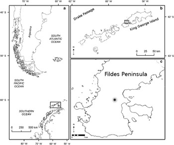
Strain INACH3013 was isolated from a soil sample collected at Fildes Peninsula (62°12'26.4''S, 58°58'28.7''W), King George Island, Antarctica, in February 2011 (Fig. 1). A modified method described by Kharel et al. (Reference Kharel, Shepherd, Nybo, Smith, Bosserman and Rohr2010) was used to isolate Streptomyces species and kill most of the non-spore-forming microbes. One gram of soil sample was weighed and suspended in 10 ml of sterile distilled water. The suspension was heated at 40°C for 30 min and serially diluted up to 103. Each dilution was spread (0.1 ml) onto the Actinomycete Isolation Agar (Difco) and incubated at room temperature for 7 days (Kharel et al. Reference Kharel, Shepherd, Nybo, Smith, Bosserman and Rohr2010).
Maps showing the sampling locations for strain INACH3013 on the a. Antarctic Peninsula, b. South Shetland Islands and c. Fildes Peninsula, King George Island. The dot indicates the exact sampling site on Fildes Peninsula (62°12'26.4''S, 58°58'28.7''W).

Morphological characterization
Morphological characterization of the strain was performed according to the guidelines of the International Streptomyces Project (ISP) (Shirling & Gottlieb Reference Shirling and Gottlieb1966). Ultrastructural characters, such as spore chain morphology, spore size and surface ornamentation of a 14-day-old cultures grown on ISP medium 2 (ISP 2) were examined using a Hitachi S-3400N (Hitachi High Technologies, Japan) conventional scanning electron microscope (SEM) in variable pressure (VP) mode. A small amount of mycelium was collected from the culture plate, mounted on an aluminium stub using carbon tape and directly analysed in VP mode. The microscope chamber was supplied with elevated gas pressure. Observations were performed at low values of electron beam intensity (3 KV). To improve the image quality, which is negatively affected by the lower beam intensity at higher magnification, we adjusted the work distances from 3 to 5 mm.
Physiological characterization
Physiological characterization was performed using API 20NE and API ZYM (Biomerieux) kits according to the manufacturer’s instructions. Carbon source utilization was evaluated using mineral ISP medium 9 (ISP 9) supplemented with different sugars as described by Shirling & Gottlieb (Reference Shirling and Gottlieb1966). Salt tolerance was tested using R2A agar medium supplemented with 1, 1.5, 2, 2.5, 3, 4, 5, 6, 8 and 10% (w/v) of NaCl incubated at 20°C. Tolerance to pH was determined using the same medium adjusted to different pH (pH 2, 3, 4, 5, 6, 7, 8, 9, 10, 11, 12 and 13) using citrate-phosphate for pH 3–6, phosphate buffer for pH 6–8 and glycine-NaOH buffer for pH 8–13, and incubated at 20°C. The effect of temperature (4, 12, 20, 28, 37, 40 and 45°C) on the rate of growth was evaluated in R2A broth medium shaken at 125 rpm. Three replicates were prepared for each incubation condition. To perform this assay, cultures with 1×10³ colony-forming units (CFU) ml-1 were incubated at the respective temperatures for 48 hours. A spread plate method was used to measure the growth in CFU ml-1. Then, 20 ul of each replicate was spread on fresh agar and incubated at room temperature until the colonies were visible. The growth rate at each temperature was calculated using:
 $$\rm {\rmu =[ln(N\, t)\,{\hbox -}\,ln(N \,0)]\, / \,ln2 }\,\left( {{\rm t{\hbox-}t \,0}} \right){\rm ,}$$
where N corresponds to the cell density at time (t) in days. The Ratkowsky square-root model:
 $$\,\surd{\rm {\rmu} =b(T }\,{\minus}\,{\rm T \,min)[1 -e\, c(T\,{\hbox -}\, T\, max)],}$$
was used to estimate the potential optimal growth temperature and maximum temperature of growth (Ratkowsky et al. Reference Ratkowsky, Lowry, McMeekin, Stokes and Chandler1983).
Genomic DNA extraction
Strain INACH3013 was grown at 20°C for 3 days on oatmeal agar. DNA was extracted using the Qiagen DNeasy Blood and Tissue kit with slight modifications. The mycelia were scraped and suspended in deionized water. The suspension was centrifuged at maximum speed and the supernatant was discarded. To suspend the cells, 400 µl of enzymatic lysis buffer (2.4% Triton X-100, 20 mM pH 8.0 Tris-HCl and 2 mM sodium EDTA) was added. The suspension was heated at 99°C for 10 min and incubated at -80°C for 15–20 min. The suspension was thawed before the addition of 20 mgml-1 of lysozyme. The suspension was incubated at 37°C for 35 min, and 25 µl of proteinase K and 200 µl of Buffer AL were added. The suspension was incubated at 56°C for 35 mins and 200 µl of absolute ethanol was added. The extraction procedure was continued according to the kit’s instructions.
Polymerase chain reaction conditions and gene sequencing
Amplification of the 16S rRNA gene was performed using a protocol recommended by the manufacturer of Taq DNA polymerase. The polymerase chain reaction (PCR) mixture consisted of 50 ng of bacterial genomic DNA, 2 µl of 10X PCR buffer, 0.4 µl of 10 mM dNTPs, 0.8 µl of forward primer (243F), 0.8 µl of reverse primer (A3R), 0.4 unit of Taq DNA polymerase (Promega, USA) and ultrapure water was added to a final volume of 25 µl. The following thermal profile was used during PCR: one cycle of 5 min at 95°C, followed by 28 cycles of 95°C for 1 min, 60°C for 30 sec and 72°C for 2 min, and a final extension step at 72°C for 10 min. The amplified PCR product (~ 1.5 kb) was separated on 1% (w/v) agarose gel, stained with 1X SYBR Safe™ (Invitrogen) and visualized under UV light. The band of interest was excised, purified with PCR Cleanup kit (Axygen) and sequenced using an AB-13730 sequencer (Macrogen, Korea).
Phylogenetic analysis
The sequence was analysed using the Basic Local Alignment Search Tool (BLAST) and phylogenetic analysis was performed using the MEGA6 software. The MUSCLE algorithm in MEGA package was used to align the 16S rRNA gene sequences obtained from the GenBank database (http://www.ncbi.nlm.nih.gov/genbank/). Aligned DNA sequences were trimmed to eliminate poorly aligned regions. The analysis involved 32 nucleotide sequences with a total of 1124 positions in the final dataset. The consensus phylogenetic tree was obtained by maximum-likelihood analysis based on the Tamura-Nei model (Tamura & Nei Reference Tamura and Nei1993). Initial tree(s) for the heuristic search were obtained automatically by applying Neighbor-Join and BioNJ algorithms to a matrix of pairwise distances estimated using the Maximum Composite Likelihood approach, and then selecting the topology with superior log-likelihood value. A discrete Gamma distribution was used to model evolutionary rate differences among sites (five categories (+G, parameter=0.1542)). The rate variation model allowed for some sites to be evolutionarily invariable (+I, 61.0117% sites). The tree topography was evaluated using bootstrap analysis of 1000 repetitions.
Antibiotic susceptibility tests
Antibiotic susceptibility testing was performed using the disc diffusion method on R2A agar medium. The following antibiotics were tested: ampicillin (25 μg), carbenicillin (100 μg), ceftazidime (30 μg), cefixime (5 μg), cefotaxime (30 μg), cefpirome (30 μg), cefpodoxime (10 μg), cephalothin (30 μg), chloramphenicol (30 μg), ciprofloxacin (10 μg), clarithromycin (15 μg), clindamycin (2 μg), erythromycin (15 μg), gentamicin (30 μg), imipenem (10 μg), lincomycin (15 μg), metronidazole (5 μg), mupirocin (5 μg), nalidixic acid (30 μg), nitrofurantoin (100 μg), novobiocin (5 μg), rifampicin (5 μg), spectinomycin (25 μg), streptomycin (25 μg), sulphonamides compound (300 μg), tetracycline hydrochloride (30 μg), trimethoprim (5 μg) and vancomycin (30 μg).
Detection of antimicrobial activity
The presence of antimicrobial activity of strain INACH3013 was tested against Staphylococcus aureus (strain from Laboratorio de Biorrecursos Antárticos, INACH collection). Staphylococcus aureus was grown in 1.3% of MH broth at 37°C for 6 hours. The culture was added into molten MH agar to reach an optical density of 0.001 at 600 nm (OD600). The molten agar was then poured into an empty petri dish and allowed to solidify. Cell-free supernatant (10 μl) of 7-day-old INACH3013 culture, previously grown in oatmeal broth (used for the cultivation of Streptomyces according to the ISP), was aliquoted and left to diffuse into the MH agar. The plate was incubated at 37°C and presence of inhibition zone was established visually after 24 hours. The same procedure was repeated to screen against 20 food pathogens including Escherichia coli (Migula) Castellani & Chalmers serotypes 0157:H7, V517 and 0125, Salmonella enterica (ex Kauffmann & Edwards) Le Minor & Popoff serovars Typhimurium and Typhi, Salmonella Biafra Van Oye & Du Pre, Salmonella braenderup Kauffmann & Juel Henningsen, Klebsiella pneumoniae (Schroeter) Trevisan 14x, Enterobacter cloacae (Jordan) Hormaeche & Edwards 22x, Enterococcus faecalis (Andrewes & Horder) Schleifer & Kilpper-Bälz, Staphylococcus haemolyticus Schleifer & Kloos, Staphylococcus equorum Schleifer et al., Vibrio parahaemolyticus (Fujino et al.) Sakazaki et al. 1808, 1896, 2053 and 2341, and Bacillus cereus Frankland & Frankland K3. The foodborne pathogens were provided by Professor Son Radu, Universiti Putra Malaysia. Listeria monocytogenes (Murray et al.) Pirie (serotype I, IV and aa) strains were provided by the Instituto de Salud Pública, Santiago, Chile (http://www.ispch.cl/).
Results
Strain isolation
The methodology used to isolate the Actinobacteria strains provided a low number of colonies. Only 25 colonies with the same morphology were obtained. All isolates were screened for antibacterial activity using the ‘Agar Spot Test’ assay against E. coli and Staphylococcus spp. Only 18 isolates produced antibacterial compounds capable of inhibiting the growth of Staphylococcus spp. but not the growth of E. coli. Strain INACH3013, which showed the largest halo zone was selected for further study.
Morphological and physiological characterizations
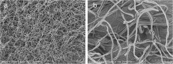
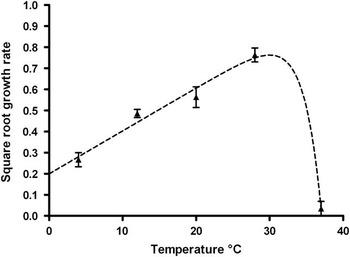
At maturity, the strain formed an extensive pad consisting of intricate grooves of aerial hyphae (Fig. 2a). At higher magnification hyphae show long, straight to rectiflexibiles (flexuous) spore chains,>20 μm long, characterized by cylindrical single spores with smooth surfaces,~587–1147 nm long and 440–556 nm in diameter (Fig. 2b). Strain INACH3013 grew well on ISP media 2, 3, 4, 5 and 7, potato dextrose agar, nutrient agar and R2A (Table I). Moderate growth was observed on ISP 6 agar. Diffusible brown pigment was produced on several media (ISP 2, 3, 4, 5, 6 and 7) and the colony colour was medium-dependent (Table I). Yellow diffusible pigments were produced on ISP 9 agar supplemented with arabinose, maltose, xylose, mannitol, galactose and raffinose. In addition, strain INACH3013 was able to grow on fructose, inositol, lactose, sodium citrate and glycerol, but not sucrose, as the sole carbon source (Table II). Dark brown diffusible pigments were produced on the potato dextrose agar. Starch hydrolysis, nitrate reduction and urea utilization were not observed. Strain INACH3013 showed positive enzymatic activity to alkaline phosphatase, acid phosphatase, naphtol-AS-BI-phosphohydrolase, esterase (C4), leucine arylamidase, valine arylamidase, beta-galactosidase, beta-glucosidase using the API ZYM system. Strain INACH3013 grew well at 10, 20 and 28°C (Fig. 3). The optimal growth temperature was 30°C, predicted by the Ratkowsky equation:
 $$\eqalignno { \surd{\rm \rmu =0}{\rm .02035(T{\plus}\!9},\!{782) [1 - {\rm e}0}{\rm .4(T{\hbox - }37} {\rm .09)]}; \cr \;\;\;\;\;\;\;\;\;\;{R}^{{\rm 2}} {\rm =0}{\rm .9502}.$$
Scanning electron micrograph of spore chains of strain INACH3013 grown in ISP2 medium for 14 days. a. Magnification=470, b. magnification=4700.

Effect of incubation temperature on the growth rate of strain INACH3013. Triangles indicate the square-root of specific growth rate (± standard deviation) calculated from growth curves at each incubation temperature. The line indicates the best-fit for the square-root of specific growth rate versus temperature function: √μ=0.02035(T+9,782) [1 – e0.4(T-37.09)].

Culture characteristics of strain INACH3013 on various media after 7 days.

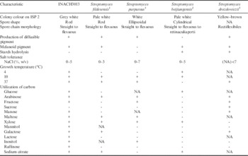
Differences in phenotypic characteristics of strain INACH3013 and the closest phylogenetic neighbours reported in literature. (+) positive activity, (-) negative activity, (NA) not available.
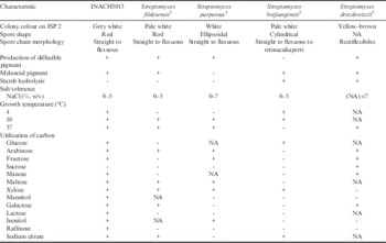
1 Li et al. Reference Li, Tian, Zhu, Yang and Li2011,
2 Li et al. Reference Li, Zhang, Xu, Cui, Lu, Xu and Jiang2002,
3 Semêdo et al. 2004.
The growth rate value estimated at optimal temperature was (μ)=0.762. The estimation of the maximal temperature for growth was 37°C. Slow growth was observed at 4°C and 37°C with an estimated growth rate of (μ)=0.2807 and 0.0336, respectively. No growth was recorded at 40°C and 45°C (Fig. 3). The salinity range was 0–5% (w/v) NaCl, with an optimum of 1.5% (w/v) NaCl. Strain INACH3013 was tolerant to a wide pH range of 5.0–12.0.
Molecular identification and phylogenetic analysis
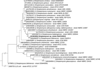
The partial 16S rRNA gene sequence (1104 nts) was compared with those available in the GenBank using BLAST. According to the BLAST analysis, this strain exhibited 99% similarity with Streptomyces fildesensis Li et al. Alignment of the partial 16S rRNA gene sequence (1124 positions with gaps) with the representative members of the Streptomyces genus stated in the species description of S. fildesensis revealed highest similarity to S. fildesensis (99.8%), followed by S. beijiangensis Li et al. (98.1%), S. drozdowiczii Semêdo et al. (97.9%) and S. purpureus (Matsumae & Hata) Goodfellow et al. (97.2%). Phylogenetic analysis based on these sequences showed that strain INACH3013 fell into a well-supported subclade with Streptomyces fildesensis, S. beijiangensis and S. purpureus LMG 19368. A phylogenetic tree based on the 16S rRNA gene sequences of strain INACH3013 and the most closely related members of the Streptomyces genus is presented in Fig. 4.
Maximum-likelihood phylogenetic tree based on 16S rRNA gene sequences (1124 base pair alignment positions including gaps) showing the relationship between strain INACH3013 and the 31 most closely related reference species. 16S rRNA gene sequence of E. coli ATCC 11775T was used as the outgroup. Numbers at nodes represent levels (%) of bootstrap support based on a maximum-likelihood analysis of 1000 resampled datasets.

Antibiotic susceptibility and resistance tests
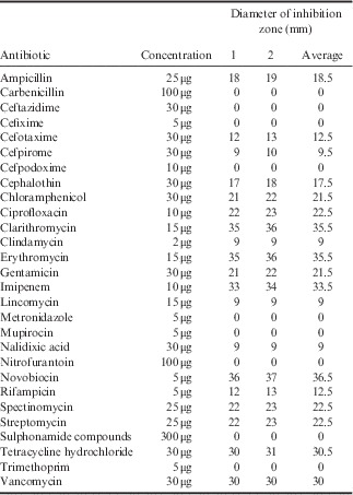
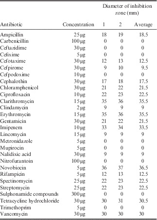
Strain INACH3013 was sensitive to 19 of the 28 antibiotics tested. Low sensitivity was detected to clindamycin, lincomycin and nalidixic acid. INACH3013 was found to be resistant to carbenicillin, ceftazidime, cefixime, cefpodoxime, metronidazole, mupirocin, nitrofurantoin, sulphonamides compound and trimethoprim (Table III).
Antibiotic susceptibility tests on strain INACH3013. The diameter of each antibiotic disc was 6 mm.
Partial characterization of antimicrobial activity
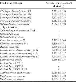
The cell-free supernatant of INACH3013 culture showed variable antimicrobial activity against a broad spectrum of different bacterial foodborne pathogens. Clear halo zones were observed around the spots aliquoted with the supernatant (10 ul). INACH3013 inhibited 15 of the 21 pathogens (Table IV). The results indicate that some of the Gram-negative bacteria were more sensitive to the antimicrobial compound than Gram-positive bacteria: Salmonella enterica serovar Typhimurium >V. parahaemolyticus 1808>Enterobacter cloacae 22x>B. cereus K3>Enterococcus faecalis > V. parahaemolyticus 2053>L. monocytogenes (serotype IV)>L. monocytogenes (serotype aa)>V. parahaemolyticus 1896>Staphylococcus haemolyticus > Listeria monocytogenes (serotype I)>Staphylococcus aureus >K. pneumoniae 14x>V. parahaemolyticus 2341>Staphylococcus equorum.
Antibacterial activity of cell-free culture supernatant from strain INACH3013 against bacterial foodborne pathogens.

Discussion
The modified method used in this study targeted spore forming soil bacteria, particularly Streptomyces. Although the temperature used in this modified method was lower than that in Kharel et al. (Reference Kharel, Shepherd, Nybo, Smith, Bosserman and Rohr2010), we managed to isolate Streptomyces strains from the soil samples from King George Island.
The isolates exhibited morphometric and ultrastructural characteristics consistent with the Streptomyces genus (Fig. 2). They also produce an ‘earthy’ smell due to the release of geosmin, which is a typical feature of Streptomyces (Gerber & Lechevalier Reference Gerber and Lechevalier1965) and BLAST analysis of the 16S rRNA gene sequence confirmed the identity of strain INACH3013 as a member of Streptomyces.
A phylogenetic tree, constructed using the 16S rRNA gene sequences of 31 Streptomyces species and E.coli as an outgroup, clustered strain INACH3013, Streptomyces fildesensis, S. beijiangensis and S. purpureus into a well-supported clade which were distinguishable from other phylogenetically related strains. Although, the evolutionary history was inferred using a 16S rRNA partial gene sequence (1104 nts) of the strain INACH3013, the tree topology that resulted was similar to the topology obtained by Li et al. (Reference Li, Tian, Zhu, Yang and Li2011) with the almost complete sequence (1440 nts).
Strain INACH3013 was isolated from soil collected within close proximity to where Streptomyces fildesensis was isolated by Li et al. (Reference Li, Tian, Zhu, Yang and Li2011) on King George Island (Fig. 1). They shared some morphological and physiological similarities, such as the spore shape, spore chain morphology, production of diffusible pigment, presence of melanoid pigment, ability to grow at 37°C and utilization of arabinose, maltose, xylose, galactose and sodium citrate (Table II). However, strain INACH3013 was clearly distinguishable from Streptomyces fildesensis by its ability to grow at temperatures as low as 4°C, utilize fructose, mannitol, lactose, inositol and raffinose as a sole carbon source and tolerance to salt concentrations up to 5% NaCl. Streptomyces fildesensis is not able to grow at 4°C and can only utilize arabinose, maltose, xylose, galactose and sodium citrate (Li et al. Reference Li, Tian, Zhu, Yang and Li2011). Streptomyces fildesensis can only tolerate salt concentrations up to 3% NaCl while the other species of the same clade, S. purpureus, S. beijiangensis and S. drozdowiczii, show slightly higher tolerances to salt (<10% NaCl). According to Larsen (Reference Larsen1986), slightly halotolerant bacteria are able to grow in up to ~1.25 M NaCl (tolerating up to 6–8%) and are frequently isolated from Antarctic soils (Moncheva et al. Reference Moncheva, Tishkov, Dimitroval, Chipeva, Antonova-Nikolova and Bogatzevska2002, Tomova et al. Reference Tomova, Gladka, Tashyrev and Vasileva-Tonkova2014b). Moncheva et al. (Reference Moncheva, Tishkov, Dimitroval, Chipeva, Antonova-Nikolova and Bogatzevska2002) reported no growth of Actinobacteria isolated from Livingston Island at salt concentrations >5%. Moreover, Tomova et al. (Reference Tomova, Gladka, Tashyrev and Vasileva-Tonkova2014b) reported that 92% of isolates from Deception Island and Galindez Island were psychrotolerant and 75% were halotolerant, all actinobacterial isolates were able to grow at 10% NaCl. Considering that psychrotolerant bacteria are characterized by growth at 0°C, poor growth at 37°C and optimum growth at 10–28°C (Jiang & Xu Reference Jiang and Xu1993), strain INACH3013 can be described as a psychrotroph rather than a psychrophile. Another psychrotroph in the same clade is Streptomyces beijiangensis, which was isolated from a low temperature habitat in Beijing, western China (Li et al. Reference Li, Zhang, Xu, Cui, Lu, Xu and Jiang2002).
The psychrotolerant and halotolerant characteristics of strain INACH3013 probably reflect some of the adaptations needed to survive in the environmental conditions of Fildes Peninsula, King George Island, which is located in the Maritime Antarctic zone. The air temperature in this region seldom exceeds 5°C (mean: 1°C) during summer (Edwards & Smith Reference Edwards and Smith1988). Furthermore, Antarctic soils are frequently enriched with nutrients and salts during the summer due to the exposure to sea spray. These tolerances represent one of the most important adaptations of terrestrial bacteria exposed to osmotic stress and desiccation in polar zones (Vincent et al. Reference Vincent, Gibson, Pienitz, Villeneuve, Broady, Hamilton and Howard-Williams2000).
Another characteristic which might be able to distinguish strain INACH3013 from Streptomyces fildesensis was the ability to produce antimicrobial compounds. However, the antimicrobial ability of Streptomyces fildesensis was neither studied nor reported by Li et al. (Reference Li, Tian, Zhu, Yang and Li2011). In this study, strain INACH3013 was found to be able to inhibit the growth of some Gram-negative bacteria including Vibrio, Enterobacter, Klebsiella and Salmonella, and some Gram-positive bacteria such as Listeria, Bacillus, Enterococcus and Staphylococcus. Antarctic Streptomyces spp. with broad-spectrum antibacterial activity against both Gram-positive and Gram-negative bacteria were previously reported by Encheva-Malinova et al. (Reference Encheva-Malinova, Stoyanova, Avramova, Pavlova, Gocheva, Ivanova and Moncheva2014). The inhibition assay was performed on different foodborne pathogenic bacteria and may not reflect the actual bacterial community in the Antarctic environment. Nevertheless, the ability to inhibit both Gram-positive and Gram-negative bacteria indicated that strain INACH3013 produced broad-spectrum antimicrobial compounds. In the natural environment, microorganisms constantly interact with one another. Thus, the ability to produce broad-spectrum antimicrobial compounds preferentially at lower temperatures (data not shown) is likely to be an advantage that allows strain INACH3013 to compete for nutrients in the environment.
Antibiotic resistance of psychrotrophic Antarctic bacteria isolated from terrestrial and aquatic samples has been reported by several research groups (De Souza et al. Reference De Souza, Nair, Bharathi and Chandramohan2006, Wong et al. Reference Wong, Tam, Alias, González, González-Rocha and Domínguez-Yévenes2011, Tomova et al. Reference Tomova, Stoilova-Disheva and Vasileva-Tonkova2014a). Strain INACH3013 was resistant to nine antibiotics, of which four were β-lactam antibiotics and the other five belonged to different antibiotic groups (metronidazole, mupirocin, nitrofurantoin, sulphonamide compounds and trimethoprim). It is known that bacteria such as Streptomyces, Mycobacterium and Nocardia, which are resistant to penicillin, have the capability to produce β-lactamase (Wallace et al. Reference Wallace, Vance, Weissfeld and Martin1978). Although strain INACH3013 was susceptible to ampicillin, the resistance to other β-lactam antibiotics, such as carbenicillin, ceftazidime, cefixime and cefpodoxime, indicate that it might have a narrow spectrum β-lactamase or a different mechanism of resistance (Poirel et al. Reference Poirel, Corvec, Rapoport, Mugnier, Petroni, Pasteran, Faccone, Galas, Drugeon, Cattoir and Nordmann2007). Multidrug resistance in Antarctic microbes has been attributed to human activity in Antarctica, which was detected by the presence of antibiotic-resistant genes in seawater samples from station sewage outlets (Miller et al. Reference Miller, Gammon and Day2009, Hernández et al. Reference Hernández, Stedt, Bonnedahl, Molin, Drobni, Calisto-Ulloa, Gomez-Fuentes, Astorga-Espana, Gonzalez-Acuna, Waldenstrom, Blomqvist and Olsena2012). However, there has been limited research on resistance genes and their distribution on land in the Antarctic. Recent research that focused on the distribution of 93 antibiotic resistance genes in samples from Chile and the Antarctic showed that microbes from glacier environments were virtually free from antibiotic resistance genes compared to those from other geographical locations (Segawa et al. Reference Segawa, Takeuchi, Rivera, Yamada, Yoshimura, Barcaza, Shinbori, Motoyama, Kohshima and Ushida2013). In addition, multiple antibiotic strains of bacteria from other pristine environments, such as the ancient Arctic soils, have been reported by Perron et al. (Reference Perron, Whyte, Turnbaugh, Goordial, Hanage, Dantas and Desai2015). The authors identified eight antibiotic resistance genes using the functional metagenomics approach. This suggests that antibiotic-resistant capabilities are naturally occurring traits in bacteria, probably employed for survival in highly competitive terrestrial environments, and not necessarily the result of exposure to clinically used antibiotics.
The demand for new natural bioactive products due to the growing global concern surrounding antibiotic resistance and the relatively low cost of genome sequencing has enabled the generation of a large database including gene sequences from over 140 Streptomyces spp. Analysis of these sequences has indicated that Streptomyces spp. express only a fraction of their biosynthetic genes under laboratory growth conditions. Different strains of the same Streptomyces species harbour at least one strain-specific biosynthetic gene cluster (Doroghazi et al. Reference Doroghazi, Albright, Goering, Ju, Haines, Tchalukov, Labeda, Kelleher and Metcalf2014, Seipke Reference Seipke2015). Therefore, strain-level diversity of secondary metabolite biosynthetic genes might indicate the possibility of discovering novel compounds in new isolates with close phylogenetic relationships (Seipke Reference Seipke2015).
In conclusion, the Antarctic Peninsula, despite human occupation in some areas, remains a largely unexploited source of cold-adapted microorganisms capable of producing valuable bioactive compounds. The current study identified a promising psychrotolerant Antarctic Streptomyces strain that might be a source of genes encoding antimicrobial compounds and enzymes with potential industrial or medical applications. It may also be used as a model to understand cold adaptation mechanisms. Further studies will be carried out to assess the optimum media and growth conditions for the production of antibiotics. Whole genome sequencing will also be conducted to look for genes with biotechnological applications.
Acknowledgements
This work was funded by the Inserción de Capital humano (No. 78111301), CONICYT, Instituto Antártico Chileno (INACH), Chile, and the Sultan Mizan Antarctic Research Foundation (YPASM), Malaysian Antarctic Research Programme (MARP), Academy of Sciences Malaysia (ASM), Ministry of Science, Technology and Innovation (MOSTI), Malaysia. The authors would like to thank personnel at the INACH for their logistical support in Antarctica and the Instituto de Salud Pública for the Listeria strains. We would also like to extend our thanks to the reviewers for their suggestions that helped to improve the quality of the manuscript.
Author contributions
Lavin, Yong and Wong conceived the study. Lavin and Yong performed the research and analyses of the data. Lavin, Yong and Wong wrote the article. De Stefano helped on SEM result interpretation and description of methodology.