Introduction
Attention-deficit hyperactivity disorder (ADHD) is a common and highly heritable neuropsychiatric disorder in childhood, with compelling evidence of involving dopaminergic neurotransmission systems (Faraone et al., Reference Faraone, Perlis, Doyle, Smoller, Goralnick, Holmgren and Sklar2005). Hence, dopamine-related genes are considered as candidate genes for ADHD. Among them, the dopamine transporter gene (DAT1) has been consistently reported to be associated with ADHD across studies (Gizer, Ficks, & Waldman, Reference Gizer, Ficks and Waldman2009; Shang, Gau, Liu, & Hwu, Reference Shang, Gau, Liu and Hwu2011). The DAT1 gene codes for the dopamine transporter (DAT), which functions to transport extracellular dopamine back into the cytoplasm of pre-synaptic dopaminergic neurons, and thereby limits the duration of synaptic activity. Although the DAT1 gene may be involved in translating the genetic risk of ADHD into a neurobiological substrate, the exact mechanisms of the DAT1 gene in the brain remain unclear.
DAT1 is mainly expressed in the striatum, and optimal dopamine signaling is critical for the function of corticostriatal loops that allow the striatum to dynamically gate and update information represented in the prefrontal cortex (Hazy, Frank, & O'Reilly, Reference Hazy, Frank and O'Reilly2007). Abnormal functional (Posner, Park, & Wang, Reference Posner, Park and Wang2014) and structural (Shang, Wu, Gau, & Tseng, Reference Shang, Wu, Gau and Tseng2013) connectivities of striatum have been detected in individuals with ADHD, and atypical functional connectivity of the corticostriatal networks is associated with ADHD severity (Cao et al., Reference Cao, Cao, Long, Sun, Sui, Zhu and Wang2009). Although converging evidence suggests that ADHD is associated with dysfunction across multiple parallel corticostriatal circuits, existing literature is heterogeneous regarding the specific abnormality in striatal and cortical regions implicated in ADHD. Some studies show decreased functional connectivity of caudate (Cao et al., Reference Cao, Zang, Sun, Sui, Long, Zou and Wang2006) and putamen (Cao et al., Reference Cao, Cao, Long, Sun, Sui, Zhu and Wang2009), while others demonstrate increased functional connectivity of caudate (Mennes et al., Reference Mennes, Vega Potler, Kelly, Di Martino, Castellanos and Milham2011). Furthermore, a recent study reports no differences in the functional connectivity of caudate and putamen between patients with ADHD and neurotypical subjects (von Rhein et al., Reference von Rhein, Oldehinkel, Beckmann, Oosterlaan, Heslenfeld, Hartman and Mennes2016).
Two important factors that should be considered regarding those mixed findings are striatal subregions and variability in genotypes. First, striatal connectivity is typically investigated using putamen and caudate as homogeneous regions. However, recent evidence suggests that the striatum may functionally segregate into smaller subregions (Di Martino et al., Reference Di Martino, Scheres, Margulies, Kelly, Uddin, Shehzad and Milham2008). For example, specific aspects of movement (Gerardin et al., Reference Gerardin, Pochon, Poline, Tremblay, Van de Moortele, Levy and Lehericy2004) and reward-related processes (O'Doherty et al., Reference O'Doherty, Dayan, Schultz, Deichmann, Friston and Dolan2004) are differentially attributed to distinct subregions of caudate and putamen. Investigating the functional connectivity of the striatum in ADHD while taking these potential subdivisions of caudate and putamen into account may enable the detection of ADHD-related effects localized within these subregions (Oldehinkel et al., Reference Oldehinkel, Beckmann, Pruim, van Oort, Franke, Hartman and Mennes2016) that may not be revealed when considering striatum as a homogeneous region. Second, genetic variations may be involved in modulating the striatal connectivity. For example, in a study of healthy young adults, 9-repeat/10-repeat heterozygotes in the 3′UTR of DAT1 demonstrate stronger functional connectivity of dorsal caudate with the insular, dorsal anterior cingulate, and dorsolateral prefrontal regions than 10-repeat homozygotes (Gordon, Devaney, Bean, & Vaidya, Reference Gordon, Devaney, Bean and Vaidya2015). During a working memory task, 10-repeat homozygotes show increased striatal connectivity relative to the 9-repeat carriers (Sambataro et al., Reference Sambataro, Podell, Murty, Das, Kolachana, Goldberg and Mattay2015). However, no data on the relationship between DAT1 genotype and striatal functional connectivity have been reported in individuals with ADHD.
The visual memory tasks include both memory and visual searches, placing high demands on effortful processing. There is growing evidence for poor performance on visual memory tasks in children with ADHD, including impairment in delayed matching to sample (DMS) (Barnett, Maruff, & Vance, Reference Barnett, Maruff and Vance2009; Rhodes, Coghill, & Matthews, Reference Rhodes, Coghill and Matthews2005). Our previous work demonstrates that visual memory assessed by the DMS task characterize a cognitive endophenotype for ADHD (Shang & Gau, Reference Shang and Gau2011). Several studies have converged to demonstrate a crucial role of dopamine in visual memory (Harmer, McTavish, Clark, Goodwin, & Cowen, Reference Harmer, McTavish, Clark, Goodwin and Cowen2001; Rhodes, Coghill, & Matthews, Reference Rhodes, Coghill and Matthews2004). For example, methylphenidate, which increases synaptic dopamine concentration by inhibiting the action of DAT to achieve the therapeutic effects in ADHD, is effective in improving performance on the DMS task in children with ADHD (Rhodes et al., Reference Rhodes, Coghill and Matthews2004). Taken together, examining the relationships between DAT1, striatum and visual memory may provide an insight into the pathways from DAT1 to ADHD.
By using haplotype-tagging SNP approaches to investigate polymorphisms across the DAT1 gene, our previous family-based association genetic work showed that the rs27048 (C)/rs429699 (T) haplotype was significantly associated with ADHD (Shang et al., Reference Shang, Gau, Liu and Hwu2011). In the present study, we examined striatal functional connectivity in youths with ADHD while taking genotypes and subdivisions within the striatum into account. We used seeds in two caudate and three putamen regions of each hemisphere, adapted from the literature of striatal functional connectivity (Di Martino et al., Reference Di Martino, Scheres, Margulies, Kelly, Uddin, Shehzad and Milham2008), and investigated the effect of the DAT1 rs27048 (C)/rs429699 (T) haplotype on the striatal functional connectivity in youths with ADHD and typically developing (TD) youths. Further, we explored whether DAT1-related alterations in striatal functional connectivity was associated with visual memory performance.
Method
Participants and procedures
We recruited 111 eligible drug-naïve youths, aged 7–16, with a clinical diagnosis of ADHD according to the DSM-IV-TR diagnostic criteria from the Department of Psychiatry, National Taiwan University Hospital, Taipei, Taiwan. They and their parents were interviewed to confirm their diagnosis of ADHD and to exclude any other psychiatric disorder by the corresponding author (SSG) using the Chinese Kiddie epidemiologic version of the Schedule for Affective Disorders and Schizophrenia (K-SADS-E) interview (Gau, Chong, Chen, & Cheng, Reference Gau, Chong, Chen and Cheng2005). In addition, 125 TD youths were recruited from similar school districts to the ADHD group. The same diagnostic procedures of the ADHD group were conducted in the TD group that they and their parents were interviewed using the Chinese K-SADS-E to ensure that they did not meet the DSM-IV-TR diagnosis of ADHD or any other psychiatric disorders.
Participants with full-scale IQ (FIQ), estimated by Wechsler Intelligence Score for Children-III, <80 and a clinical diagnosis of systemic disease, neurological disorders, autism spectrum disorder, or any other major psychiatric disorders were excluded from the study. Informed consent was obtained from participants and their parents after they had received detailed information about experimental purpose and administration. The procedure was approved by the Research Ethics Committee (IRB ID: 200903062R; ClinicalTrials.gov number, NCT00916851) of the National Taiwan University Hospital, Taiwan before study implementation.
The measure of ADHD symptoms
The Chinese version of the Swanson, Nolan, and Pelham, version IV scale (SNAP-IV)
The SNAP-IV, a 26-item parent-rated scale, consists of Inattention and Hyperactivity/Impulsivity, and Opposition, corresponding to the core symptoms of DSM-IV-TR ADHD and oppositional defiant disorder, respectively (Swanson et al., Reference Swanson, Kraemer, Hinshaw, Arnold, Conners, Abikoff and Wu2001). The items of the SNAP-IV are rated on a 4-point Likert scale, with scores of 0–3 representing: ‘not at all,’ ‘just a little,’ ‘quite a bit,’ and ‘very much.’ The severity of ADHD symptomatology is represented by sum scores of Inattention and Hyperactivity/Impulsivity subscales (Item 1–18). The Chinese SNAP-IV Parent Form has good validity and reliability (Gau et al., Reference Gau, Shang, Liu, Lin, Swanson, Liu and Tu2008).
Measures of visual memory
We used the DMS task from the Cambridge Neuropsychological Test Automated Battery (CANTAB) to index the visual memory of all participants.
DMS
The DMS task assesses the participant's ability to remember the visual features of a complex and abstract target stimulus in a four-choice delayed recognition memory paradigm (Egerhazi, Berecz, Bartok, & Degrell, Reference Egerhazi, Berecz, Bartok and Degrell2007). At the outset of each trial, a sample pattern appears in the center of the screen for 4.5 s. At the simultaneous matching condition, the sample pattern remains on-screen when four-choice patterns appear. At the delayed condition, a delay of 0, 4 or 12 s is introduced between covering the sample pattern and showing the choice patterns. The participant is instructed to touch the pattern that matches the sample. After three practice trials, there are 20 counterbalanced test trials in a pseudo-random order, including five simultaneous trials and five trials at each of the three delay intervals. This ensures comparable encoding across all task conditions, as the participant cannot predict which task condition will follow and all conditions are identical until after stimulus presentation. Two indices are reported: (1) the number of correct responses in the total trials, and (2) the probability of an error following a correct response.
Genotyping
The primers are designed by the platform of National Genotyping Center of Academia Sinica (http://lims.ngc.sinica.edu.tw/service/) using GenePipe (http://genepipe.ibms.sinica.edu.tw/seqtool/pages/getSeq.jsp) to retrieve SNP flanking sequences. The SNP genotyping of rs27048 and rs429699 is performed by the method of matrix-assisted laser desorption/ionization-time of flight mass spectrometry (MALDI-TOF MS) (Rodi, Darnhofer-Patel, Stanssens, Zabeau, & van den Boom, Reference Rodi, Darnhofer-Patel, Stanssens, Zabeau and van den Boom2002). Briefly, a DNA fragment (100–300 bp) encompassing the SNP site is amplified using polymerase chain reaction (PCR) in GeneAmp 9700 thermocycler (Applied Biosystems, Foster City, CA, USA) according to the manufacturer's instructions. Then the unincorporated deoxynucleotide triphosphate of the PCR reaction mixture is neutralized by treating with shrimp alkaline phosphatase (SAP). The probe, Thermo Sequenase (Amersham Pharmacia, Piscataway, NJ, USA), is added then for primer extension after SAP activity is inactivated. After amplification, different extension products are differentiated by mass through MALDI-TOF. This genotyping method has been successfully applied in molecular genetic studies of ADHD (Shang, Chiang, & Gau, Reference Shang, Chiang and Gau2015; Shang & Gau, Reference Shang and Gau2014; Shang et al., Reference Shang, Gau, Liu and Hwu2011), as it fulfills criteria such as accuracy of SNP detection, sensitivity to score SNPs using a small amount of template throughput capacity, and cost-effectiveness. The Hardy-Weinberg equilibrium is tested for both SNPs using the goodness of fit χ2 test.
MRI data acquisition
Image data were obtained on a 3 T-system scanner (Siemens Magnetom Tim Trio) with a 32-channel phased-arrayed head coil. All participants were verbally instructed to remain still with their eyes closed to complete a 6-min RS-fMRI scan. Wakefulness was checked at the end of the scan. The imaging parameters were 180 echo-planar imaging (EPI) volumes; TR = 2000 ms; TE = 24 ms; flip angle = 90°; field of view (FOV) = 256 × 256 mm2; matrix size = 64 × 64; 34 axial slices acquired in an interleaved ascending order; slice thickness = 3 mm; voxel size = 4 × 4 × 3 mm3; imaging plane being parallel to the anterior commissure–posterior commissure (AC–PC) image plane. For spatial normalization, a high-resolution T1-weighted anatomical image is also acquired (MPRAGE, TR = 2000 ms; TE = 2.98 ms; TI = 900 ms; flip angle = 9°; FOV = 256 × 256 mm2; matrix size = 256 × 256; isotropic voxel size = 1 mm).
Preprocessing (detailed in online supplementary information)
Standardized imaging preprocessing was performed using Statistical Parametric Mapping (SPM12, http://www.fil.ion.ucl.ac.uk/spm) and DPARSF toolbox (rfmri.org/DPARSF). The first five functional volumes were removed to allow for scanner calibration. Each participant's slice-timed and realigned EPI data were first co-registered to their T1 image, normalized to a Montreal Neurological Institute template via the gray matter segment, resampled to isotropic 3 mm voxel, then smoothed with a 6 mm full-width at the half-maximum Gaussian kernel. The smoothed fMRI data were first denoised for motion using Independent Component Analysis-based strategy for Automatic Removal of Motion Artifacts (ICA-AROMA) (Pruim et al., Reference Pruim, Mennes, van Rooij, Llera, Buitelaar and Beckmann2015). Other non-neural noises were further removed through a component-based approach (Behzadi, Restom, Liau, & Liu, Reference Behzadi, Restom, Liau and Liu2007). First three principal components of signals from white matter and cerebrospinal fluid, and linear detrending were included as regressors in the first-level denoising regression model. The denoised fMRI data were band-pass filtered (0.01–0.08 Hz).
Head motion
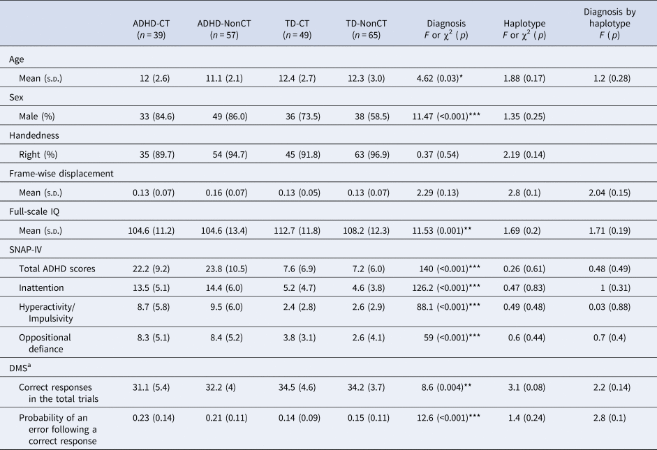
The in-scanner head motion level was estimated by mean frame-wise displacement (FD) derived with Jenkinson's relative root mean square algorithm (Jenkinson, Bannister, Brady, & Smith, Reference Jenkinson, Bannister, Brady and Smith2002). Participants with mean FD exceeding 1 s.d. (0.14 mm) above the sample mean (0.19 mm) were excluded from further analysis. Using this criterion, 26 participants (15 ADHD and 11 TD) were excluded, yielding a final sample of 210 participants. There was no significant Diagnosis, Genotype, or Diagnosis × Genotype interaction effect on the mean FD (Table 1).
Demographics, ADHD symptomatology, and DMS performance between the four groups

ADHD, attention deficit hyperactivity disorder; TD, typically developing; SNAP-IV, the Swanson, Nolan, and Pelham, version IV scale; DMS, Delayed Matching to Sample.
a Controlled for age, sex, and full-scale IQ.
*p < 0.05, **p < 0.01, ***p < 0.001.
Seed selection and functional connectivity analysis
We used 10 previously validated seed regions of interest in the bilateral striatum (Di Martino et al., Reference Di Martino, Scheres, Margulies, Kelly, Uddin, Shehzad and Milham2008, Reference Di Martino, Kelly, Grzadzinski, Zuo, Mennes, Mairena and Milham2011). Caudate seeds included the dorsal caudate (DC) and the superior ventral striatum (VSs). Owing to the limited spatial coverage, the inferior ventral striatum was not included as a seed. Putamen seeds included the dorsal-caudal putamen (dcP), dorsal-rostral putamen (drP), and ventral-rostral putamen (vrP). Each spherical seed covered 7 voxels in 3 mm isotropic voxel space (radius = 4 mm; online Supplementary Table S2 for coordinates of the seeds). Functional connectivity was calculated by correlating the seed time-series with the time course of all other voxels. The resulting Pearson's correlation coefficients were Fisher-z transformed to conform to normality assumptions for second-level analyses.
Statistical analyses
The Hardy-Weinberg equilibrium was tested for both SNPs using the goodness of fit χ2 test. For the genetic analysis, the two bi-allelic polymorphisms at rs27048 and rs429699 were processed and analyzed. The rs27048 (C)/rs429699 (T) haplotype was previously described (Shang & Gau, Reference Shang and Gau2014; Shang et al., Reference Shang, Gau, Liu and Hwu2011). The genotypes of the DAT1 variant were divided into two groups according to the presence of the rs27048 (C) /rs429699 (T) haplotype: the CT group (homozygous and heterozygous carriers of the CT haplotype) and the Non-CT group (CT haplotype non-carriers). Among the 96 youths with ADHD, 30 were diagnosed as the combined subtype (12 ADHD-CT and 18 ADHD-NonCT), while another 66 were diagnosed as the inattentive subtype (27 ADHD-CT and 39 ADHD-NonCT).
A two-way ANOVA including factors of diagnosis (ADHD v. TD) and genotype (CT v. non-CT) based on the full factorial design in SPM12 was calculated to illuminate the effects of DAT1 and ADHD on the striatal functional connectivity. Age, sex, FIQ, and mean FD of each participant were included as covariates. All significant clusters in neuroimaging-related statistical analyses were corrected for multiple comparisons at the cluster level by controlling topological family-wise error (FWE) calculated based on Gaussian Random Field theory, using a cluster-forming voxel-level height threshold of p < 0.005 and a spatial extent threshold that ensured a cluster-wise FWE at p < 0.05.
Partial correlations were calculated to associate the functional connectivity of clusters, which showed significant Genotype × Diagnosis interaction effects, with DMS indexes. Potential confounding factors, including age, sex, mean FD and FIQ, were controlled in the models. To accommodate multiple testing in the correlation analyses, we used false discovery rate (FDR) procedure within each of the four groups and an FDR-adjusted q value threshold of 0.05 to determine statistical significance.
Results
Demographics and clinical characteristics
Detailed demographic characteristics of all the participants are presented in Table 1. Youths with ADHD were younger and had lower FIQ, and fewer correct responses in the total trials, and a higher probability of an error following a correct response in DMS than TD youths (Table 1). Males were more predominant in youths with ADHD than TD youths. There were no significant differences in the distribution of subtypes between the ADHD-CT and ADHD-NonCT groups (p = 0.93). Online Supplementary Table S1 presents the genotypic distributions of the two SNPs. Each of the genotypes for the variants fitted the values expected based on the Hardy-Weinberg equilibrium (p > 0.05). No differences in the genotypic frequencies of rs27048 and rs429699 were observed between the ADHD and TD groups, as well as the frequencies of the CT haplotype.
For youths with ADHD, there were no significant differences in age, gender, handedness, ADHD severity, and the performance of the DMS task between the ADHD-CT and ADHD-NonCT groups.
Striatal functional connectivity
Online Supplementary Fig. S1 depicts within-group surface maps of the striatal functional connectivity using one-sample t tests. Each group demonstrated extensive but distinct regions significantly associated with BOLD fluctuations in the striatal seeds. The observed networks in the two TD groups, regardless of CT presence, largely corresponded with striatal networks as reported in the existing literature (Di Martino et al., Reference Di Martino, Scheres, Margulies, Kelly, Uddin, Shehzad and Milham2008; Reference Di Martino, Kelly, Grzadzinski, Zuo, Mennes, Mairena and Milham2011). That is, DC showed positive relationships with regions implicated in cognitive control, including the anterior cingulate, prefrontal cortex, precentral-postcentral gyri, superior temporal gyri, and precuneus (online Supplementary Fig, S1A, B). dcP and drP seeds exhibited positive connectivity with sensorimotor areas (online Supplementary Fig, S1C, D, E, F). vrP seeds had positive connections with the insula, anterior midcingulate, and superior temporal gyrus (online Supplementary Fig. S1G, H). VSs seeds showed positive relationships with the paralimbic regions and ventromedial prefrontal cortex (online Supplementary Fig. S1I, J).
Main effects of ADHD
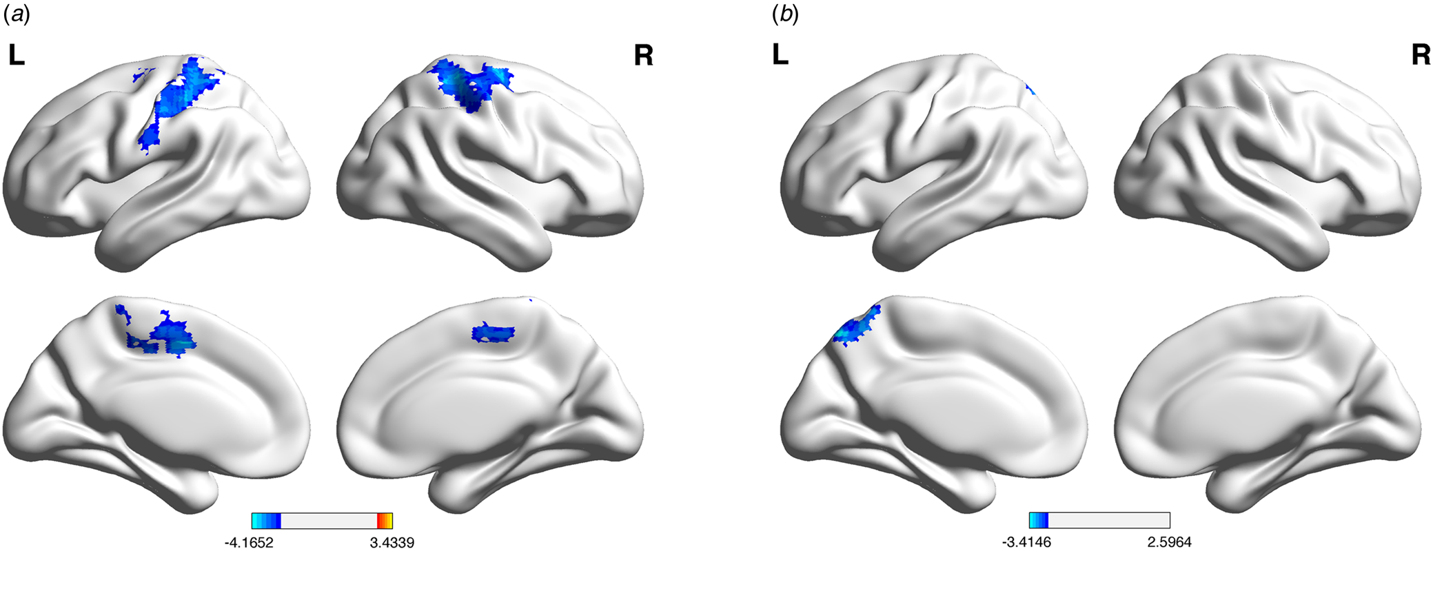
Youths with ADHD, relative to TD youths, showed significant hypoconnectivity of the left DC with bilateral sensorimotor clusters (Fig. 1a; coordinates −12, −3, 51; cluster size 28 161 mm3), and hypoconnectivity of the left drP with the left precuneus (Fig. 1b; coordinates −9, −72, 54; cluster size 3645 mm3).
Main effects of diagnostic status on striatal functional connectivity. Relative to the TD group, the ADHD group demonstrated (a) hypoconnectivity of the left DC with bilateral sensorimotor clusters (coordinates −12, −3, 51; cluster size 28 161 mm3) and (b) hypoconnectivity of the left drP with the left precuneus (coordinates −9, −72, 54; cluster size 3645 mm3). All clusters survived cluster-level Gaussian Random Field corrected p < 0.05, with cluster-height threshold p < 0.005. DC, dorsal caudate; drP, dorsal-rostral putamen.

Main effects of the CT haplotype
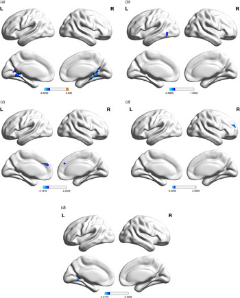
There are significant effects of the DAT1 CT haplotype on striatal functional connectivity in both ADHD and TD groups. For all the participants, youths with the CT haplotype showed significant hypoconnectivity of the right dcP with the bilateral lingual gyri and calcarine regions (Fig. 2a; left: coordinates −9, −48, −6; cluster size 7884 mm3; right: coordinates 24, −45, 6; cluster size 8181 mm3). For the ADHD group, youths with the CT haplotype showed significant hypoconnectivity of the left DC with the left middle temporal gyrus (Fig. 2b; coordinates −39, −54, −6; cluster size 3267 mm3), hypoconnectivity of the right DC with the bilateral superior frontal gyri-anterior cingulate (Fig. 2c; coordinates 0, 42, 30; cluster size 4050 mm3), and hypoconnectivity of the right VSs with the right superior frontal gyrus (Fig. 2d; coordinates 18, 51, 12; cluster size 2727 mm3). For the TD group, youths with the CT haplotype showed significant hypoconnectivity of the right dcP with the left lingual gyrus and calcarine region (Fig. 2e; coordinates −15, −54, 0; cluster size 3456 mm3).
Main effects of the CT haplotype on striatal functional connectivity. (a) For all the participants, youths with the CT haplotype showed hypoconnectivity of the right dcP with the bilateral lingual gyri and calcarine regions (left: coordinates −9, −48, −6; cluster size 7884 mm3; right: coordinates 24, −45, 6; cluster size 8181 mm3). For the ADHD group, youths with the CT haplotype showed (b) hypoconnectivity of the left DC with the left middle temporal gyrus (coordinates −39, −54, −6; cluster size 3267 mm3), (c) hypoconnectivity of the right DC with the bilateral superior frontal gyri (coordinates 0, 42, 30; cluster size 4050 mm3), and (d) hypoconnectivity of the right VSs with the right superior frontal gyrus (coordinates 18, 51, 12; cluster size 2727 mm3). (e) For the TD group, youths with the CT haplotype showed significant hypoconnectivity of the right dcP with the left lingual gyrus and calcarine region (coordinates −15, −54, 0; cluster size 3456 mm3). All clusters survived cluster-level Gaussian Random Field corrected p < 0.05, with cluster-height threshold p < 0.005. DC, dorsal caudate; dcP, dorsal-caudal putamen; VSs, superior ventral striatum.

Effects of CT haplotype × ADHD interactions
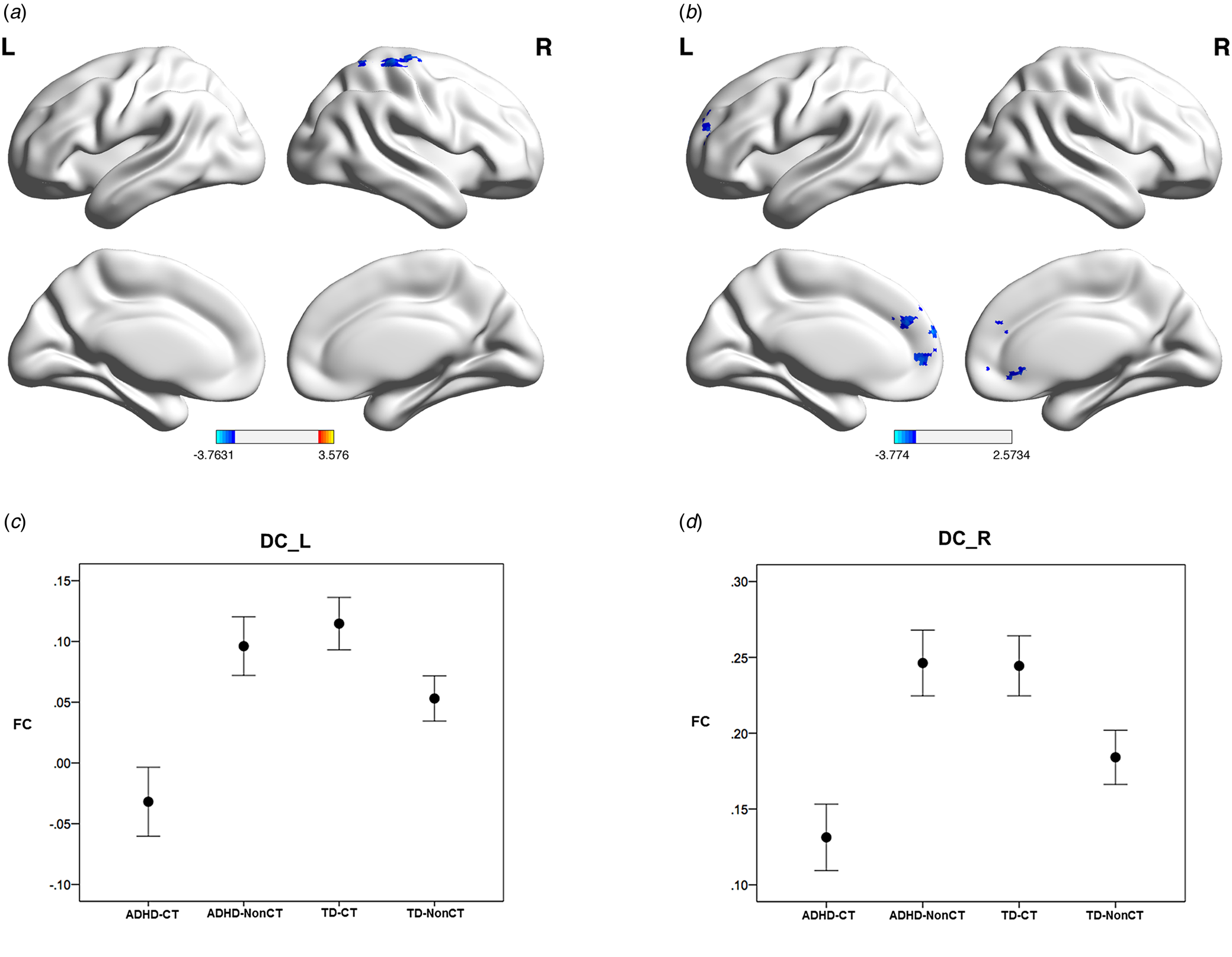
Significant interactions were found in the connectivity between the left DC and the right sensorimotor cluster (Fig. 3a; peak coordinates 36, −72, 45; cluster size 3969 mm3), and between the right DC and the left dorsolateral prefrontal-bilateral anterior cingulate clusters (Fig. 3b; peak coordinates 0, 42, 30; cluster size 8343 mm3). Figures 3c and 3d show interactional distributions of these connections for the DAT1 CT haplotype and ADHD. In the ADHD group, the strengths of bilateral DC connectivity decreased with the CT haplotype; while in the TD group, the reverse pattern was found.
Effects of interaction of diagnosis × CT haplotype on striatal functional connectivity. Significant interactions were found in (a) the connectivity between the left DC and the right sensorimotor cluster (peak coordinates 36, −72, 45; cluster size 3969 mm3); and (b) the connectivity between the right DC and the left dorsolateral prefrontal-bilateral anterior cingulate cluster (peak coordinates 0, 42, 30; cluster size 8343 mm3). All clusters survived cluster-level Gaussian Random Field corrected p < 0.05, with cluster-height threshold p < 0.005. (c) Functional connectivity between the left DC and the right sensorimotor cluster across the four groups; (d) Functional connectivity between the right DC and the left dorsolateral prefrontal-bilateral anterior cingulate cluster across the four groups. Each error bar indicates 95% confidence interval. DC, dorsal caudate; FC, functional connectivity.

Notably, based on the left DC seed, the right sensorimotor cluster showing DAT1 CT haplotype by ADHD interactions partially overlapped with the cluster showing ADHD effects (center coordinates of the overlapping cluster 31, −31, 65; overlapping size 1968 mm3). Based on the right DC seed, the DLPFC-anterior cingulate cluster showing CT haplotype by ADHD interactions also partially overlapped with the cluster exhibiting genotype effects in the ADHD group (center coordinates of the overlapping cluster −6, 44, 27; overlapping size 2592 mm3).
Correlations between striatal functional connectivity and visual memory
Table 2 shows the correlations between functional connectivity and visual memory for the ADHD-CT group. Lower functional connectivity between the left DC and the right sensorimotor cluster was significantly correlated with fewer correct responses in the total trials (r = 0.38, q = 0.04) and a higher probability of an error following a correct response (r = −0.44, q = 0.03) in DMS. No significant correlations between functional connectivity of bilateral DC and measures of DMS were observed in the ADHD-NonCT (online Supplementary Table S3), TD-CT (online Supplementary Table S4), and TD-NonCT (online Supplementary Table S5) groups.
Correlations between visual memory and dorsal caudate functional connectivity in the ADHD-CT group

ADHD, attention deficit hyperactivity disorder; l, left; r, right; b, bilateral; DC; dorsal caudate; SM, sensorimotor; DLPF, dorsolateral prefrontal; AC, anterior cingulate.
a Adjusted by false discovery rate.
*q < 0.05.
Effects of IQ and age
After we removed IQ from the covariates and repeated the main imaging analyses (Supplementary Information), we identified an additional hypoconnectivity of the right dcP with bilateral sensorimotor clusters in the effects of the CT haplotype, while the connectivity findings of the left DC and the left drP in the effects of ADHD, and the right DC in the effects of CT haplotype × ADHD interactions were no longer significant. The other two significant clusters of the right dcP in the effects of the CT haplotype and the left DC in the effects of CT haplotype × ADHD interactions overlapped with those in the previous analyses (online Supplementary Figs S2 and S3).
In the next step, we added the age-squared item into the covariates of imaging analyses to model the effects of age more accurately (Supplementary Information). We found that all the significant clusters from the main analyses, which have controlled for IQ, remained after the analyses with IQ removed from the covariates (online Supplementary Figs S4 and S5).
Discussion
In the present study, we investigated the effects of the DAT1 rs27048 (C)/rs429699 (T) haplotype on striatal functional connectivity in ADHD and TD youths. We observed that the CT haplotype exhibited significant interaction effects with ADHD diagnosis on the functional connectivity in the networks of bilateral DC, and that DAT1-affected DC functional connectivity was associated with visual memory performances in youths with ADHD. Our findings provide evidence to support that the DAT1 rs27048 (C)/rs429699 (T) haplotype may play an important role in the altered functional connectivity of DC in youths with ADHD.
Effects of ADHD on visual memory and striatal functional connectivity
Our finding that youths with ADHD had fewer correct responses in the total trials and a higher probability of an error following a correct response in DMS than TD youths supports a relationship between ADHD and impaired visual memory (Rhodes et al., Reference Rhodes, Coghill and Matthews2004; Shang & Gau, Reference Shang and Gau2011).
Compared to TD youths, youths with ADHD showed hypoconnectivity between the left DC-bilateral sensorimotor clusters and the left drP-left precuneus. The DC is part of the executive attention system (Castellanos & Proal, Reference Castellanos and Proal2012) via connections with the prefrontal cortex and motor cortex (Draganski et al., Reference Draganski, Kherif, Kloppel, Cook, Alexander, Parker and Frackowiak2008; Robinson et al., Reference Robinson, Laird, Glahn, Blangero, Sanghera, Pessoa and Fox2012). Children with ADHD often show difficulties with sensorimotor systems, paralleling difficulties with higher-order cognitive control (Mostofsky, Newschaffer, & Denckla, Reference Mostofsky, Newschaffer and Denckla2003). Impairments associated with ADHD are reflected in sensorimotor function in biologically meaningful ways (Mostofsky & Simmonds, Reference Mostofsky and Simmonds2008), and a large-scale resting-state fMRI study demonstrates atypical functional connectivity patterns in sensorimotor regions in children with ADHD (Fair et al., Reference Fair, Nigg, Iyer, Bathula, Mills, Dosenbach and Milham2012). Notably, although our finding of altered dorsal caudate-sensorimotor interactions extends the existing literature on striatal-cortical circuits in ADHD (Hong et al., Reference Hong, Harrison, Fornito, Sohn, Song and Kim2015; Posner et al., Reference Posner, Rauh, Gruber, Gat, Wang and Peterson2013), this identified sensorimotor cluster from diagnosis contrasts overlapped with the postcentral cluster showing significant CT haplotype by Diagnosis interaction. Such finding implies that the observed functional connectivity associated with ADHD in the literature might present a more complex picture if the genetic makeup data is available (Klein et al., Reference Klein, Onnink, van Donkelaar, Wolfers, Harich, Shi and Franke2017).
The putamen is involved in the modulation of the balance between goal-directed and habitual modes of action control (FitzGerald, Friston, & Dolan, Reference FitzGerald, Friston and Dolan2012). Decreased functional connectivity of DC and dorsal putamen has been reported in children with ADHD (Hong et al., Reference Hong, Harrison, Fornito, Sohn, Song and Kim2015; Posner et al., Reference Posner, Rauh, Gruber, Gat, Wang and Peterson2013). Our finding of decreased dorsal putamen-precuneus connections is consistent with an earlier report (Cao et al., Reference Cao, Cao, Long, Sun, Sui, Zhu and Wang2009), suggesting altered intrinsic interactions between striatal and attentional control systems are implicated in ADHD.
Distribution of the CT haplotype between the ADHD and TD groups
In our previous family-based genetic study with a larger sample size (n = 906), we found the significant association of the CT haplotype with ADHD (Shang et al., Reference Shang, Gau, Liu and Hwu2011). In the present imaging genetics study, the sample size (n = 210) may not achieve sufficient power to detect causal risk genetic variant for ADHD, which is diagnosed based on behavioral phenotypes. In contrast, genes may have a high penetrance on neuroimaging measures, which are able to provide larger effects than behavioral phenotypes (Mier, Kirsch, & Meyer-Lindenberg, Reference Mier, Kirsch and Meyer-Lindenberg2010).
Effects of the CT haplotype on striatal functional connectivity in youths with ADHD
While there are no significant differences in the severity of ADHD symptoms and performance on the DMS task between the ADHD-CT and ADHD-NonCT groups, our imaging genetic findings highlight differential striatal connectivity related to this CT haplotype. Compared with the ADHD-NonCT group, the ADHD-CT group showed reduced functional connectivity of bilateral DC and the right VSs. The association between the DAT1 CT haplotype and functional connectivity within ADHD raises the possibility that the CT haplotype may contribute to the heterogeneity of ADHD through modulation of brain function (Wahlstedt, Thorell, & Bohlin, Reference Wahlstedt, Thorell and Bohlin2009). In contrast, the TD-CT group showed reduced functional connectivity of the right dcP in comparison to the TD-NonCT group. Although the exact function of the CT haplotype is unknown, the distinct patterns of striatal functional connectivity profiles imply that the CT haplotype may modulate neural activity through different molecular pathways between the ADHD and TD groups.
Interaction effects of the CT haplotype and ADHD on striatal functional connectivity and visual memory
We found a significant CT haplotype × ADHD effect in the functional connectivity patterns of bilateral DC with the smallest functional connectivity value in the ADHD-CT group. The interaction of the CT haplotype and ADHD followed a crossover pattern, indicating that the effects of the DAT1 CT haplotype on the strengths of bilateral DC connectivity differ between the ADHD and TD groups. Our findings suggest that youths with ADHD may engage the DC networks in a different way than TD youths regarding DAT1 CT haplotype.
Furthermore, for the ADHD-CT group, the left DC-right sensorimotor connectivity was correlated positively with the correct responses in the total trials and negatively with a probability of an error following a correct response in DMS. In other words, decreased functional connectivity of the left DC was associated with decreased visual memory performance in youths with ADHD who were the carriers of the CT haplotype. The caudate nucleus is involved in visuospatial processing and may play an important role in visual memory (Davis, Filoteo, Kesner, & Roberts, Reference Davis, Filoteo, Kesner and Roberts2003). Previous studies report that reduced dopaminergic activity in the caudate nucleus is associated with impaired visual memory (Jokinen et al., Reference Jokinen, Bruck, Aalto, Forsback, Parkkola and Rinne2009), and caudate nucleus atrophy is correlated with poor visual memory function (Bartres-Faz et al., Reference Bartres-Faz, Junque, Serra-Grabulosa, Lopez-Alomar, Moya, Bargallo and Clemente2002). Our findings suggest that genetically determined hypoconnectivity of DC may contribute to impaired performance on the visual memory tasks in youths with ADHD. Taken together, our finding of gene-brain-behavior linkage within one study is supported by several previous studies describing dopamine-caudate (Jokinen et al., Reference Jokinen, Bruck, Aalto, Forsback, Parkkola and Rinne2009), caudate-visual memory (Bartres-Faz et al., Reference Bartres-Faz, Junque, Serra-Grabulosa, Lopez-Alomar, Moya, Bargallo and Clemente2002), and dopamine-visual memory (Rhodes et al., Reference Rhodes, Coghill and Matthews2004) relationships.
Several limitations of this study should be considered when interpreting our findings. First, we included a wide age range of subjects, potentially confounding our data with multiple stages of brain development. Due to the small number of samples in the subgroups after stratification by age, we did not conduct further imaging analyses in the age-stratified data. Although age was included as a nuisance covariate to statistically control for its confounding effect, future studies with a narrower age range are warranted. Second, our findings may not be able to be generalized to the community-based ADHD population due to the referred sample with strict inclusion and exclusion criteria set in the present study. Third, the present imaging-genetic-behavioral approach in a cross-sectional cohort is still inherently observational and correlational. Future studies with additional levels and integration of novel methodologies are needed (Vilor-Tejedor, Caceres, Pujol, Sunyer, & Gonzalez, Reference Vilor-Tejedor, Caceres, Pujol, Sunyer and Gonzalez2017). Fourth, due to the small number of subjects in the subgroups stratified by subtype, we did not conduct further imaging analyses to examine the relationship among ADHD subtypes, the CT haplotype, and striatal functional connectivity. Fifth, there is still an active debate whether IQ needs to be included as a covariate in the investigations of ADHD, and the optimal approach may vary depending on the specific research question (Willcutt, Doyle, Nigg, Faraone, & Pennington, Reference Willcutt, Doyle, Nigg, Faraone and Pennington2005). Previous resting-state fMRI studies have shown significant associations between brain functional connectivity and intelligence (Pamplona, Santos Neto, Rosset, Rogers, & Salmon, Reference Pamplona, Santos Neto, Rosset, Rogers and Salmon2015; Song et al., Reference Song, Zhou, Li, Liu, Tian, Yu and Jiang2008), which could argue in favor of including IQ as a covariate in our imaging analyses for the striatal functional connectivity. Further imaging genetics studies are warranted to examine the genetically determined alterations in the striatal functional connectivity across different IQ subgroups in youths with ADHD. Sixth, the sample sizes in the present study are slightly imbalanced, with 88 in the CT group and 122 in the Non-CT group. Future imaging genetics studies with larger equal samples are needed to prevent the imbalance problems. Seventh, given the small sample sizes of homozygous carriers of the CT haplotype (six in the ADHD-CT group and four in the TD-CT group), future studies are warranted to examine the differential neurobiological effects between the heterozygous and homozygous carriers of the CT haplotype in a larger sample of youths with ADHD. Lastly, to reach the balance between reproducibility and the inflation of type I errors (Chen, Lu, & Yan, Reference Chen, Lu and Yan2018), the current study attempts to find the middle ground with a cluster-forming threshold at p < 0.005. We acknowledge there is no consensus on this issue in resting-state fMRI connectivity literature (Chen et al., Reference Chen, Lu and Yan2018; Eklund, Nichols, & Knutsson, Reference Eklund, Nichols and Knutsson2016). Although the current exploration, as the first attempt in the related field, may facilitate further hypothesis generation, the results based on the cluster correction still need cautious interpretation.
Counteracting the limitations, the current findings are strengthened by several features of this study. First, stimulant medication has been reported to alter striatal functional connectivity in youths with ADHD (Rubia et al., Reference Rubia, Halari, Cubillo, Mohammad, Brammer and Taylor2009). Our present study recruit drug-naïve youths with ADHD to avoid the potential confounding effects of medications. Second, given that the allele frequency distribution of the DAT1 polymorphisms varies significantly across ethnicity, the ethnically homogeneous sample of our present study is optimal for examining genetic effects on the striatal functional connectivity. Third, integration of specific neuroimaging, neuropsychological, and genetic assessments within one study could enhance our understanding of the neurobiological basis of ADHD.
In conclusion, our present study indicates that the DAT1 haplotype rs27048 (C)/rs429699 (T) may affect the functional connectivity of the left DC in youths with ADHD, resulting in impaired performance on the DMS task. By demonstrating linkages between DAT1, altered striatal functional connectivity, and the DMS performance, our present work may contribute to a model of visual memory enabled by the dopamine-related striatal regions in youths with ADHD.
Supplementary material
The supplementary material for this article can be found at https://doi.org/10.1017/S0033291719003830.
Acknowledgements
The research reported in this article was supported by grants NSC96-2628-B-002-069-MY3, NSC98-2314-B-002-051 -MY3, and NSC99-2321-B-002-037 from National Science Council (Taiwan) and grants NHRI-EX98-9407PC and NHRI-EX100-0008PI from National Health Research Institute (Taiwan). The data management and manuscript preparation were partially supported by grants NHRI-EX105-10404PI and NHRI-EX106-10404PI from National Health Research Institute (Taiwan) and Chen-Yung Foundation. SNP genotyping was performed by the National Genotyping Center of Academia Sinica, Taiwan, and by the support from the Department of Medical Research, National Taiwan University Hospital, Taipei, Taiwan.
Conflict of interest
None.