Implications
Intestinal immune system plays a critical role in human and animal health. The mammalian target of rapamycin (mTOR) signaling pathway has been proved to be involved in the immune responses induced by lipopolysaccharide (LPS) in several cells, leading to the hypothesis that supplementation of leucine could have consequences for the intestine of chicken embryos, which may affect the regulation in vitro cultural system. All the results may give the information about the understanding of the cross-talk between mTOR pathway and the intestinal inflammatory responses in chicken embryos.
Introduction
Secretory immunoglobulin A (sIgA), which is the most abundant immunoglobulin in mucosal secretions, plays an important role in the intestinal mucosal immune system and the maintenance of mucosal homeostasis. Meanwhile, sIgA plays an essential role in the maintenance of an appropriate intestinal microbiota, which is required for local and systemic immune homeostasis (Cerutti and Rescigno, Reference Cerutti and Rescigno2008). In chickens, IgA secreted by the intestine is similar to mammalian sIgA and is associated with a protein homologous to a mammalian secretory component of 350 000 daltons (Watanabe et al., Reference Watanabe, Kobayashi and Isayama1975).
Normal intestinal mucosa contains abundant IgA-secreting cells, which are generated from B cells after activation of naive B cells by the B cell receptor in gut-associated lymphoid tissues. A significant proportion of this specific anti-commensal IgA induction is through a pathway that is independent of T cell help and of follicular lymphoid tissue organization (Macpherson et al., Reference Macpherson, Gatto, Sainsbury, Harriman, Hengartner and Zinkernagel2000).
The mTOR, which is a highly conserved and ubiquitously expressed signaling molecule, is involved in cell growth and proliferation and various cellular functions. Mammalian target of rapamycin can sense metabolic signals and exert anabolic effects by stimulating protein synthesis and ribosomal biogenesis, enhancing cell proliferation and promoting cell survival (Fielhaber et al., Reference Fielhaber, Carroll, Dydensborg, Shourian, Triantafillopoulos, Harel, Hussain, Bouchard, Qureshi and Kristof2012). Mammalian target of rapamycin transfers its regulating effect by stimulating the phosphorylation of the downstream target protein p70 ribosomal S6 kinase (p70S6K). The mTOR signaling pathway has been shown to play an important role in the intestinal tract (Sampson et al., Reference Sampson, Davis, Grogg and Zheng2016). In chickens, the mTOR signaling pathway, as in mammals, senses the changes of nutrient supply and takes part in the regulation of cell proliferation and differentiation (Duchéne et al., Reference Duchéne, Audouin, Crochet, Duclos, Dupont and Tesseraud2008). Leucine stimulates protein synthesis by regulating gene expression via activation of the mTOR signaling pathway in skeletal muscle, small intestinal and whole body (Duan et al., Reference Duan, Li, Li, Tang, Kong, Feng, Anthony, Watford, Hou, Wu and Yin2016; Wessels et al., Reference Wessels, Kluge, Hirche, Kiowski, Schutkowski, Corrent, Bartelt, König and Stangl2016).
Mammalian target of rapamycin also regulates the differentiation and the functionality of immune cells. Emerging evidence has indicated that the mTOR pathway involved in the immunity, and its effect on regulating nuclear factor-κB (NF-κB) depended on the cell type or model (Weichhart et al., Reference Weichhart, Costantino, Poglitsch, Rosner, Zeyda, Stuhlmeier, Kolbe, Stulnig, Hörl, Hengstschläger, Müller and Säemann2008; Fielhaber et al., Reference Fielhaber, Carroll, Dydensborg, Shourian, Triantafillopoulos, Harel, Hussain, Bouchard, Qureshi and Kristof2012). In LPS-stimulated murine macrophages, the cross-talk between the mTOR and NF-κB pathways has been shown to be involved in the LPS-induced response (Dos Santos et al., Reference Dos Santos, Delattre, De Longueville, Bult and Raes2007). Mammalian target of rapamycin increased the phosphorylation of I Kappa B Kinase to activate NF-κB in lung epithelial cells (BEAS-2B cells) (Hou et al., Reference Hou, Chiu, Yeh and Yeh2012). The Akt/mTOR cascade modulates LPS-induced cytokine production differentially in peripheral blood mononuclear cells (Schaeffer et al., Reference Schaeffer, Arbabi, Garcia, Knoll, Cuschieri, Bulger and Maier2011). In human monocyte cells, blocking the mTOR pathway suppresses the expression of chemokines by modulating the p38-mitogen-activated protein kinase (MAPK) and NF-κB p65-mediated signaling pathways (Lin et al., Reference Lin, Chang, Hung, Kuo, Hwang, Chen, Hung and Lin2014).
In Caco-2 cells, TNF-ɑ regulation of the secretory component expression is mediated through a phosphatidylinositol 3-kinase (PI3K)/AKT/mTOR signaling pathway (Liu et al., Reference Liu, Wang, Jiang and Ding2013). In newborn piglets, Rotavirus infection at D5 of life resulted in activation of mTOR, but mTOR inhibition did not affect the clinical course of bowel recovery (Rhoads et al., Reference Rhoads, Corl, Harrell, Niu, Gatlin, Phillips, Blikslager, Moeser, Wu and Odle2007). Recently, we showed that rapamycin, which is an mTOR inhibitor, suppressed the development of small intestine and IgA production in the small intestinal tract, suggesting that the mTOR signaling pathway is involved in the development and immune function of the small intestinal tract in broiler chickens (Liu et al., Reference Liu, Zhao, Fan, Liu, Jiao, Wang, Sun and Lin2015). Hence, we hypothesized that the mTOR signaling pathway is involved in the LPS-induced immune responses of the intestinal tract.
In the present study, the in vitro cultural system of intestinal tissue from chicken embryo was employed to test the effect of LPS on intestinal inflammatory response in the present of leucine. sIgA concentrations and cytokine gene expression was determined. The phosphorylation levels of mTOR, NF-κB and p38 MAPK were also measured.
Material and methods
Ethics statement
All study procedures were approved by the Animal Care and Use Committee of Shandong Agricultural University and were in accordance with the Guidelines for Experimental Animals established by the Ministry of Science and Technology (Beijing, China).
In vitro intestinal tissue culture
Specific pathogen free embryonated chicken eggs were obtained and incubated in the laboratory incubator (38±0.5°C, 60% humidity) with daily rotation. According to approved ethical standards, 19-day-old embryos were euthanized by cervical dislocation then dissected. Small intestines were separated sterilely and cut into segments of up to 1-cm length and then cultured in uncoated 24-well cell culture plates (Corning, NY, USA) with ~2 segments/well. The culture conditions were as follows: 37.5°C in a 5% CO2 incubator in Dulbecco’s modified Eagle’s F12 Ham medium (DMEM-F12; Gibico, Grand Island, NY, USA) containing 2% Fetal Bovine Serum (FBS; Gibico, Grand Island, NY, USA), insulin (10 µg/ml), epidermal growth factor (20 ng/ml; Sigma Aldrich, St. Louis, MO, USA), and penicillin (100U/ml) – streptomycin (0.1 mg/ml) (P1400; Salarbio, Beijing, China).
Lipopolysaccharide treatment
The intestinal tissues were cultured in DMEM-F12 with LPS at dosages of 0, 0.5, 1 and 2 μg/ml (Baker et al., Reference Baker, Wang, Mackman and Luyendyk2009) for 2 h. After treatment, the intestinal tissue (n=12) was collected, snap frozen in liquid nitrogen, and stored at −80°C for further analysis. The culture solution (n=12) was collected for the measurement of sIgA.
Leucine treatment
The intestinal tissue was cultured in DMEM-F12 and subjected to five treatments: negative control without any treatment, positive control subjected to 2 μg/mL LPS treatments for 2 h, and leucine treatment at a dose of 10, 20 and 40 mM (Wang et al., Reference Wang, Yang, Wang, Jiao, Song and Lin2016) in the presence of 2 μg/ml LPS. The tissues and supernatant (n=12) were collected for further analysis as mentioned above.
Measurement of secretory immunoglobulin A concentration by ELISA
Immunoplates (Costar; Corning, NY, USA) were coated by adding 100 μl of goat anti-chicken IgA (A30-103A; Bethyl Laboratories, Montgomery, TX, USA) dissolved in phosphate buffer solution (PBS, pH 9.6) and incubated at 37°C for 1 h. Each well was washed and blocked with 100 μL of 1% bull serum albumin (BSA) in PBS overnight. After washing, 100 μL of the samples and chicken reference serum (RS10-102-3; Bethyl Laboratories, Montgomery, TX, USA) were added and the plate was incubated at 37°C for 1 h. Thereafter, unbound materials were washed away and 100 μL of horse radish peroxidase (HRP)-conjugated goat anti-chicken IgA (A30-103P-35; Bethyl, Bethyl Laboratories, Montgomery, TX, USA) solutions in 1% BSA of PBS was added. After the plate was incubated at 37°C for 1 h and washed, 100 μl of TMB (CW0050; CWBIO, Beijing, China) was added and the plate was allowed to stand at 37°C for 15 min. After 50 μl of 2.5 M H2SO4 was added to each well to stop the reaction, the absorbance at 450 nm was measured using a Titertek Multiskan Plus MKII (Flow Laboratories, McLean, VA, USA).
Messenger RNA expression analysis by quantitative real-time PCR
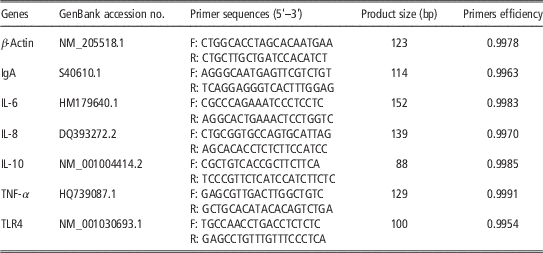
Total RNA was extracted from the intestinal tissue using Trizol reagent (Invitrogen, Grand Island, NY, USA) according to the manufacturer’s instructions, and all reactions were performed on ice. Total RNA, 1 µg, was used for reverse transcription with PrimeScript RT reagent kit (04897030001; Roche, Pleasanton, CA, USA) to prepare the complementary DNA. Primer sequences and the melting curves of all the primers are shown in Table 1 and Supplementary Figure S1, respectively.
Gene-specific primer sequences used for gene transcription analysis in chicken

IgA=immunoglobulin A; IL=interleukine; TNF-α=tumor necrosis factor-α; TLR4=Toll-like receptor 4.
Quantitative real-time PCR was conducted using SYBR Green I Dye (04913914001; Roche Pleasanton, CA, USA) on the QuantStudio 5 Real-time PCR System (Applied Biosystems, Foster, CA, USA). The housekeeping gene β-actin, was used as the internal standard for the PCR reaction (△C
T
). Relative messenger RNA (mRNA) abundance was calculated using the
 $$2^{{{\minus}\Delta \Delta C_{T} }} $$
method. Therefore, all gene transcription results were reported as the n-fold difference relative to the calibrator.
Western blot analysis
After the intestinal tissue culture was lysed and homogenized, total proteins were extracted and measured with a BCA Protein Assay Kit (Beyotime, Nantong, Jiangsu, China). For the western blotting analysis, equal protein amounts (35 µg/well) were denatured for 10 min at 100°C and then separated with 7.5 and 12% SDS-PAGE. The protein was transferred onto polyvinylidene fluoride microporous membrane (Millipore, Bedford, MA,USA) at 200 mA, 4°C for 2 h. After 1 h blocking in blocking solution (Beyotime, Nantong, Jiangsu, China) at room temperature, the membranes were incubated at 4°C overnight in primary antibodies against phospho-NF-κB p65 (Ser536) (93H1), phosphor-p38 MAPK (Thr180/Tyr182), phospho-c-Jun NH2-terminal kinase (JNK) (Tyr185) (81E11), phospho-p70S6 Kinase (Thr389) (108D2), phospho-mTOR (Ser2448) rabbit mAb (Cell Signaling Technology Inc. Danvers, MA, USA), total NF-κB p65 (C22B4), p38 MAPK, JNK (56G8), p70S6 Kinase (49D7), mTOR rabbit mAb (Cell Signaling Technology Inc. Danvers, MA, USA), and β-actin mouse monoclonal (AA128; Beyotime, Nantong, Jiangsu, China), respectively. The proteins were detected using either HRP-labeled goat anti-mouse IgG (H+L) secondary antibody or goat anti-rabbit IgG (H+L)-HRP-conjugated secondary antibody (Beyotime, Nantong, Jiangsu, China). And then the blots were visualized and analysed on a FUSION device (FUSION FX; Vilber Lourmat).
Statistical analysis
All experiments were performed in duplicate. And all the data were analysed using Statistical Analysis Software (version 8e; SAA Institute Inc., Cary, NC, USA). Before analysis, all data were examined for normality by examination of histograms and normal distribution plots (UNIVARIATE procedure). A one-way ANOVA model was used to estimate the main effect of treatment. Means for the treatments were compared using a Duncan’s test and were considered to be significantly different when P<0.05.
Results
Effect of lipopolysaccharide treatment on secretory immunoglobulin A concentrations and gene expression
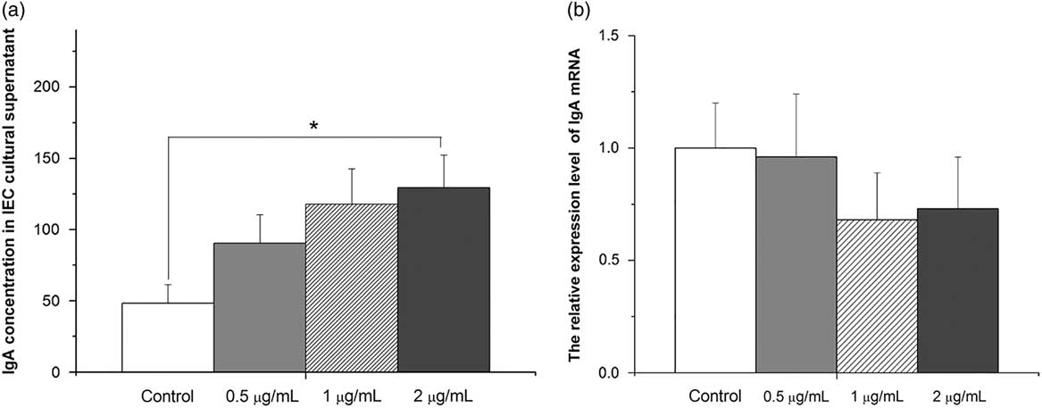
The sIgA concentration in culture solution of intestinal tissues was increased by LPS treatment in a dose-depend manner (Figure 1a). The sIgA concentration in the 2 µg/ml LPS group was higher than that in the control group (P<0.05). In contrast, the mRNA level of IgA in intestinal tissue was not influenced by LPS treatment (P>0.05, Figure 1b).
Effect of lipopolysaccharide (LPS) treatment (0, 0.5, 1 and 2 µg/ml) on secretory immunoglobulin A (sIgA) concentration (ng/ml) in tissue culture solution (a) and the relative expression level of IgA messenger RNA (mRNA) (b). The culture solution and intestinal tissue of chicken embryos were collected after LPS (0, 0.5, 1 and 2 µg/ml) treatment for 2 h to test the sIgA concentration and IgA mRNA. The data were subjected to one-way ANOVA analysis. The values indicate the mean ± SEM (n=12). * Means are significantly different (P< 0.05). IEC=intestinal epithelial cell.

Effect of lipopolysaccharide treatment on toll-like receptor 4 and cytokine expression
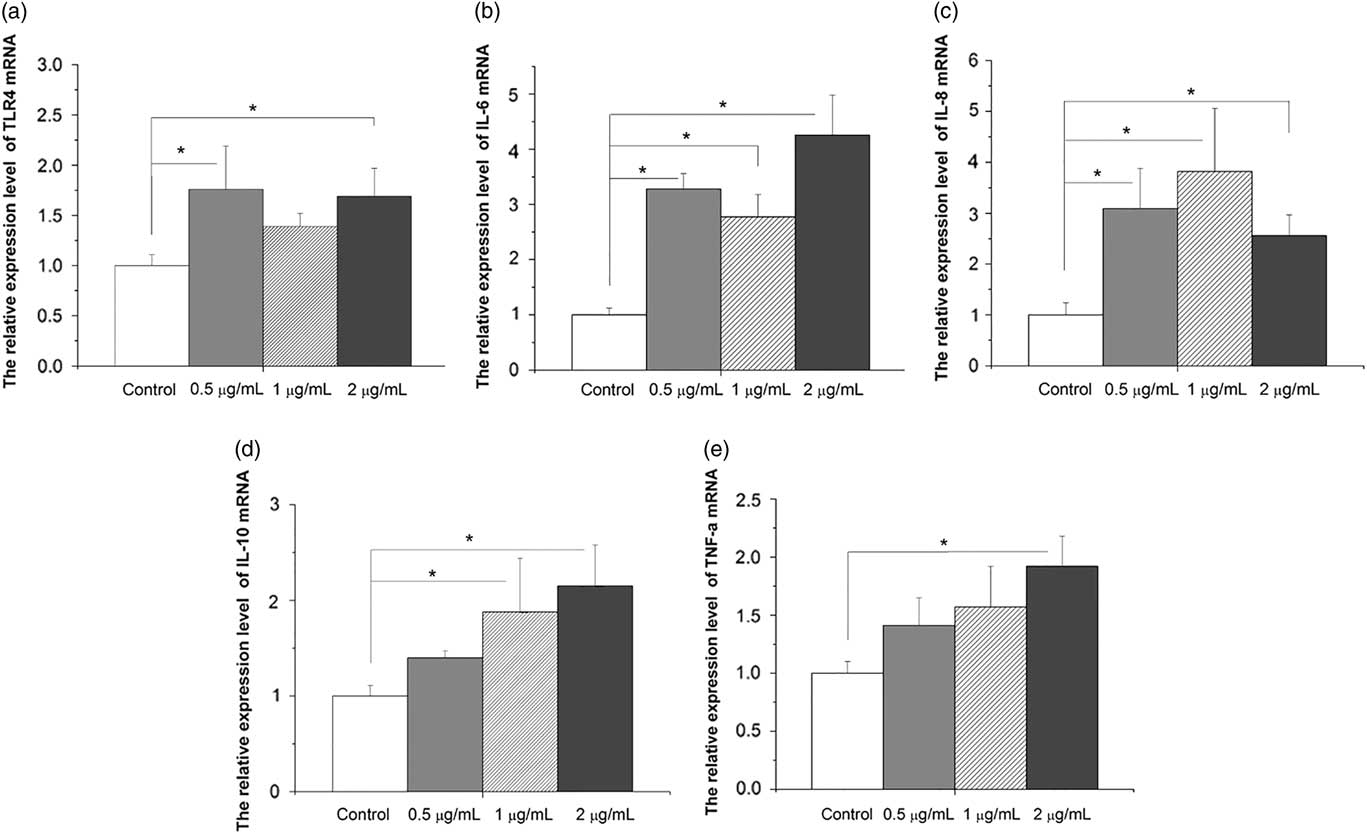
The mRNA expression levels of Toll-like receptor (TLR) 4 (P<0.05), interleukine (IL)-6 (P<0.05), IL-8 (P<0.05), IL-10 (P<0.05) and tumor necrosis factor-α (TNF-α) (P<0.05) were increased by LPS treatment (Figure 2a to e).
Effect of lipopolysaccharide (LPS) treatment (0, 0.5, 1 and 2 µg/ml) on messenger RNA (mRNA) levels of Toll-like receptor (TLR) 4 (a), interleukine (IL)-6 (b), IL-8 (c), IL-10 (d) and tumor necrosis factor-α (TNF-α) (e) in intestinal culture. The intestinal tissue of chicken embryos were collected after LPS treatment for 2 h to measure the TLR4 and the cytokines mRNA expression level. The data were subjected to one-way ANOVA analysis. The values indicate the mean ± SEM (n=12). * Means are significantly different (P<0.05).

The effect of lipopolysaccharide treatment on the phosphorylation of the mammalian target of rapamycin, mitogen-activated protein kinase and nuclear factor-κB
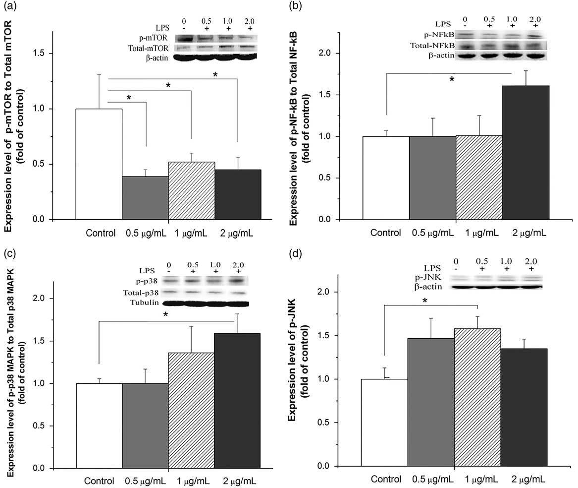
Compared with the control group, LPS treatment decreased the phosphorylation level of mTOR in all three dosages (P<0.05, Figure 3a). In contrast, the phosphorylation of NF-κB p65 was increased by LPS treatment only in the 2 µg/ml dose group (P<0.05, Figure 3b) compared with the control, and there was no difference among the other groups (P>0.05). Compared with the control, LPS treatment at a dose of 2 µg/ml increased the phosphorylation level of p38 MAPK (P<0.05, Figure 3c). The phosphorylation of JNK was increased by LPS treatment (P<0.05) and the 2 µg/ml LPS group had a higher level of p-JNK compared with the control group (Figure 3d).
Effect of lipopolysaccharide (LPS) treatment (0, 0.5, 1 and 2 µg/ml) on the protein phosphorylation level of the mammalian target of rapamycin (mTOR) (a), nuclear factor-κB p65 (NF-κB p65) (b), p38 (c) and Jun N-terminal kinase (JNK) (d) mitogen-activated protein kinase (MAPK). The intestinal tissue of chicken embryos were collected after LPS treatment for 2 h to measure the mTOR, NF-κB p65, p38 and JNK MAPK expression. The data were subjected to one-way ANOVA analysis. The values indicate the mean±SEM (n=12). * Means are significantly different (P<0.05).

Leucine treatment on lipopolysaccharide-induced secretory immunoglobulin A and inflammatory responses
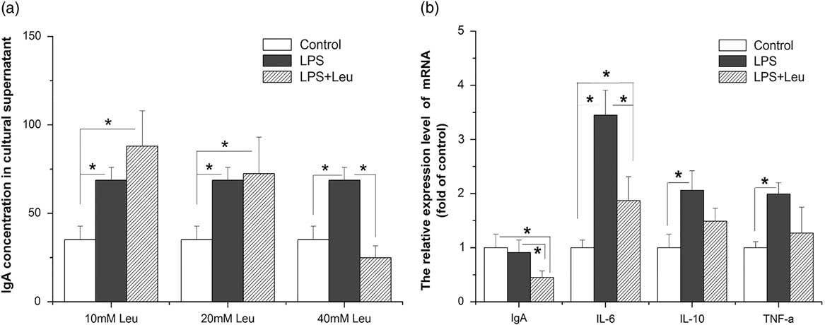
Lipopolysaccharide treatment increased sIgA concentrations in the culturing solution (P<0.05). Leucine supplementation decreased sIgA content at the dose of 40 mM (P<0.05; Figure 4a). However, there was no detectable effect of leucine treatment at a dose of 10 or 20 mM (P>0.05). The mRNA levels of IL-6, IL-10 and TNF-α were upregulated by LPS treatment (P<0.05, Figure 4b). In contrast, leucine supplementation suppressed the effect of LPS on IL-6 (P<0.05). Compared with the control, there was no significant difference in the mRNA level of IL-10 and TNF-ɑ in the LPS + leucine group (P>0.05, Figure 4b).
Effect of leucine treatment (10, 20 and 40 mM, 2 h) on secretory immunoglobulin A (IgA) concentration (a) and on the messenger RNA (mRNA) levels of IgA, interleukine (IL)-6, IL-10 and tumor necrosis factor-α (b, 40 mM leucine) in the presence of lipopolysaccharide (LPS) (2 µg/ml). The tissues and supernatant of chicken embryos were collected for mRNA analysis after LPS (LPS) and 10, 20 and 40 mM leucine treatment. The data were subjected to one-way ANOVA analysis. The values indicate the mean±SEM (n=12). * Means are significantly different (P<0.05).

The effect of Leucine treatment on the phosphorylation of the mammalian target of rapamycin, p70 ribosomal S6 kinase, mitogen-activated protein kinase and nuclear factor-κB induced by lipopolysaccharide
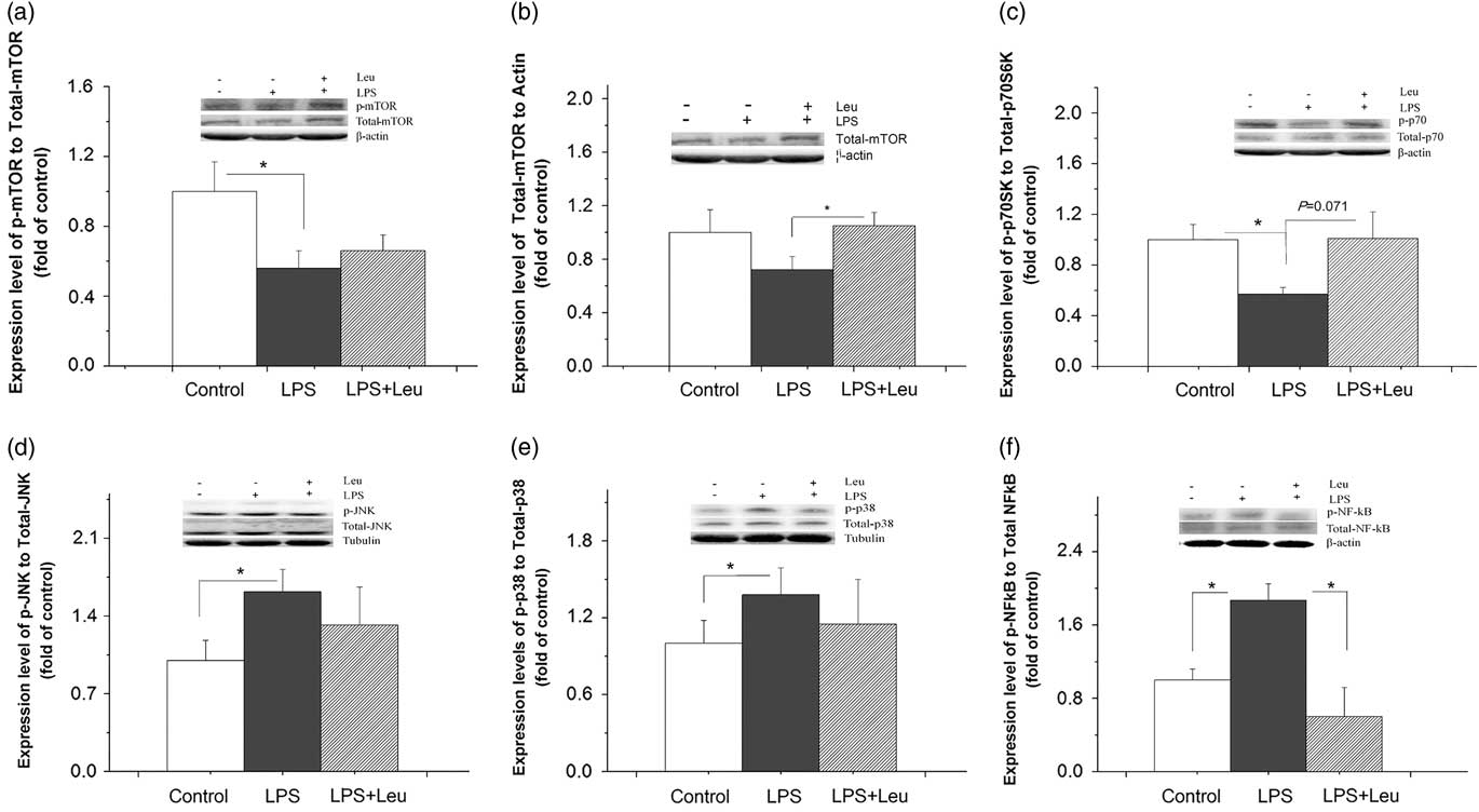
Compared with control, LPS suppressed the phosphorylation level of mTOR (P<0.05) and p70S6K (P<0.05, Figure 5a and c), but had no significant influence on total mTOR level (P>0.05, Figure 5b). Compared with LPS treatment, the total mTOR level was increased (P<0.05, Figure 5b), the phosphorylated mTOR was not changed (P>0.05, Figure 5a), and the phosphorylated p70S6K levels tended to be increased by leucine+LPS treatment (P=0.071, Figure 5c).
Effect of leucine treatment (40 mM) on the lipopolysaccharide (LPS) (2 µg/ml) induced protein phosphorylation of the mammalian target of rapamycin (mTOR) (a) and total mTOR (b), the phosphorylation of the p70 ribosomal S6 kinase (p70S6K) (c), Jun N-terminal kinase (JNK) (d), p38 (e) mitogen-activated protein kinase (MAPK) and nuclear factor-κB p65 (NF-κB p65) (f). The intestinal tissue of chicken embryos were collected after lipopolysaccharide (LPS) and 40 mM leucine treatment to measure the mTOR, p70S6K, JNK, p38 MAPK and NF-κB p65 expression. The data were subjected to one-way ANOVA analysis. The values indicate the mean±SEM (n=12). * Means are significantly different (P<0.05).

Lipopolysaccharide treatment upregulated the phosphorylation level of JNK (P<0.05, Figure 5d) and p38 MAPK (P<0.05, Figure 5e), compared with control. In contrast, there were no significant difference among the control, LPS and leucine-LPS groups (P<0.05). The phosphorylation level of NF-κB p65 was increased by LPS treatment (P<0.05) and restored to normal level in the presence of leucine (P<0.05, Figure 5f) (Supplementary Figure S2).
Discussion
In the present study, the role of the leucine in the inflammatory responses of the small intestinal tract was determined in vitro. The results showed that LPS stimulated the secretion of sIgA, the expression of TLR4, IL-6, IL-8, IL-10 and TNF-α and down-regulated the phosphorylation of mTOR, whereas upregulating the phosphorylation of NF-κB, p38 MAPK and JNK. In contrast, leucine supplementation reversed the effect of LPS on sIgA secretion, gene transcription for IL-6, and the phosphorylation of NF-κB p65. These results suggested that leucine could alleviate the LPS-induced immune stimulating effect in the intestinal tract of chicken embryos.
Nuclear factor-κB and mitogen-activated protein kinase pathways are associated with lipopolysaccharide-induced immune responses
In this study, sIgA secretion increased in a dose-dependent manner after LPS challenge. In contrast, the mRNA level of IgA was not significantly affected by LPS treatment. In previous studies, Ig secretion could be regulated post-transcriptionally (Sitia et al., Reference Sitia, Neuberger and Milstein1987; Lebman et al., Reference Lebman, Lee and Coffman1990). In LPS-stimulated B cell cultures, IL-2 resulted in a 5- to 10-fold increase in IgA secretion in the present of TGF-β, without a change in neither the proportion nor the total number of sIgA+ cells and a change in neither the ratio of secreted to membrane form of ɑ-mRNA nor the steady state level of ɑ-mRNA, suggesting that IgA secretion is controlled post-transcriptionally (Lebman et al., Reference Lebman, Lee and Coffman1990). The present result may imply that the LPS-induced elevation of sIgA concentrations is regulated at translation and/or secretion level in LPS-challenged intestinal tract.
In intestinal mucosa, TLRs are able to recognize and sense specific molecular patterns and initiate subsequent immune responses (Nemazee et al., Reference Nemazee, Gavin, Hoebe and Beutler2006). In intestinal epithelial cell lines expressing TLRS, TLR4 appears to mediate LPS stimulation of specific intracellular signal transduction pathways (Cario et al., Reference Cario, Rosenberg, Brandwein, Beck, Reinecker and Podolsky2000). By binding to TLR4 on the plasma membrane, LPS triggers the TLR4-NF-κB signaling pathway in many types of epithelial cells thereby resulting in the up- or down-regulation of gene families (Lu et al., Reference Lu, Yeh and Ohashi2008; Austenaa et al., Reference Austenaa, Carlsen, Hollung, Blomhoff and Blomhoff2009). Consistent with previous studies in mammals, the upregulated transcription of TLR4 in intestinal tissue indicates that TLR4 is involved in the LPS-induced immune response in the intestinal tissue of chicks, as well as that in mammals.
By the recognition of TLR4, LPS activates two main intracellular signaling pathways: the MAPK/NF-κB and the PI3K/Akt signaling pathway (Ci et al., Reference Ci, Ren, Xu, Li, Yu, Song, Wang, Li and Deng2010). The increased phosphorylation of NF-κB p65 indicates that LPS activates the NF-κB signaling pathway. As the abundant of nuclear NF-κB was not measured in this study, this result should be interpreted with caution. We then measured the phosphorylation levels of p38 MAPK and JNK and the results showed the activation of p38 MAPK and JNK. Collectively, these results suggest that LPS activates the MAPK/NF-κB signaling pathway through the recognition of TLR4. This result is consistent with studies in mammals where NF-κB and the MAPK signaling pathways play an important role in LPS-induced immune response (Ci et al., Reference Ci, Ren, Xu, Li, Yu, Song, Wang, Li and Deng2010).
In RAW264.7 macrophages, LPS-induced production of inflammatory cytokines by activating the MAPK/NF-κB signaling pathway (Ci et al., Reference Ci, Ren, Xu, Li, Yu, Song, Wang, Li and Deng2010). The activation of NF-κB has been shown to induce expression of TNF-α (Huang et al., Reference Huang, Xiao, Tan, Xiao, Wang, Yin, Duan, Huang, Yang and Yin2016). In the present study, the gene expression of pro-inflammatory cytokines such as TNF-α, IL-6, and IL-8 was measured as well as IL-10, which is a pleiotropic cytokine. In line with previous studies (Moraes et al., Reference Moraes, Ribeiro, Santin and Klasing2016; To et al., Reference To, Lau, Woo, Lau, Chan, Chan, Zhang, Chen and Yuen2016), the marked upregulation of gene expression for TNF-α, IL-10, IL-6, IL-8 indicated the stimulated cytokine expression in the in vitro intestinal tissue. The simultaneous activation of the MAPKs/NF-κB signaling pathway should be responsible for the LPS-induced immune response. However, this conclusion should be explained with caution as the circulating cytokines levels were not measured directly in this study.
Leucine relieve lipopolysaccharide-induced immune response via the mammalian target of rapamycin signaling pathways
Mammalian target of rapamycin/p70S6K pathway plays a central role in the regulation of cell proliferation, growth and survival (Sarbassov et al., Reference Sarbassov, Ali and Sabatini2005; Krämer et al., Reference Krämer, Wang-Rosenke, Scholl, Binder, Loof, Khadzhynov, Kawachi, Shimizu, Diekmann, Budde, Neumayer and Peters2008). Mammalian target of rapamycin also plays an important role in the mediation of innate and adaptive immunity (Liu et al., Reference Liu, Li, Song, Liu, Wang, Chang, Wu, Chen, Zhao, Zhang and Wei2012). Hence, we assumed that the mTOR/p70S6K signaling pathway is involved in the regulation of sIgA synthesis. The present results showed that LPS challenge markedly blocked the mTOR/p70S6K pathway by suppressing the phosphorylation level of mTOR and p70S6K. In vivo, block mTOR/p70S6K with rapamycin suppressed IgA content in jejunal mucosa of chicks (Liu et al., Reference Liu, Zhao, Fan, Liu, Jiao, Wang, Sun and Lin2015). Although, the pathway was differently influenced, these results imply that mTOR/p70S6K is involved in IgA synthesis. Moreover, as the phenomena occurs concomitant with the activation of the MAPKs/NF-κB pathway, this result may imply that mTOR/p70S6K is associated with LPS-induced immune responses in the intestinal tract.
In this study, we activated mTOR pathway by leucine supplementation. Although the ratio of phosphorylate mTOR to total mTOR was not significantly changed, the total mTOR level was increased by LPS+leucine treatment compared with LPS group. Furthermore, the phosphorylation level of p70S6K was increased by leucine supplementation compared with LPS group (177.2%, P=0.071), indicating that leucine supplementation may relieve the suppressive effect of LPS on mTOR/P70S6K signaling.
The augmented sIgA secretion by LPS was suppressed by leucine at a dose of 40 mM, suggesting that leucine could lessen LPS-induced sIgA secretion. Although IgA mRNA didn’t change by LPS stimulation, it was down-regulated by leucine treatment, which was consistent with the results of sIgA and some cytokines. The stimulating effect of leucine on the translation and/or the secretion of IgA remains to be elucidated in future. In accordance with the change in mTOR/p70S6K pathway, the elevated phosphorylation level of NF-κB p65 by LPS was completely restored to a normal level while the phosphorylation level of JNK (18.5%) and p38 MAPK (16.7%) was concurrently partially recovered by leucine supplementation, suggesting that leucine suppresses IgA secretion by the blockage of NF-κB/MAPK pathway. This result was in line with the work of Tan et al. (Reference Tan, Yin, Kong, Li, Li, Gao, Li, Huang and Wu2010), who reported that arginine increased phosphorylated mTOR and p70S6K-1 while reducing the phosphorylated levels of NF-κB p65 in LPS-treated IPEC-1 cells (Tan et al., Reference Tan, Yin, Kong, Li, Li, Gao, Li, Huang and Wu2010).
The increased expression of IL-6 by LPS was down-regulated by leucine supplementation, which was consistent with the change in NF-κB pathway. However, the expression of IL-10 and TNF-α were not significant changed by leucine treatment, in contrast to that in macrophages from mice. In peritoneal macrophages, inhibition of the mTOR pathway with rapamycin inhibited LPS induction of IL-10 mRNA and protein and enhanced the expression of TNF-ɑ (Baker et al., Reference Baker, Wang, Mackman and Luyendyk2009). Moreover, block of mTOR suppressed the release of IL-10 by via the transcription factor STAT3 (Weichhart et al., Reference Weichhart, Costantino, Poglitsch, Rosner, Zeyda, Stuhlmeier, Kolbe, Stulnig, Hörl, Hengstschläger, Müller and Säemann2008). In chicken, however, there is few studies focus on the regulating effect of mTOR on cytokine expression. Recently, it was reported that the early response (4 to 48 h) of chickens to Salmonella infection was pro-inflammatory, fueled by glycolysis and mTOR-mediated protein synthesis, whereas by the later phase (4 to 5 days), the local environment had undergone an immune-metabolic reprogramming to an anti-inflammatory state driven by AMP kinase (AMPK) directed oxidative phosphorylation (Kogut et al., Reference Kogut, Genovese, He and Arsenault2016). The result implies that mTOR and AMPK, respectively, play a role in the regulating metabolic supporting for anti-inflammatory cytokine production at different time phase. And the regulating effect of mTOR on IL-10 and TNF-α in intestinal tract of chicken embryos needs to be studied further.
The mTOR and NF-κB signaling pathways could cross-talk when regulating the expression of inflammatory cytokines. Mammalian target of rapamycin has a role of ‘speed breaker’ in suppressing NF-κB activation (Mendes et al., Reference Mendes, Candi, Vansteenbrugge, Pignon, Bult, Boudjeltia, Munaut and Raes2009). By stimulating NF-κB, mTOR potentiates cell survival responses (Ghosh et al., Reference Ghosh, Tergaonkar, Rothlin, Correa, Bottero, Bist, Verma and Hunter2006). In the present study, the phosphorylation level of p70S6K tended to be increased by leucine supplementation (P=0.071), suggesting that the p70S6K pathway is activated in the present experiment, in line with previous results (Sarbassov et al., Reference Sarbassov, Ali and Sabatini2005; Krämer et al., Reference Krämer, Wang-Rosenke, Scholl, Binder, Loof, Khadzhynov, Kawachi, Shimizu, Diekmann, Budde, Neumayer and Peters2008). The simultaneously suppressed NF-κB expression induced by leucine treatment implied that leucine relieved the LPS-induced immune activation via NF-κB pathway. In our in vivo study, the block of mTOR by rapamycin inhibited the secretion of IgA in intestinal tract of chickens (Liu et al., Reference Liu, Zhao, Fan, Liu, Jiao, Wang, Sun and Lin2015). Moreover, the mTOR pathway has been proved to be a pivotal negative regulator of NF-κB signaling in myeloid innate immune cells of human (Dos Santos et al., Reference Dos Santos, Delattre, De Longueville, Bult and Raes2007). In peritoneal macrophages of mice, inhibition of mTOR by rapamycin promoted production of pro-inflammatory cytokines via activation of NF-κB (Baker et al., Reference Baker, Wang, Mackman and Luyendyk2009). However, the cross-talk between mTOR/p70S6K and NF-κB remains to be elucidated in future via the silence or over-expression of mTOR/p70S6K or NF-κB. Collectively, the present results suggest that leucine supplementation could suppress the intestinal inflammation response induced by LPS challenge via NF-κB pathway. The mTOR/p70S6K pathway may involve in the regulation of intestinal immune function of chicken embryos.
Conclusion
In conclusion, LPS showed significant inflammatory action in intestinal tissue of chicken embryo as a promising model of in vitro regulation via NF-κB signaling pathway activation. And leucine could alleviate LPS-induced immune response by evoking mTOR/p70S6K signaling pathway, which may involve in regulating the NF-κB pathway with its downstream inflammation cytokine expression during LPS exposure in chicken embryos.
Acknowledgments
The authors thank Ms M. Zhao for reagent and RT-PCR technical assistance. The present study was supported by the National Key Research Program of China (2016YFD05005100), National Natural Science Foundation of China (31272467, 31472114), Taishan Scholars Program (No. 201511023), and Taian Municipal Sci-tech Development Program (2016NS1090).
Supplementary material
To view supplementary material for this article, please visit https://doi.org/10.1017/S1751731117003342