INTRODUCTION
Dorsal root ganglia (DRG) – anatomy
The DRG, also known as ‘spinal sensory ganglia’, are clusters of peripheral sensory neurons arranged in a series of segments at the intervertebral foramen (IVF) in the vertebral column. The number of ganglia in a given species corresponds to the number of segments of the spinal cord. In addition to its own thick connective tissue capsule, DRG are further covered by the spinal dura mater that extends into the IVF. However, DRG are largely considered as being located ‘outside the blood-brain barrier’.
DRG – development
Developmentally, neurons and glial cells of the DRG are derived from the neural crest cells that arise from the dorsolateral aspect of the developing neural tube and they migrate to ventrolateral aspect of the spinal cord (Pannese, Reference Pannese1981). Neurogenin transcription factors, ngn1 and ngn2, are thought to play a vital role during the early migration phase of the neural crest cells and subsequent early developmental stage of DRG (Ma et al., Reference Ma, Fode, Guillemot and Anderson1999; Perez et al., Reference Perez, Rebelo and Anderson1999). In addition, peripheral future targets of these developing neuroblasts also play key roles in differentiation and patterning of DRG. For example; (a) the molecules secreted by paraxial mesodermal mesenchymal cells are essential for segmentation of spinal cord and DRG (Kalcheim and Teillet, Reference Kalcheim and Teillet1989), (b) the inducting forces secreted by the notochord and/or differentiating neural tube itself may influence DRG development (Halpern et al., Reference Halpern, Thisse, Ho, Thisse, Riggleman and Trevarrow1995; Talbot et al., Reference Talbot, Trevarrow, Halpern, Melby, Farr and Postlethwait1995). Ungos et al. (Reference Ungos, Karlstrom and Raible2003) have demonstrated that sonic hedgehog protein is a candidate for mediating local signalling required for specification of DRG precursors. In addition to these and other unknown factors, neurotrophins and a diffusible endogenous gaseous signalling molecule, nitric oxide (NO), appear to play an important role in DRG development from embryonic stages through to adulthood (Bredt and Snyder, Reference Bredt and Snyder1994; Memberg and Hall, Reference Memberg and Hall1995; Lewin and Barde, Reference Lewin and Barde1996; Thippeswamy et al., Reference Thippeswamy, McKay, Quinn and Morris2005a).
NO signalling pathway
NO is a gaseous free radical capable of diffusing across the cell membrane. NO synthase (NOS) catalyses the formation of NO from the substrate, l-arginine. There are three well characterised NOS isoforms; neuronal NOS (nNOS) produced in neurons and in some astrocytes, inducible NOS (iNOS) produced mainly by immune cells, non-neuronal cells, and occasionally by neurons, and endothelial NOS (eNOS) by the endothelium lining the blood vessels (reviewed by Garthwaite, Reference Garthwaite2008). NO binds to soluble guanylyl cyclase (sGC), often considered as the receptor for NO, to generate cyclic guanosine monophosphate (cGMP) from guanosine triphosphate (GTP), the downstream of which activates protein kinase G and/or nucleotide-gated ion channels (Roy et al., Reference Roy, Halvey and Garthwaite2008). However, NO can also directly influence cell function via S-nitrosylation of cellular proteins (Nott et al., Reference Nott, Watson, Robinson, Crepaldi and Riccio2008). Selective inhibition of nNOS or iNOS versus eNOS may provide useful tools for investigating biological functions of NO and a novel therapeutic approach to various diseases (Boer et al., Reference Boer, Ulrich, Klein, Mirau, Haas and Baur2000).
OBJECTIVE
We have recently found that non-neuronal cells are a potential source of NO in addition to neurons in the DRG. They produce iNOS following peripheral injury or when cultured, and the number of phagocytic cells also increases in axotomised DRG. In this paper, a brief summary of these findings and our past work and the findings from others on NO-mediated neuron-satellite glia signalling in axotomised DRG is presented and discussed.
NO-nerve growth factor (NGF) INTERACTION IN DRG
During early embryonic development (embryonic day 12, E12) of mouse or rat almost all of the DRG neurons produce nNOS (Bredt and Snyder, Reference Bredt and Snyder1994; Thippeswamy et al., Reference Thippeswamy, McKay, Quinn and Morris2005). Review of the literature and our experiments suggests that neurotrophins and their receptors, both within the DRG and their peripheral targets, play major roles in the normal function of the nervous system. There is considerable evidence for their roles in development and retrograde neurotrophic signal transduction mechanism (Schäfers et al., Reference Schäfers, Geis, Brors, Yaksh and Sommer2002). As development progresses, the number of neurons that contain nNOS decreases gradually to <5% in adult rat DRG (Thippeswamy et al., Reference Thippeswamy, McKay, Morris, Quinn, Wong and Murphy2005b).
During the early 1990s, it became known that peripheral nerve injury increases expression of nNOS mRNA (Verge et al., Reference Verge, Xu, Xu, Wiesenfeld and Hokfelt1992) and protein (Zhang et al., Reference Zhang, Verge, Wiesenfeld, Ju, Bredt and Synder1993) in small-to-medium sized DRG neurons, which were thought to be peptidergic neurons expressing tyrosine kinase A (trkA) receptor for NGF. This led to a hypothesis that a peripheral source of NGF may suppress nNOS expression in a subset of DRG neurons. Using dissociated DRG cultures, we demonstrated that NGF-deprivation increases the number of nNOS producing neurons (Thippeswamy and Morris, Reference Thippeswamy and Morris1997a). This suggests that depriving adult DRG neurons of NGF either by axotomy or by neutralising endogenous sources of NGF, produced from satellite glia cells, with NGF antibodies in culture induces stress in DRG neurons. Neurons stressed this way increase NO production by upregulating nNOS synthesis (Thippeswamy and Morris, Reference Thippeswamy and Morris1997a; Thippeswamy et al., Reference Thippeswamy, Jain, Mumtaz and Morris2001b) due to increased intracellular calcium levels (Tong et al., Reference Tong, Eichler and Rich1996). This lead to a further hypothesis that increased NO in stressed DRG neurons could be an adaptive response. We subsequently tested the effect of inhibiting NO production on morphology of these neurons in vivo and in vitro. Postnatal DRG neurons do not require NGF for survival in culture or in vivo unlike embryonic or neonatal DRG neurons (Luizzi and Tedeschi, Reference Liuzzi and Tedeschi1991; Tong et al., Reference Tong, Eichler and Rich1996; Thippeswamy et al., Reference Thippeswamy, McKay, Quinn and Morris2005a; Ernsberger, Reference Ernsberger2009). However, as mentioned above, NGF-deprivation of postnatal DRG neurons enhances NO production as revealed by nitrite assay (Thippeswamy et al., Reference Thippeswamy, McKay, Quinn and Morris2005a), increased nNOS+ neurons (Thippeswamy et al., Reference Thippeswamy, Jain, Mumtaz and Morris2001a) and increased cGMP expression in the satellite glia cells (Thippeswamy et al., Reference Thippeswamy, Jain, Mumtaz and Morris2001b, Reference Thippeswamy, Haddley, Corness, Howard, McKay and Beaucourt2007a). cGMP is produced in response to NO reaction with sGC (Koesling et al., Reference Koesling, Russwurm, Mergia, Mullershausen and Friebe2004; Russwurm and Koesling, Reference Russwurm and Koesling2004). NOS blockers treatment suppresses nitrite levels and cGMP synthesis (Thippeswamy et al., Reference Thippeswamy, Jain, Mumtaz and Morris2001a, Reference Thippeswamy, Haddley, Corness, Howard, McKay and Beaucourt2007a) suggesting that the increased nNOS expression is indeed mediating NO and cGMP production. We have also confirmed these observations using dominant negative vectors targeting nNOS or sGC in vivo and in vitro to intervene in the NO–cGMP pathway (Thippeswamy et al., Reference Thippeswamy, McKay, Morris, Quinn, Wong and Murphy2005b, Reference Thippeswamy, Haddley, Corness, Howard, McKay and Beaucourt2007a).
ENDOGENOUS NO IS REQUIRED FOR SURVIVAL OF AXOTOMISED/NGF-DEPRIVED DRG NEURONS
We have demonstrated that blocking the NO–cGMP pathway in NGF-deprived DRG cultures causes apoptotic death via bax and caspase 3 (Thippeswamy et al., Reference Thippeswamy, Jain, Mumtaz and Morris2001b) and, in vivo, abnormal neuronal morphology resembling apoptosis was observed. Further detailed analysis revealed that both nNOS+ neurons and neurons that do not contain nNOS are affected following NO-cGMP intervention. DRG neurons in vivo do not express cGMP whereas their satellite glia cells do. However, NGF-deprived cultured DRG neurons do express cGMP and blockade of sGC causes neuronal death (Thippeswamy and Morris, Reference Thippeswamy and Morris1997b, Reference Thippeswamy and Morris2001b). These studies lead to a further hypothesis that endogenous NO produced from stressed neurons signals for support from the surrounding glial cells. To test this hypothesis, Schwann cells were isolated and grown in vitro and were stimulated with NO donor, NONOate derivative dimethyl-triamino-benzidine NO (DETA/NO). Such NO-stimulated glial cells produce neurotrophic factors, NGF and neurotrophin3. In order to test whether this supports stressed neurons, NGF-dependent neonatal DRG neurons were co-cultured with NO-stimulated glial cells (Thippeswamy et al., Reference Thippeswamy, McKay, Morris, Quinn, Wong and Murphy2005b). Neonatal DRG neurons are dependent on NGF for survival and when they were cultured without NGF ~80% neurons died within 48 h (Thippeswamy et al., Reference Thippeswamy, McKay, Quinn and Morris2005a). However, these neurons survived when co-cultured with NO stimulated satellite glia (without NGF added to the culture medium), thus confirming glial neurotrophic support in response to the NO signal. To test whether this mechanism is mediated by cGMP, satellite glia cells were pre-treated with sGC blocker, 1H-[1, 2, 4] oxadiazolo [4, 3-a] quinoxalin-1-one or a dominant negative vector for sGC before being treated cells with DETA/NO. When neonatal DRG neurons were co-cultured with these glial cells, neurons did not survive. TUNEL staining and gel electrophoresis of DNA extracted from these cultures showed typical ladder formation suggesting that the NO–cGMP–neurotrophins pathway protects neurons (Thippeswamy et al., Reference Thippeswamy, McKay, Morris, Quinn, Wong and Murphy2005b) in a paracrine fashion. Subsequently, we further demonstrated that increased nNOS synthesis in axotomised DRG neurons induces another lately identified neuroprotective molecule, activity-dependent neuroprotective protein in satellite glia cells (Thippeswamy et al., Reference Thippeswamy, Howard, Cosgrave, Arora, McKay and Quinn2007b). Overall, these experiments support our hypothesis that NO produced from stressed neurons signals the satellite glia to produce neurotrophic factors.
Since NOS or sGC inhibition causes death of both nNOS+ and nNOS− neurons, we hypothesised that the NO–cGMP pathway could also function in an autocrine fashion in neurons. Based on the published literature we knew that production of neuropeptides such as galanin increases in axotomised DRG neurons (Zhang et al., Reference Zhang, Aman and Hökfelt1995; Elliott-Hunt et al., Reference Elliott-Hunt, Marsh, Bacon, Pope, Vanderplank and Wynick2004; Pirondi et al., Reference Pirondi, Fernandez, Schmidt, Hokfelt, Giardino and Calza2005) and that galanin too functions as a neuroprotective peptide (Bacon et al., Reference Bacon, Kerr, Holmes, Gaston and Wynick2007; Thippeswamy et al., Reference Thippeswamy, Haddley, Corness, Howard, McKay and Beaucourt2007a). Incidentally, a significant number of trkA containing nNOS+ DRG neurons also upregulates galanin following axotomy, and nNOS inhibition selectively suppresses galanin in some of these neurons. This suggests that the vast majority of NGF-responsive nNOS neurons are peptidergic. Blocking endogenous NO in NGF-deprived DRG cultures suppressed galanin promoter activity (Thippeswamy et al., Reference Thippeswamy, Haddley, Corness, Howard, McKay and Beaucourt2007a). Consistent with this, axotomised/NGF-deprived DRG neurons expressed high levels of nNOS and galanin. Further, using NOS blockers, or adenoviral vectors expressing dominant-negative either for nNOS or sGC in vivo and in vitro, we have shown that the NO–cGMP pathway induces endogenous galanin in DRG neurons. It is also proposed that satellite glia cells too contain galanin (Hu and McLachlan, Reference Hu and McLachlan2003). However, whether it is synthesised by glia or taken up following local release by neurons is not clear. These results demonstrate that the NO–cGMP pathway or NO directly regulate galanin in stressed DRG neurons as a protective mechanism in an autocrine fashion.
ADULT NEUROGENESIS IN DRG?
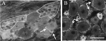
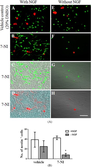
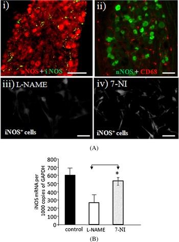
Neurons are post-mitotic, and there is very little evidence for adult neurogenesis in the DRG. Clusters of proliferating and reactive satellite glia cells were found in the interstitial spaces of neurons in the axotomised DRG and, incidentally, these cells were also nNOS+ (Fig. 1). Since all early differentiated rat DRG neurons during embryonic development (E12) contain nNOS, we speculated that these proliferating cells in the adult DRG are potential stem cells and could differentiate into neurons if an adaptive response of neurons to axotomy fails. Using nestin and bromodeoxyuridine (BrdU) immunostaining combined with neuronal or Schwann cell marker, we confirmed that these proliferating cells in culture are indeed stem cells that could differentiate into neurons or Schwann cells (Arora et al., Reference Arora, Cosgrave, Howard, Bubb, Quinn and Thippeswamy2007). Using cultured satellite glia cells in the presence of acidic fibroblast growth factor and BrdU incorporation, we have shown that NO and neuronal restrictive silencer transcription factor (NRSF, also termed REST), a master regulator of neuronal genes in non-neuronal cells (Palm et al., Reference Palm, Belluardo, Metsis and Timmusk1998; Ballas et al., Reference Ballas, Grunseich, Lu, Speh and Mandel2005), regulate neurogenesis. Increased levels of NRSF/REST suppress neurogenesis whereas lower levels have the opposite effect. With respect to NO, it has a dual effect on neurogenesis that depends on the source of NO and the stage of neuronal differentiation (Gouge et al., Reference Gouge, Marshburn, Gordon, Nunley and Huet-Hudson1998; Moreno-Lopez et al., Reference Moreno-Lopez, Noval, González-Bonet and Estrada2000; Romero-Grimaldi et al., Reference Romero-Grimaldi, Moreno-López and Estrada2008). For example, nNOS inhibition promotes differentiation and migration whereas iNOS-mediated NO production could be toxic to newly differentiated immature neurons (Packer et al., Reference Packer, Stasiv, Benraiss, Chmielnicki, Grinberg and Westphal2003; Gutièrrez-Mecinas et al., Reference Gutièrrez-Mecinas, Crespo, Blasco-Ibáñez, Nácher, Varea and Martínez-Guijarro2007). NO donor, DETA/NO treatment of satellite glia cell cultures results in a significant increase in NRSF/REST mRNA expression, whereas L-NAME suppressed its mRNA below the control basal level (Arora et al., Reference Arora, Cosgrave, Howard, Bubb, Quinn and Thippeswamy2007). Axotomy or NGF-deprivation of DRG neurons-satellite glia co-culture increases NO production by neurons and treating them with the pan NOS inhibitor L-NAME in vitro or nNOS-specific blocker 7-nitroindazole (7NI) in vivo, causes a significant increase in nestin+ cell numbers (Arora et al., Reference Arora, Cosgrave, Howard, Bubb, Quinn and Thippeswamy2007). This may suggest that NOS inhibition in general promotes neurogenesis by suppressing REST/NRSF in neuronal stem cells such as nestin+ cells. However, surprisingly, blocking neuronal source of NO alone by 7NI in satellite glia–neuron co-culture decreased the number of nestin+ cells compared with control cultures (Fig. 2). This might be due to selective loss of a population of nestin+ cells, especially those with extensive processes resembling early differentiated neurons, through iNOS-mediated NO toxicity. QPCR analysis of these cultures for iNOS mRNA, and immunostaining for its protein (Fig. 3) revealed a significant increase in these cultures (7NI-treated) compared with L-NAME treated suggesting a possible source of iNOS. Overall these results suggest that NO produced from different isoforms of NOS may have different effects on nestin+ cell numbers and postnatal neurogenesis in DRG.
Sections of axotomised DRG immunostained for nestin (A) and nNOS (B). Clusters of nestin+ cells are marked with dotted lines in A and isolated nestin+ cells are indicated by arrows. nNOS immunostaining revealed that the clusters of proliferating satellite glia cells produce nNOS (arrows in B). Cells indicated with stars are nNOS+ neurons. Scale bar: 100 µm.

7NI decreases the number of nestin+ cells in NGF-deprived sateliite glia-DRG neuron co-cultures. (A) Photomicrographs of cultures showing double immunostaining for nestin (green in all) and a neuronal marker, PGP 9.5 (red in all). Cultures were treated with vehicle control (20% DMSO) or 7NI in the presence of NGF (A–D) and without NGF (E–H). Bright field image of cultures merged with images taken from different channels (C,G,D,H). Scale bars: all 100 μm. (B) Quantification of nestin+ cells in DRG cultures treated with 7NI and vehicle control (20% DMSO). In cultures treated with 7NI (with NGF), there was no significant difference in number of nestin+ cells when compared to vehicle control. However, there was a significant decrease in the number of nestin+ cells in cultures without NGF when treated with 7NI compared to vehicle control (*p < 0.01).

NO source in axotomised DRG and in DRG neuron-satellite glia co-culture treated with L-NAME or 7NI. (A) In addition to neuronal source of NO by nNOS (red cells in i), green cells in ii)) there were numerous iNOS+ and CD68+ (macrophage marker) cells in axotomised DRG. This may explain why 7NI and TRIM, both specific inhibitors of nNOS, have different effects compared to the general NOS blocker L-NAME on nNOS immunoreactive neuron numbers in axotomised/NGF-deprived DRG. (iii) and (iv) represent cultures immunostained for iNOS. There were more numbers of iNOS+ cells in 7NI (iv) than in L-NAME-treated culture (iii) which seems to correlate with iNOS mRNA transcripts analysed by qPCR (B) from parallel cultures (*p < 0.05). Scale bar: 100 μm.

INDUCIBLE NOS IN DRG – DIFFERENTIAL EFFECTS OF nNOS-SPECIFIC BLOCKERS VERSUS GENERAL NOS BLOCKER
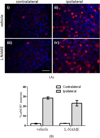
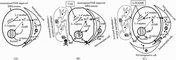
Axotomised DRG showed numerous iNOS+ non-neuronal cells and macrophages (CD68+ cells), also a potential source of iNOS, in the interstitial space of neurons (Fig. 3). This implies that in addition to neuronal NO, non-neuronal NO may also contribute to any axotomy-induced adaptive response. However, it has been shown that excessive amounts of NO produced from the iNOS-mediated mechanism is cytotoxic (Tran et al., Reference Tran, Hardin-Pouzet, Verge and Owens1997). In the absence of endogenous neuronal NO, the compensatory mechanism for cell survival may be compromised due to a shift towards the iNOS-mediated NO production which is toxic. We have previously shown that nNOS block in vivo either with nNOS inhibitor, 1-(2-trifluoromethylphenyl) imidazole (TRIM) (Thippeswamy et al., Reference Thippeswamy, Jain, Mumtaz and Morris2001) or with 7NI causes death of a subpopulation of DRG neurons (Thippeswamy et al., Reference Thippeswamy, Haddley, Corness, Howard, McKay and Beaucourt2007a). Therefore, it is plausible that the pan NOS inhibitor, L-NAME, treatment of axotomised rats did not induce degenerative changes in DRG neurons (Fig. 4A). Indeed, there was no statistically significant difference in nNOS+ neuron numbers (Fig. 4B) as opposed to nNOS-specific inhibitors (Thippeswamy et al., Reference Thippeswamy, Jain, Mumtaz and Morris2001, Reference Thippeswamy, Haddley, Corness, Howard, McKay and Beaucourt2007a). In this scenario, it seems that L-NAME effectively blocks all sources of NO production, including cytotoxic iNOS. However, in NGF-deprived dissociated DRG cultures, both L-NAME and nNOS blockers caused a significant reduction in nNOS+ neurons (Thippeswamy and Morris, Reference Thippeswamy and Morris1997b). This different effect of L-NAME on DRG neurons in culture and in vivo needs further investigation. This discrepancy could be due to: (i) selective loss of a subpopulation of DRG neurons in culture that are dependent on specific trophic factors, and these factors may be at suboptimum levels or absent in the culture medium; (ii) culture medium may dilute neuropeptides/trophic factors produced by glia cells that are not responsive to NO (for example, glia cells that do not produce sGC) as a compensatory mechanism (Fig. 5C); (iii) the proximity of NO-sensitive and NO-insensitive satellite glia cells to nNOS+ neurons in culture was not taken into account in those studies (Thippeswamy and Morris, Reference Thippeswamy and Morris1997b); (iv) the effect of NOS inhibition-induced neurogenesis (Packer et al., Reference Packer, Stasiv, Benraiss, Chmielnicki, Grinberg and Westphal2003), in vivo, to replace dead neurons in axotomised DRG; and (v) L-NAME dose used in vivo (50 mg/kg) may not be optimum to cause similar effects as seen in cultures. However, a higher dose of L-NAME (>50 mg) tested in vivo caused respiratory and cardiac distress and increased mortality in rats. Overall, these observations suggest that different sources of NO have different effects on axotomised DRG cells in culture and in vivo.
L-NAME treatment of axotomised rats did not significantly affect DRG neuron morphology (A) or neuron number (B) ipsilateral to nerve injury. Red labelled cells are nNOS+ neurons and nuclei are labelled blue (DAPI). As expected the number of nNOS positive neurons increased following nerve injury, however, unlike nNOS blockers, TRIM (Thippeswamy et al., Reference Thippeswamy, Jain, Mumtaz and Morris2001) and 7NI (Thippeswamy et al., Reference Thippeswamy, Howard, Cosgrave, Arora, McKay and Quinn2007b), L-NAME did not affect neuronal morphology and number. Scale bar: 100 µm. ANOVA result showed no significant difference between treatments (ipsi vehicle versus ipsi L-NAME p = 0.181).

Schematic diagrams depicting the effects of axotomy/NGF-deprivation and NOS inhibition on DRG neurons. (A) Axotomy/NGF deprivation increases neuronal NO production, and cGMP in both neurons and satellite glia cells that contain NO-responsive sGC. The downstream of the NO–cGMP pathway results in the production of neurotrophic factors by glia and appropriate receptors by neurons (Thippeswamy et al., Reference Thippeswamy, McKay, Morris, Quinn, Wong and Murphy2005b). Axotomy causes macrophage migration and also induces activation of fibroblasts in the DRG (not depicted in A). (B) Neuronal NOS inhibitor (7NI) blocks nNOS-mediated NO production, thus further depriving DRG neurons of tropic support from NO-responsive glia cells. Axotomy induces iNOS in activated fibroblasts and invading macrophages in the DRG. NO produced in excess from these sources causes neuronal death in DRG and also decreases the number of nestin+ cells as shown in Fig. 2. (C) L-NAME, the general NOS inhibitor, prevents NO production from both iNOS- and nNOS-mediated mechanism. Therefore, following NOS inhibition by L-NAME, it is plausible that an NO-independent compensatory mechanism may protect neurons via VIP or galanin or activity-dependent neuroprotective protein (Thippeswamy et al., Reference Thippeswamy, Haddley, Corness, Howard, McKay and Beaucourt2007a, Reference Thippeswamy, Howard, Cosgrave, Arora, McKay and Quinnb) from satellite glia cells that are not NO-responsive (depicted by an upward arrow and a star in (C)).

SUMMARY
In conclusion, the NO produced from different cell types via different isoforms of NOS has specific effects on neuronal survival in axotomised DRG. In particular, blocking the endogenous neuronal source of NO triggers neurodegeneration through iNOS-mediated NO. This may also suggest that the neuronal NO signals satellite glia in axotomised DRG to neutralise cytotoxic effect of iNOS by inducing neurotrophic factors in the glial cells.
ACKNOWLEDGEMENTS
M. Bradman is funded by BBSRC (DTA) CASE studentship supported by Pfizer, UK. The Pain Relief Foundation also contributed to our costs of consumables. We would also like to thank Dr. P.C. Emson for providing the nNOS antibody used here.
Statement of interest
None.