




No CrossRef data available.
Article contents
Identification and characterization of cross-reactive antigens from Neospora caninum and Toxoplasma gondii
Published online by Cambridge University Press: 22 December 2004
Abstract
Murine monoclonal antibodies (mAbs) against Neospora caninum tachyzoites were produced to identify the cross-reactive antigens between N. caninum and Toxoplasma gondii. Ten mAbs recognizing cross-reactive antigens of both parasites were obtained and tentatively classified into 6 different groups based on their reactivity patterns in an indirect fluorescent antibody test and Western blot analysis. Three mAbs in group 1 recognized antigens located on the surface of parasites with molecular masses ranging from 28 to 76 kDa; one mAb in group 2 recognized antigens located on interior organelles of parasites with a molecular mass of 50 kDa; one mAb in group 3 recognized antigens located on interior organelles of parasites with molecular masses of 35 kDa and 14 kDa; three mAbs in group 4 recognized antigens located on interior organelles with a molecular mass of 64 kDa; one mAb in group 5 recognized antigens located on the surface of parasites with an unknown molecular mass; one mAb in group 6 recognized antigens located on the apical end of parasites with an unknown molecular mass. The mAbs in groups 1, 2, 3, and 5 showed inhibitory effects on the growth of the two parasites in vitro in a dose-dependent manner. A cDNA expression library prepared from N. caninum tachyzoite mRNA was immunoscreened with the mAb panel. Three kinds of proteins, protein disulfide isomerase (PDI), heat-shock protein 70 (HSP70), and ribosomal protein 1 (RP1), were identified as cross-reactive antigens recognized by mAbs in groups 2, 3, and 4, respectively. Some of the proteins could be useful in developing vaccines or drugs for controlling the diseases caused by the two parasites.
Keywords
Information
- Type
- Research Article
- Information
- Copyright
- © 2005 Cambridge University Press
INTRODUCTION
Neospora caninum and Toxoplasma gondii, two closely related apicomplexan parasites, can infect many warm-blooded vertebrates (Dubey et al. 1998, 1999). T. gondii has long been recognized as an important pathogen in humans and animals. However, compared with T. gondii, N. caninum was recently regarded as a major cause of bovine neonatal mortality and abortion (Dubey & Lindsay, 1996). Because these two organisms have nearly identical morphology and can cause similar pathology and disease, N. caninum has often been incorrectly identified as T. gondii (Dubey et al. 1999). Both neosporosis and toxoplasmosis caused by N. caninum and T. gondii, respectively, have been recognized as economically important diseases with considerable impact on the livestock industry.
Since N. caninum was identified as an aetiological agent of neosporosis and was found to be close to T. gondii, a great deal of research has been conducted to determine the differences and the nature of the relationship between N. caninum and T. gondii (Lindsay, Speer & Toivio-Kinnucan, 1993; Baszler et al. 1996; Howe et al. 1998; Howe & Sibley, 1999). For a better understanding of the relationship between the two parasites and for the development of future control measures, detailed information about cross-reactive antigens for the infection process in N. caninum and T. gondii is necessary. In addition, the characterizations of the common antigens of N. caninum and T. gondii are important pre-requisites for detailed analyses of the common mechanisms of parasite adhesion to, and invasion of, host cells.
The objectives of the present study were to identify and characterize cross-reactive antigens, which can be responsible for important functions during parasite-host interactions, by using monoclonal antibodies. Several mAbs recognizing both N. caninum and T. gondii tachyzoites were produced, and 3 kinds of proteins, protein disulfide isomerase (PDI), heat-shock protein 70 (HSP70), and ribosomal protein 1 (RP1), were identified as cross-reactive antigens by using the mAb panel.
MATERIALS AND METHODS
Culture and purification of parasites
The NC-1 strain of N. caninum and the RH strain of T. gondii were cultured in Vero cell monolayers in minimum essential medium (MEM) supplemented with 8% (v/v) fetal bovine serum (FBS) and 50 μg/ml kanamycin at 37 °C in a 5% CO2 air environment. For the purification of tachyzoites, the parasites were scraped from the flask and then passed through a 27G needle and a 5·0 μm filter (Millipore, USA). They were then washed in phosphate-buffered saline (PBS). The pellet was stored at −30 °C until use.
Production and purification of monoclonal antibodies
N. caninum tachyzoite lysate was used as an antigen. The primary immunization of BALB/c mice (8 weeks old) consisted of an intraperitoneal injection of the antigen (1×10⁸ tachyzoites/mouse) with Freund's complete adjuvant. Successive immunizations were done intraperitoneally twice with the same antigen with Freund's incomplete adjuvant. The mouse with the highest antibody titre against N. caninum tachyzoites, as indicated by an indirect fluorescent antibody test (IFAT), was selected for hybridoma production. Mouse spleen cells were fused with SP2 myeloma cells, and hybridomas were cultured in a GIP medium (Wako, Tokyo, Japan) with hypoxantine, aminopterin, and thymidine (HAT) (Sigma, MO, USA) in 96-well plates. Two to three weeks after fusion, screening for hybridoma-producing antibodies was performed by IFAT and Western blot analysis. Hybridoma-producing mAbs against N. caninum and T. gondii tachyzoites were identified and cloned 3 times by limiting dilution. Ascitic fluid was produced in incomplete Freund's adjuvant-primed BALB/c mice. Purification of the mAbs was performed by 50% (w/v) ammonium sulphate precipitation. The isotype of the mAbs was determined using a mouse mAb isotyping kit (Amersham Biosciences, NJ, USA).
Indirect fluorescent antibody test
Both purified tachyzoites and parasites grown in Vero cells were analysed by IFAT. For purified tachyzoites, N. caninum and T. gondii tachyzoites (5×10⁴) were mounted on slides, dried and fixed with acetone before use. For parasites in Vero cells, monolayers of Vero cells grown on 14-well glass slides were infected with freshly purified N. caninum and T. gondii tachyzoites (800 cells/well), respectively. After 2–3 days incubation at 37 °C with 5% CO2, infected cells were fixed with 3% paraformaldehyde in PBS for 30 min and were then permeabilized with 0·3% Triton X-100 in PBS for 30 min. The slides were subsequently incubated in blocking solution (PBS containing 1% BSA and 50 mM glycine) for 30 min. Both slides with purified parasites and parasites grown in Vero cells were incubated with hybridoma-cultured medium, followed by incubation with a goat anti-mouse immunoglobulins (IgG+IgA+IgM) Alexa-488 antibody (Molecular Probes, OR, USA) diluted 1[ratio ]500 in blocking solution. The location of the antigens on parasites reacted with mAbs was observed with fluorescent microscopy and confocal laser microscopy.
SDS-PAGE and Western blot analysis
Purified N. caninum and T. gondii tachyzoites were suspended in PBS and mixed with sample buffer (62·5 mM Tris-HCl, pH 6·8, 2% SDS, 5% β-mercaptoethanol, 10% glycerol, 0·02% bromophenol blue) under reducing conditions. The samples were run on 15% polyacrylamide gels. After SDS-PAGE, the proteins in the gel were electrically transferred onto a membrane. The membrane was blocked with PBS containing 3% skim milk and then incubated with a hybridoma-cultured medium at 37 °C for 60 min. Then, the membrane was washed 3 times with PBS and then incubated with peroxidase-conjugated goat anti-mouse IgG, IgA, and IgM (Southern Biotechnology, AL, USA) diluted 1[ratio ]500 in PBS containing 3% skim milk. After washing with PBS 3 times, the bands on the membrane were visualized by incubation with 3′3-diaminobenzidine.
In vitro assay for the inhibitory effects of mAbs on the growth of the parasites
Tachyzoites (1×10⁴) of N. caninum and T. gondii expressing green fluorescent protein (GFP) (Nishikawa et al. 2003; Kaneko et al. 2004; Xuan et al., manuscript in preparation) were incubated with purified mAbs at different concentrations of 0·5, 0·1, 0·05, and 0·01 mg/ml at 37 °C for 1 h. Isotype-matched mouse mAbs (IgG1, IgG2a, IgG2b, or IgM) against Babesia caballi (Ikadai et al. 1999) were used as a control. After incubation, the parasites were added to confluent monolayers of Vero cells grown in 24-well plates. After incubation at 37 °C for a further 1 h, infected Vero cells were covered with 1% low-melting agarose gel diluted in MEM supplemented with 1% FBS. After 48 h, the number of plaques of each well was counted with fluorescent microscopy. The percentage inhibition was calculated by the following formula: [1−the number of parasites' plaques in tested mAb treatment wells/the number of parasites' plaques in control wells]×100%. Each mAb was tested in triplicate in each inhibitory assay, and the assays were repeated twice.
Construction and immunoscreening of the N. caninum cDNA library and cDNA sequencing
Total RNA was prepared from N. caninum tachyzoites (1×10⁹) by use of the acid guanidinium thiocyanate-phenol-chloroform extraction method (Chomczynski & Sacchi, 1987), and then poly(A)+ RNA was purified with Oligotex-dT 30 (Takara, Tokyo, Japan). The cDNA was synthesized by using a Zap-cDNA synthesis kit, ligated to an Uni-ZAP XR vector, and packaged by using a Gigapack III packaging system according to the manufacturer's instructions (Stratagene, CA, USA). The N. caninum cDNA library (107 PFU) was screened with hybridoma-cultured medium, and the in vivo excision of the cDNA inserts from lambda phage to plasmid pBluescriptSK(+) was done according to the manufacturer's instructions (Stratagene). Both strands of the cDNA were sequenced by using the ABI Prism Dye Terminator Cycle Sequencing Ready Reaction Kit, and then analysed with an ABI3700 DNA sequencer (Applied Biosystems, CA, USA). Sequence data were analysed by computer programme (MacVector, Oxford Molecular, CA, USA).
Nucleotide sequence Accession numbers
Nucleotide sequence data reported in this article are available in the GenBank, EMBL and DDBJ databases under the Accession numbers AB178220, AB178286, and AB178287.
RESULTS
Production of mAbs recognizing both N. caninum and T. gondii tachyzoites
A total of 384 wells from 5 microplates containing hybridomas were subjected to screening of mAbs recognizing cross-reactive antigens by IFAT with purified tachyzoites. Culture media from all 384 wells were IFAT-positive for N. caninum tachyzoites. Of 384 wells, 10 wells (10/384, 2·6%) were IFAT-positive for T. gondii tachyzoite. After cloning of hybridoma cells by limiting dilution, 10 mAbs recognizing cross-reactive antigens between the two parasites were obtained and subjected to further analyses. These 10 mAbs were tentatively classified into 6 different groups based on the results of IFAT and Western blot analyses (see below), and their properties are summarized in Table 1.

Localization of antigens on parasites recognized by mAbs
The localization of antigens on N. caninum and T. gondii tachyzoites recognized by mAbs was analysed by IFAT with both purified tachyzoites and parasites grown in Vero cells (Table 1 and Fig. 1). The mAbs in groups 1 and 5 reacted with the surface of both parasites (Fig. 1A and E). Group 2 and 3 mAb-coated tachyzoites of both parasites exhibited clearly scattered fluorescent dots inside the parasites, and most of the fluorescent dots were around the surface of the tachyzoites (Fig. 1B and C). The fluorescence exhibited by the tachyzoites that had been reacted with mAbs in group 4 was different from the reactive patterns observed in group 2 and group 3 mAbs: it seemed to be suffused inside the tachyzoites (Fig. 1D). One mAb in group 6 reacted with the apical end of the two parasites (Fig. 1F).

Fig. 1. Localization of the cross-reactive antigens on Neospora caninum and Toxoplasma gondii tachyzoites recognized with mAbs by IFAT with both purified tachyzoites (left) and parasites grown in Vero cells (right). Tachyzoites reacted with 9D12 in group 1 (A), 9E8 in group 2 (B), 10F7 in group 3 (C), 10A10 in group 4 (D), 10G6 in group 5 (E), 10F10 in group 6 (F), respectively.
Western blot analysis of antigens recognized by mAbs
Western blot analysis was performed to identify the antigens recognized by mAbs. As shown in Fig. 2 and Table 1, mAbs in group 1 reacted with numerous major proteins with molecular masses ranging from 28 to 76 kDa in the two parasites; mAbs in group 2 recognized a 50 kDa protein; mAbs in group 3 recognized 35 kDa and 14 kDa proteins; mAbs in group 4 recognized a 64 kDa protein. The proteins that had been reacted with mAbs in IFAT in groups 5 and 6 were not detectable by Western blot analysis in both reducing (Fig. 2) and non-reducing (data not shown) conditions.

Fig. 2. Western blot analysis of the cross-reactive antigens of Neospora caninum (A) and Toxoplasma gondii (B) recognized with mAbs. Purified tachyzoite lysates were run on SDS-PAGE gels, transferred into nitrocellulose membranes and probed with 9D12 in group 1 (lane 1), 9E8 in group 2 (lane 2), 10F7 in group 3 (lane 3), 11A10 in group 4 (lane 4), 10G6 in group 5 (lane 5), and 10F10 in group 6 (lane 6), respectively.
Inhibitory effects on growth of N. caninum and T. gondii tachyzoites
In order to detect whether the mAbs affect the growth of the parasites by inhibiting the invasion or proliferation of parasites in vitro, the parasites were pre-treated with mAbs and then infected to Vero cells. The number of parasites that proliferated in Vero cells was calculated to analyse the inhibitory effects. As shown in Table 2, mAbs in groups 1, 2, 3, and 5 had more than a 90% inhibitory effect on the growth of the two parasites at a 0·5 mg/ml concentration compared with the control mAbs. The different lower concentration of mAbs from 0·01 to 0·1 mg/ml was used in the inhibition assay to examine if the inhibition effects were dependent on the concentration of the mAbs. The results showed that the inhibition rate decreased corresponding to the reduction in the concentration of mAbs.

Identification of genes encoding cross-reactive antigens
A cDNA expression library prepared from the mRNA of N. caninum tachyzoites was immunoscreened with mAbs recognizing cross-reactive antigens between N. caninum and T. gondii. In total, 32 positive clones were obtained by immunoscreening of the cDNA library with mAbs in groups 2 (15 clones), 3 (7 clones), and 4 (10 clones). The clones with the longest cDNA in each group were chosen for sequencing. The amino acid sequences deduced from the nucleotide sequences show high homology with the PDI of T. gondii (GenBank Accession number AB178220), the RP1 of Plasmodium falciparum (GenBank Accession number AB178286), and the HSP70 of T. gondii (GenBank Accession number AB178287) (Table 3).

DISCUSSION
Several reports have shown that there are serological cross-reactions and protective cross-reactions between N. caninum and T. gondii (Kasper & Khan, 1998; Lindsay et al. 1998; Nishikawa et al. 2002). These works suggested that the two parasites might possess cross-reactive antigens important for host immunity against the two parasites. However, only a few cross-reactive antigens have been reported to date: a 42 kDa protease of T. gondii, which is a rhoptry protein, has been confirmed to show immunological cross-reactivity with N. caninum (Ahn et al. 2001); the nucleoside triphosphate hydrolase (NTPase) of N. caninum, which is a dense granule protein, has been described as showing antigenic cross-reactivity with the NTPases of T. gondii (Asai et al. 1998); another antigen, the BAG5 of T. gondii, which is a bradyzoite-specific antigen, has shown cross-reactivity with the bradyzoites of N. caninum (McAllister et al. 1996). On the other hand, antigens sharing common epitopes involving a protective immunity have not been identified.
In the present study, we focused on the search for cross-reactive antigens between N. caninum and T. gondii with mAbs against N. caninum tachizoites. Ten mAbs recognizing the cross-reactive antigens of both parasites were obtained and classified into 6 different groups based on their reactivity patterns in IFAT and Western blot analysis. After immunoscreening of a cDNA library of N. caninum with the mAbs, 3 kinds of proteins, PDI, HSP70, and RP1, were identified as cross-reactive antigens.
PDI is a multifunctional protein of the endoplasmic reticulum (RE) known to be involved in the formation, breakage, and rearrangements of disulfide bonds during the folding of secreted proteins. PDI not only catalyses the folding and isomerization of proteins, but it also acts either as a chaperone or an anti-chaperone depending on its concentration. It is also essential for assisting unfolded or incorrectly folded proteins to attain their native state (Gilbert, 1998). The functions of PDI in protozoan parasites, such as T. gondii, Leishmania major, and Plasmodium falciparum, have been described (Florenta et al. 2000; Achour et al. 2002; Meek et al. 2002a,b). Conserved regions of PDI are targeted by the natural IgA antibody in humans infected with T. gondii, which would be involved in defence against pathogens (Meek et al. 2002b). In the present study, mAbs recognizing the PDI of N. caninum and T. gondii showed inhibitory effects on the growth of both parasites. In addition, the specific inhibitors of PDI, such as zinc bacitracin and tocinoic acid, have shown inhibitory effects on the growth of L. major in vitro (Achour et al. 2002). Therefore, the targeting of PDI would offer new opportunities for the prevention and chemotherapeutic strategies for the control of N. caninum and T. gondii infections.
Ribosomal protein is a structural protein that plays an essential role in the ribosomes. The sera against the ribosomal phosphoprotein P0 of the human malarial parasite P. faliparum (PfP0) inhibited the invasion of erythrocytes by merozoites in a concentration-dependent manner (Singh et al. 2002). The same antibodies protected mice against a challenge with a virulent strain of Plasmodium (Singh et al. 2002). RP1, one of the ribosomal proteins, might have potential protective immunogenicity for N. caninum and T. gondii infections, since the mAb recognizing RP1 had significant growth inhibitory effects on the growth of the two parasites.
The HSP70 of protozoan parasites, such as Leishmania, Trypanosoma, and Plasmodium, is believed to play a fundamental role during infection and has been demonstrated to possess antigenic properties (Rico, Real & Soto, 1998). It has been proposed that HSP70 of T. gondii may have potential as an antigen for vaccines and may protect a host against T. gondii infection (Yang et al. 1997; Chen et al. 2000; Mun et al. 2000). Further work should be performed in order to determine the protective effects against both N. caninum and T. gondii infections.
Generally, protein on the surface of intracellular pathogens is believed to play a critical role in infection, since it represents the initial interaction between the pathogen and the host cell surface molecules and the host immune response. All mAbs in groups 1 and 5, which recognize the surface antigens of the two parasites, had significant inhibitory effects on the growth of the parasites in vitro in a dose-dependent manner. This result suggested that the conserved surface antigens of the two parasites would play an important role in infections. On the other hand, mAb 9E8 in group 2 and 10F7 in group 3, which respectively reacted with PDI and RP1 proteins located inside the parasites, also had significant inhibitory effects on the growth of the parasites. It is important to examine how an antibody can react with an antigen located inside a live parasite. The mechanism is still unknown. However, it is possible that T. gondii PDI exits the endoplasmic reticulum bound to its targets, such as SAG1, and relates to surface proteins functioning as a chaperone for these membrane proteins (Hehl et al. 2000), since the binding of PDI-specific IgA antibodies to the exterior of parasites clearly demonstrated the presence of PDI on the surface (Meek et al. 2000). PDI or RP1 may also participate in adhesion to host cells during the initial phase of infection.
In this study, we could not identify the genes encoding any surface proteins or apical protein recognized with mAbs in groups 1, 5, and 6, from the cDNA expression library in E. coli. The mAbs in group 1 recognized numerous proteins with molecular weights ranging from 28 to 76 kDa. These proteins might be multiple different glycoproteins that possess a carbohydrate epitope that is recognized by the mAbs. On the other hand, the mAbs in groups 5 and 6 did not recognize any proteins by Western blot analysis, although they showed clear fluorescent staining on the parasites by IFAT. Therefore, the antigens recognized with mAbs in groups 5 and 6 were speculated to share conformational epitopes. Because neither glycoprotein nor conformational protein can be expressed in E. coli, further immunoscreening using a cDNA expression library in eukaryotic cells needs to be carried out.
This study was supported by a grant from The 21st Century COE Program (A-1), Ministry of Education, Culture, Sports, Science, and Technology, Japan, and Grants-in-Aid for Scientific Research from the Japan Society for the Promotion of Science.
References
REFERENCES
ACHOUR, Y. B.,
CHENIK, M.,
LOUZIR, H. &
DELLAGI, K. (
2002).
Identification of a Disulfide Isomerase protein of Leishmania major as a putative virulence factor.
Infection and Immunity
70,
3576–3585.CrossRefGoogle Scholar
AHN, H. J.,
SONG, K. J.,
SON, E. S.,
SHIN, J. C. &
NAM, H. W. (
2001).
Protease activity and host cell binding of the 42-kDa rhoptry protein from Toxoplasma gondii after secretion.
Biochemical and Biophysical Research Communications
187,
630–635.CrossRefGoogle Scholar
ASAI, T.,
HOWE, D. K.,
NAKAJIMA, K.,
NOZAKI, T.,
TAKEUCHI, T. &
SIBLEY, L. D. (
1998).
Neospora caninum: tachyzoites express a potent Type-I nucleoside triphosphate hydrolase, but lack nucleoside diphosphate hydrolase activity.
Experimental Parasitology
90,
277–285.CrossRefGoogle Scholar
BASZLER, T. V.,
KNOWLES, D. P.,
DUBEY, J. P.,
GAY, J. M.,
MATHISON, B. A. &
McELWAIN, T. F. (
1996).
Serological diagnosis of bovine Neosporosis by Neospora caninum monoclonenal antibody-based competitive inhibition ELISA.
Journal of Clinical Microbiology
34,
1423–1428.Google Scholar
CHEN, M.,
AOSAI, F.,
MUN, H. E.,
NOROSE, K.,
HATA, H. &
YANO, A. (
2000).
Anti-HSP70 autoantibody formation by B-1 cells in Toxoplasma gondii-infected mice.
Infection and Immunity
68,
4893–4899.CrossRefGoogle Scholar
CHOMCZYNSKI, P. &
SACCHI, N. (
1987).
Single-step method of RNA isolation by acid guanidinium thiocyanate-phenol-thymidylate extraction.
Analytical Biochemistry
162,
156–159.CrossRefGoogle Scholar
DUBEY, J. P. &
LINDSAY, D. S. (
1996).
A review of Neospora caninum and Neosporosis.
Veterinary Parasitology
67,
1–59.CrossRefGoogle Scholar
DUBEY, J. P.,
ROMAND, S.,
HILALI, M.,
KWOK, O. C. &
THULLIRZ, P. (
1998).
Seroprevalence of antibodies to Neospora caninum and Toxoplasma gondii in water buffaloes (Bubalus bubalis) from Egypt.
International Journal for Parasitology
28,
527–529.CrossRefGoogle Scholar
DUBEY, J. P.,
VENTURINI, M. C.,
VENTURINI, L.,
McKINNEY, J. &
PECORARO, M. (
1999).
Prevalence of antibodies to Sarcocystis neurona, Toxoplasma gondii and Neospora caninum in horses from Argentina.
Veterinary Parasitology
86,
59–62.CrossRefGoogle Scholar
FLORENTA, I.,
MOURAY, E.,
ALI, F. D.,
DROBECQ, H.,
GIRAULT, S.,
SCHREVEL, J.,
SERGHERAERT, C. &
GRELLIER, P. (
2000).
Cloning of Plasmodium falciparum protein disulfide isomerase homologue by affinity purification using the antiplasmodial inhibitor 1,4-bis{3-[N-(cyclohexyl methyl)amino]propyl}piperazine.
FEBS Letters
484,
246–252.CrossRefGoogle Scholar
GILBERT, H. F. (
1998).
Protein disulfide isomerase.
Methods in Enzymology
290,
26–50.CrossRefGoogle Scholar
HEHL, A. B.,
LEKUTIS, C.,
GRIGG, M. E.,
BRADLEY, P. J.,
DUBREMETZ, J. F.,
ORTEGA-BARRIA, E. &
BOOTHROYD, J. C. (
2000).
Toxoplasma gondii homologue of plasmodium apical membrane antigen 1 is involved in invasion of host cells.
Infection and Immunity
68,
7078–7086.CrossRefGoogle Scholar
HOWE, D. K.,
CRAWORD, A. C.,
LINDSAY, D. S. &
SIBLEY, L. D. (
1998).
The p29 and p35 immunodominant antigens of Neospora caninum tachyzoites are homologous to the family of surface antigens of Toxoplasma gondii.
Infection and Immunity
66,
5322–5328.Google Scholar
HOWE, D. K. &
SIBLEY, L. D. (
1999).
Comparision of the major antigens of Neospora caninum and Toxoplasma gondii.
International Journal for Parasitology
29,
1489–1496.CrossRefGoogle Scholar
IKADAI, H.,
TAMAKI, Y.,
XUAN, X.,
IGARASHI, I.,
KAWAI, S.,
NAGASAWA, H.,
FUJISAKI, K.,
TOYODA, Y.,
SUZUKI, N. &
MIKAMI, T. (
1999).
Inhibitory effect of monoclonal antibodies on the growth of Babesia caballi.
International Journal for Parasitology
29,
1785–1791.CrossRefGoogle Scholar
KANEKO, Y.,
TAKASHMA, Y.,
XUAN, X.,
IGARASHI, I.,
NAGASAWA, H.,
MIKAMI, T. &
OTSUKA, H. (
2004).
Natural IgM antibodies in sera from various animals but not the cat kill Toxoplasma gondii by activating the classical complement pathway.
Parasitology
128,
123–129.CrossRefGoogle Scholar
KASPER, L. H. &
KHAN, I. A. (
1998).
Antigen-specific CD8+ cells protect against lethal toxoplasmosis in mice infected with Neospora caninum.
Infection and Immunity
66,
1554–1560.Google Scholar
LINDSAY, D. S.,
LENZ, S. D.,
DYKSTRA, C. C.,
BLAGBURN, B. L. &
DUBEY, J. P. (
1998).
Vaccination of mice with Neospora caninum: response to oral challenge with Toxoplasma gondii oocysts.
The Journal of Parasitology
84,
311–315.CrossRefGoogle Scholar
LINDSAY, D. S.,
SPEER, C. A. &
TOIVIO-KINNUCAN, M. A. (
1993).
Use of infected cultured cells to compare ultrastructurral features of Neospora caninum from dogs and Toxoplasma gondii.
America Journal of Veterinary Research
54,
103–106.Google Scholar
McALLISTER, M. M.,
PARMLEY, S. F.,
WEISS, L. M.,
WELCH, V. J. &
McGUIRE, A. M. (
1996).
An immunohistochemical method for detecting bradyzoite antigen (BAG5) in Toxoplasma gondii-infected tissues cross-reacts with a Neospora caninum bradyzoite antigen.
The Journal of Parasitology
82,
354–355.CrossRefGoogle Scholar
MEEK, B.,
BACK, J. W.,
KLAREN, V. N. A.,
SPEIJER, D. &
PEEK, R. (
2002 a).
Protein disulfide isomerase of Toxoplasma gondii is targeted by mucosal IgA antibodies in human.
FEBS Letters
522,
104–108.Google Scholar
MEEK, B.,
BACK, J. W.,
KLAREN, V. N. A.,
SPEIJER, D. &
PEEK, R. (
2002 b).
Conserved regions of protein disulfide isomerase are targeted by natural IgA antibodies in human.
International Immunology
14,
1291–1301.Google Scholar
MEEK, B.,
KLAREN, V. N.,
VAN HAERINGEN, N. J.,
KIJLSTRA, A. &
PEEK, R. (
2000).
IgA antibodies to Toxoplasma gondii in human tears.
Investigative Ophthalmology and Visual Science
41,
2584–2590.Google Scholar
MUN, H. S.,
NOROSE, K.,
AOSAI, F.,
CHEN, M. &
YANO, A. (
2000).
A role of carboxy-terminal region of Toxoplasma gondii-heat shock protein 70 in enhancement of T. gondii infection in mice.
The Korean Journal of Parasitology
38,
107–110.CrossRefGoogle Scholar
NISHIKAWA, Y.,
CLAVERIA, F. G.,
FUJISAKI, K. &
NAGASAWA, H. (
2002).
Studies on serological cross-reaction of Neospora caninum with Toxoplasma gondii and Hammondia heydorni.
Journal of Veterinary Medicine Science
64,
161–164.CrossRefGoogle Scholar
NISHIKAWA, Y.,
XUAN, X.,
MAKALA, L.,
VIELEMEYER, O.,
JOINER, K. &
NAGASAWA, H. (
2003).
Characterization of Toxoplasma gondii engineered to express mouse interferon-gamma.
International Journal for Parasitology
33,
1525–1535.CrossRefGoogle Scholar
RICO, A. I.,
REAL, G. D. &
SOTO, M. (
1998).
Characterization of the immunostimulatory properties of Leishmania infantum Hsp70 by fusion to the Escherichia coli maltose-binding protein in normal and nu/nu BALB/c mice.
Infection and Immunity
66,
347–352.Google Scholar
SINGH, S.,
SEHGAL, A.,
WAGHMARE, S.,
CHAKRABORTY, T.,
GOSWAMI, A. &
SHARMA, S. (
2002).
Surface expression of the conserved ribosomal protein P0 on parasite and other cells.
Molecular and Biochemical Parasitology
119,
121–124.CrossRefGoogle Scholar
YANG, T. H.,
AOSAI, F.,
NOROSE, K.,
MUN, H. S. &
YANO, A. (
1997).
Heat shock cognate protein 71-associated peptides function as an epitope for Toxoplasma gondii-specific CD4+ CTL.
Microbiology and Immunology
41,
553–561.CrossRefGoogle Scholar

Table 1. Properties of mAbs against both N. caninum and T. gondii
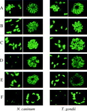
Fig. 1. Localization of the cross-reactive antigens on Neospora caninum and Toxoplasma gondii tachyzoites recognized with mAbs by IFAT with both purified tachyzoites (left) and parasites grown in Vero cells (right). Tachyzoites reacted with 9D12 in group 1 (A), 9E8 in group 2 (B), 10F7 in group 3 (C), 10A10 in group 4 (D), 10G6 in group 5 (E), 10F10 in group 6 (F), respectively.

Fig. 2. Western blot analysis of the cross-reactive antigens of Neospora caninum (A) and Toxoplasma gondii (B) recognized with mAbs. Purified tachyzoite lysates were run on SDS-PAGE gels, transferred into nitrocellulose membranes and probed with 9D12 in group 1 (lane 1), 9E8 in group 2 (lane 2), 10F7 in group 3 (lane 3), 11A10 in group 4 (lane 4), 10G6 in group 5 (lane 5), and 10F10 in group 6 (lane 6), respectively.
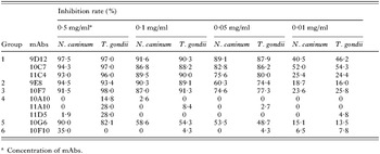
Table 2. Inhibition effects on growth of N. caninum and T. gondii tachyzoites
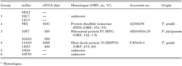
Table 3. cDNA clones encoding cross-reactive antigens between N. caninum and T. gondii
 You have
Access
- 38
- Cited by