Introduction
Patent ductus arteriosus closure is usually a straightforward transcatheter procedure. Reference Malekzadeh-Milani, Padovani and Sivakumar1,Reference Gałeczka, Kapałka, Chojnicki, Krawiec, Tyc and Fiszer2 However, prior surgical ligation may create external or intraluminal barriers that complicate device delivery.
We report a 1.5-month-old infant (2.9 kg) with Edwards syndrome, a large ventricular septal defect, and persistent patent ductus arteriosus after pulmonary artery banding and surgical ductal ligation. The pulmonary artery banding had been intentionally applied loosely, with a peak gradient of 40 mmHg, anticipating future weight gain. However, combined with the additional pulmonary flow through the residual patent ductus arteriosus, the shunt remained haemodynamically significant and required closure.
Cardiac catheterisation for the residual patent ductus arteriosus closure was performed under general anaesthesia. Although the presence of the pulmonary artery banding was taken into consideration, its relatively low gradient (40 mmHg) suggested that antegrade passage through the pulmonary artery would be feasible. Therefore, a femoral venous approach with 4F access was intentionally chosen to avoid arterial puncture in this low-weight infant.
Angiography, supported by transthoracic echocardiography, revealed a 2.5 mm residual patent ductus arteriosus with a left-to-right shunt. Based on these findings, an Amplatzer Piccolo Occluder 4/2 mm (Abbott) was initially selected for implantation.
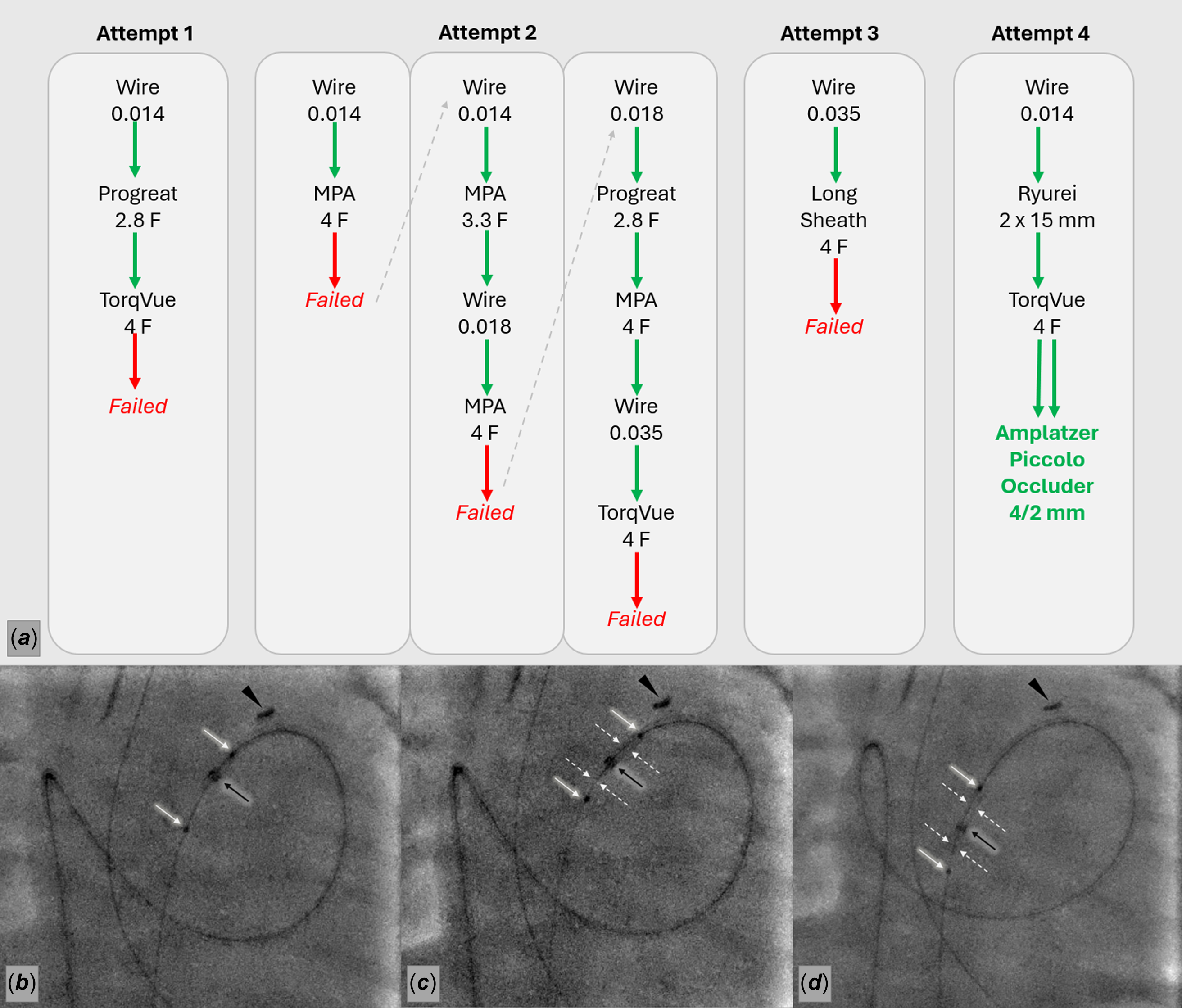
Despite the expected simplicity of the procedure, device delivery proved unexpectedly challenging, and multiple bailout strategies were sequentially attempted to overcome equipment mismatch (Figure 1).
Stepwise strategy (Panel a ) and balloon-assisted bailout for residual ductus arteriosus (DA) closure (Panels b – d ). ( a ) Schematic workflow of four sequential strategies, showing three failed attempts (telescoping, wire and catheter escalation, and long sheath) and the final successful balloon-assisted technique. ( b ) Introduction of the 2 × 15 mm Ryurei (Terumo) coronary balloon across the residual DA (black arrow—TorqVue (Terumo) tip at the obstructed DA entry point; black arrowhead—surgical clip on pulmonary artery banding (PAB); white arrows—proximal and distal coronary balloon markers). ( c ) Hand inflation of the coronary balloon within the DA, with the proximal balloon segment docked inside the TorqVue tip and the distal segment spanning the residual DA (black arrow—TorqVue tip at the level of DA obstruction; black arrowhead—PAB surgical clip; white arrows—coronary balloon markers; white dashed arrows—outline of the inflated coronary balloon). ( d ) Advancement of the inflated coronary balloon—TorqVue catheter assembly across the residual DA into the descending aorta, enabling subsequent device delivery (black arrow—TorqVue tip advanced into the descending aorta; black arrowhead—PAB surgical clip; white arrows—balloon markers; white dashed arrows—outline of the inflated coronary balloon).

Figure 1. Long description
Panel A: A flowchart depicting four sequential strategies for patent ductus arteriosus closure. The first attempt involves using a 0.014 wire, Progreat 2.8 F catheter, and TorqVue 4 F, which fails. The second attempt uses a 0.014 wire, MPA 4 F catheter, and escalates to a 0.018 wire and MPA 3.3 F, then to a 0.035 wire and TorqVue 4 F, all of which fail. The third attempt uses a 0.035 wire and Long Sheath 4 F, which also fails. The fourth attempt successfully uses a 0.014 wire, Ryurei 2 x 15 mm balloon, and TorqVue 4 F, followed by the deployment of an Amplatzer Piccolo Occluder 4/2 mm. Panel B: An X-ray image showing the introduction of the 2 x 15 mm Ryurei coronary balloon across the residual ductus arteriosus. The TorqVue tip is at the obstructed ductus arteriosus entry point, and a surgical clip is visible on the pulmonary artery banding. The proximal and distal coronary balloon markers are indicated. Panel C: An X-ray image showing the hand inflation of the coronary balloon within the ductus arteriosus. The proximal balloon segment is docked inside the TorqVue tip, and the distal segment spans the residual ductus arteriosus. The TorqVue tip is at the level of the ductus arteriosus obstruction, and the outline of the inflated coronary balloon is visible. Panel D: An X-ray image showing the advancement of the inflated coronary balloon-TorqVue catheter assembly across the residual ductus arteriosus into the descending aorta, enabling subsequent device delivery. The TorqVue tip is advanced into the descending aorta, and the outline of the inflated coronary balloon is visible.
Attepmt 1—Telescoping strategy
Initial antegrade patent ductus arteriosus crossing with a 0.014″ coronary guidewire was straightforward. However, advancement of the TorqVue 4F (Abbott) delivery system was unsuccessful, as the support provided by hte 0.014″ guidewire proved insufficient. To minimise equipment mismatch while maximising guidewire support, several telescoping configurations were sequentially attempted using 0.014″ guidewires of varying stiffness, a Progreat 2.8F (Terumo) microcatheter and the TorqVue 4F delivery system. Despite these efforts, advancement of the delivery sheath across the patent ductus arteriosus remained impossible (Figure 1( a )—Attempt 1).
Attepmt 2—Stepwise wire and catheter escalation
To improve guidewire support and comply with standard TorqVue delivery requirements, a stepwise escalation strategy was adopted (Figure 1( a )—Attempt 2).
A 4F multipurpose catheter could not be advanced across the residual patent ductus arteriosus over the initial 0.014″ wire. Therefore, a smaller 3.3F multipurpose catheter (Mongoose, PediaVascular) was successfully advanced over the 0.014″ wire, allowing exchange for a reinforced 0.018″ wire (Terumo). This was expected to provide sufficient support for advancement of the 4F multipurpose catheter, enabling subsequent exchange to a standard 0.035″ guidewire. However, this apptoach was also unsuccessful.
Therefore, returning to the 0.018″ wire, a telescoping configuration using a Progreat 2.8F microcatheter and 4F multipurpose catheter finally enabled successful passage of the 4F multipurpose catheter. This allowed exchange to a 0.035″ guidewire in accordance with standard TorqVue delivery practice. Despite the improved guidewire support and standard delivery configuration, advancement of the TorqVue 4F delivery system again failed at the same level of the patent ductus arteriosus. (Figure 1( a )—Attempt 2).
Attempt 3—Long sheath strategy
With a stabile 0.035″ guidewire position achieved, a long 4F Flexor sheath (Cook), compatible with a 0.035” guidewire and suitable for Amplatzer Piccolo delivery, was selected as an alternative delivery system. Owing to its relatively soft profile and elimination of guidewire—catheter mismatch, successful passage across the patent ductus arteriosus was anticipated. However, the 4F Flexor sheath again failed to cross the obstruction (Figure 1( a )—Attempt 3).
The persistence of the obstacle despite escalation to 0.035″ guidewire and the use of multiple delivery systems strongly suggested the presence of a fixed mechanical barrier, most likely related to the previous surgical ligation.
Attempt 4—Balloon-assisted TorqVue advancement
As a bailout strategy, a 2 × 15 mm Ryurei coronary balloon (Terumo) was advanced across the patent ductus arteriosus over a 0.014″ guidewire across. The TorqVue 4F delivery catheter was positioned at the level of the obstruction and docked at the balloon’s proximal marker. Gentle manual inflation of the balloon filled the distal tip of the TorqVue delivery sheath, allowing the balloon-catheter assembly to cross the obstruction and enter the descending aorta. The balloon was then withdrawn, allowing successful deployment of the Amplatzer Piccolo Occluder 4/2 mm (Figure 1( a )—Attempt 4; Figure 1 b – d ; Supplementary Movie 1).
Discussion
A small 2.0 mm coronary balloon was deliberately selected and inflated only gently, without any intention of dilating the duct. Given the previous surgical ligation, external compression of the duct or partial penetration of the ductal wall by the surgical ligature could not be excluded. Therefore, forceful balloon dilatation was intentionally avoided because of the potential risk of ductal injury, rupture, or mediastinal bleeding.
A primary retrograde transaortic approach was not initially selected because the patient weighed only 2.9 kg, and a venous-only strategy was considered preferable to avoid femoral arterial puncture. Although the presence of the pulmonary artery band was taken into account, its intentionally loose configuration and low pressure gradient suggested that antegrade passage would be feasible.
During the procedure, conversion to a retrograde transaortic approach was considered. However, after repeated failures despite sequential guidewire, multiple catheter configurations, and a long sheath, the obstruction appeared to represent a fixed mechanical barrier, most likely related to the previous surgical ligation. Consequently, a similar difficulty was anticipated from the aortic side while exposing the patient to the additional arterial access.
Limitations
This report does not propose a novel approach to patent ductus arteriosus closure. Rather, it demonstrates that, in surgically altered ducts, procedural success may depend on recognising a fixed mechanical obstacle and overcoming subtle equipment mismatch when standard delivery techniques fail.
Conclusion
Even apparently simple patent ductus arteriosusclosures may become unexpectedly complex after surgery. This case highlights the importance of stepwise problem-solving at the catheterisation table, where multiple strategies were exhausted before success was achieved. Balloon-assisted sheath advancement proved to be a simple and effective bailout technique, underscoring the importance of flexibility and procedural adaptability in fragile infants.
Learning points
-
Patent ductus arteriosus closure after surgical ligation may present unexpected technical challenges.
-
Failure of delivery despite escalation from 0.014″ to 0.035″ guidewires may indicate a fixed postsurgical obstruction rather than insufficient guidewire support.
-
Balloon-assisted sheath advancement can provide an effective bailout when standard delivery techniques fail.
Supplementary material
The supplementary material for this article can be found at https://doi.org/10.1017/S1047951126123804.
Competing interests
The authors declare no conflict of interest.