INTRODUCTION
The largest snapping shrimp genus, Alpheus Fabricius, 1798, is represented by at least 40 species in the western Atlantic (e.g. Chace, Reference Chace1972; Christoffersen, Reference Christoffersen1979; Knowlton & Keller, Reference Knowlton and Keller1985; Martínez-Iglesias et al., Reference Martínez-Iglesias, Ríos and Carvacho1997; Wicksten & McClure, Reference Wicksten and McClure2003; A. Anker, personal observation). However, many of them, especially species that have been reported to occur also in the eastern Pacific and/or eastern Atlantic, are actually species complexes (Knowlton & Mills, Reference Knowlton and Mills1992; Knowlton et al., Reference Knowlton, Weigt, Solorzano, Mills and Bermingham1993; Anker, Reference Anker2001). One of these problematic species is A. cristulifrons Rathbun, Reference Rathbun1900, the only member of the A. crinitus Dana, 1852 group outside the Indo-West Pacific.
Alpheus cristulifrons was actually a new name given by Rathbun (Reference Rathbun1900) for Pocock's specimens originally reported as A. obeso-manus Dana, 1852 from Fernando do Noronha, Brazil (Pocock, Reference Pocock and Ridley1890). Rathbun (Reference Rathbun1900) did not provide species diagnosis, simply referring to Pocock's description of specimens from Fernando do Noronha and listing additional specimens from Maceió, Alagoas, Brazil. Later, Rathbun (Reference Rathbun1901) provided a short diagnosis of A. cristulifrons on the basis of specimens from Puerto Rico, but also listing specimens from Fernando do Noronha and Maceió. This fact complicates the designation of type specimens and the type locality of A. cristulifrons. Kim & Abele (Reference Kim and Abele1988) listed Fernando do Noronha as the type locality of A. cristulifrons. Therefore, Pocock's original specimens, reported as A. obeso-manus (Pocock, Reference Pocock and Ridley1890) and deposited in the Natural History Museum, London (NHM), should be regarded as original syntypes of A. cristulifrons (see below).
Crosnier & Forest (Reference Crosnier and Forest1966) reported A. cristulifrons from São Tomé and Principe in the Gulf of Guinea, eastern Atlantic Ocean, providing a detailed description and illustrations; all their specimens were deposited in the Muséum National d'Histoire Naturelle, Paris (MNHN). These authors also examined ten specimens that Pocock (Reference Pocock and Ridley1890) reported from Fernando do Noronha and two specimens from Puerto Rico identified by Rathbun as A. cristulifrons, deposited in the National Museum of Natural History, Smithsonian Institution, Washington, DC (USNM), and confirmed that all these specimens belong to the same species, A. cristulifrons. However, they also noticed that the eastern and western Atlantic specimens differ in the presence/absence of a distodorsal spine on the propodus of the third and fourth pereiopods, and in the sub-balaeniceps condition of the male minor chela. Based on these observations, Crosnier & Forest (Reference Crosnier and Forest1966) suggested that A. cristulifrons may be a subspecies or even a synonym of one of the ‘variable’ Indo-West Pacific species.
Christoffersen (Reference Christoffersen1979) examined specimens from Brazil and found that the absence/presence of distodorsal spine on the propodus of the third and fourth pereiopod was too inconsistent to be used as a basis for separation of eastern and western Atlantic forms into distinct species. This author listed further variable characters, such as the relative length of the scaphocerite and the marked sexual dimorphism in the shape and size of the major chela. However, Christoffersen noticed that the Brazilian specimens were consistently different from the African specimens in the shorter rostral carina and the relative length of the first and second segments of the antennular peduncle.
Wicksten (Reference Wicksten1983) reported A. cristulifrons—to our knowledge for the first time—from the eastern Pacific. Kim & Abele (Reference Kim and Abele1988) provided a detailed description and illustrations of A. cristulifrons based on specimens from Las Perlas Islands, Panama, and summarized the records of this species in the eastern Pacific. These authors noticed that in the eastern Pacific specimens, the fingers of the minor cheliped are not balaeniceps in both sexes, and that the ischium of the third pereiopod lacks a spine (cf. Kim & Abele, Reference Kim and Abele1988, figure 18).
Ramos & Von Prahl (Reference Ramos and Von Prahl1989) described A. utriensis Ramos & Von Prahl, Reference Ramos and Von Prahl1989 based on specimens from Ensenada Utría (Utria Sound), Pacific coast of Colombia. They contrasted A. utriensis with the western/eastern Atlantic A. cristulifrons, but did not refer to Kim & Abele's (1988) study (of which they were unaware at the time of the manuscript submission). Ramos & Von Prahl stated that A. utriensis can be separated from A. cristulifrons by the presence of ‘marked branchiostegal and infraorbital furrows’ on the carapace, and the absence of spines on the carpus of the third pereiopod. Only two important differences are noticeable between A. cristulifrons sensu Kim & Abele (Reference Kim and Abele1988) and A. utriensis. The first is the presence of a spine on the ischium of the third pereiopod in A. utriensis (apparently absent in A. cristulifrons sensu Kim & Abele, but see below). The second lies in the relative width of the merus and carpus of the third pereiopod, which, according to Ramos & Von Prahl's drawings, appears to be distinctly more slender in A. utriensis than in A. cristulifrons sensu Kim & Abele, Reference Kim and Abele1988. Both A. cristulifrons and A. utriensis were listed as distinct species in the most recent checklist of the eastern Pacific caridean shrimps (Wicksten & Hendrickx, Reference Wicksten, Hendrickx and Hendrickx2003).
Knowlton & Mills (Reference Knowlton and Mills1992) compared colour patterns of ‘A. cristulifrons Pacific’ and ‘A. cristulifrons Caribbean’ and found that the two forms ‘can be readily distinguished by several pattern differences’, including the presence of conspicuous, alternating yellow-brown bands on the antennae in the Pacific form, which are lacking in the Caribbean form. Knowlton et al. (Reference Knowlton, Weigt, Solorzano, Mills and Bermingham1993) and Williams et al. (Reference Williams, Knowlton, Weigt and Jara2001) found that the two forms are genetically different and reproductively incompatible, and thus should be recognized as distinct species.
We were able to collect and photograph alive numerous specimens of A. cristulifrons (sensu lato) on the Pacific and Caribbean coasts of Panama, as well as on São Tomé Island. One of us (A.A.) also examined a large collection of specimens from Atol das Rocas, Brazil, less than 200 km away from Fernando do Noronha, the type locality of A. cristulifrons, as well as two specimens from older collections from Brazil, including one from Fernando do Noronha. Additional specimens from Belize, Guadeloupe, Costa Rica and the Dominican Republic, some with photographs, were also included. This material covers almost the entire range of the A. cristulifrons species complex in all three oceanic provinces: eastern Pacific and western and eastern Atlantic. We also examined the female holotype of A. utriensis deposited in the crustacean collection of the Allan Hancock Foundation in Los Angeles (AHF 842).
At least 20 randomly selected specimens from the Atol das Rocas, Dominican Republic, Belize, Caribbean and Pacific coasts of Panama, and São Tomé were examined for the presence of a distodorsal spine on the propodus of the third to fourth pereiopods; the presence of ventral spines on the carpus of the third pereiopod; the balaeniceps condition of the fingers of the male minor cheliped; the segment ratio of the antennular peduncle; the presence of a ventrolateral spine on the ischium of the third and fourth pereiopod; the presence of carapace furrows; and a number of other morphological characters. We found that two characters, namely the presence/absence of a distodorsal spine on the propodus of the third and fourth pereiopods, and the ratio of segments of the antennular peduncle, are inconsistent or ambiguous, and do not reliably separate the eastern Pacific, western and eastern Atlantic populations. The ‘branchiostegal and infraorbital furrows’, used as a diagnostic key character in the description of A. utriensis, are also present in specimens of A. cristulifrons from the western Atlantic and eastern Pacific (cf. Kim & Abele, Reference Kim and Abele1988, figure 18a), thus eliminating one of the major differences used by Ramos & Von Prahl (Reference Ramos and Von Prahl1989) to separate these two species. We also found that the ischium of the third pereiopod always bear a ventrolateral spine, very strong in the eastern and western Atlantic specimens, less strong in the eastern Pacific specimens. At our request, Dr Rafael Lemaitre (USNM) re-examined this feature in two specimens of A. cristulifrons sensu Kim & Abele (Reference Kim and Abele1988) (USNM 237743) and confirmed that the ischium of the only third pereiopod present in the sample (loose) is indeed unarmed, however, the ischium of the very similar fourth pereiopod has a spine. The presence of a spine on the fourth pereiopod indicates that it should be normally present on the third pereiopod, as in many other Alpheus species (A. Anker, personal observation); its absence on this particular leg may be due to an injury or a slight genetic abnormality. This observation eliminated the most important difference between A. utriensis and A. cristulifrons sensu Kim & Abele (Reference Kim and Abele1988).
In A. utriensis, according to the drawings of Ramos & Von Prahl (Reference Ramos and Von Prahl1989, figure 1D), the appendix masculina appears to be unusually robust and elongate. According to our observations, this condition occurs in all species of the A. cristulifrons complex. Furthermore, in females of A. cristulifrons, the walking legs are generally more slender than in males, nearing the width–length ratio illustrated for the female holotype of A. utriensis (Ramos & Von Prahl, Reference Ramos and Von Prahl1989, figure 2E). All these facts suggest that A. utriensis and A. cristulifrons sensu Kim & Abele (Reference Kim and Abele1988) are the same species. This initial hypothesis was finally confirmed by the examination of the holotype of A. utriensis (AHF 842). The figures of A. utriensis by Ramos & Von Prahl were apparently drawn without a camera lucida and are therefore not accurate, especially in respect to article proportions.
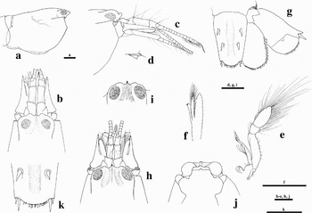
Alpheus cristulifrons Rathbun, Reference Rathbun1900, (a–g) adult male from Atol das Rocas, Brazil (MNRJ 17899); (h–k), ovigerous female (h), young male (i) and adult male (j & k) from Atol das Rocas, Brazil (MNRJ 20111): (a) carapace, lateral view; (b) frontal region, dorsal view; (c) same, lateral view; (d) first segment of antennular peduncle, tooth on ventromesial carina, lateral view; (e) right third maxilliped, lateral view; (f) endopod of right second pleopod, mesial view; (g) telson and right uropod, dorsal view; (h) frontal region, dorsal view; (i) frontal margin of carapace, dorsal view; (j) carapace, frontal view; and (k) telson, detail of posterior margin. Scale bars: 1 mm.

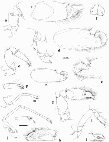
Alpheus cristulifrons Rathbun, Reference Rathbun1900, adult male (a–m) and ovigerous female (n–p) from Atol das Rocas, Brazil (MNRJ 17899): (a) left (male major) cheliped, coxa to carpus, mesial view; (b) same, lateral view; (c) same, chela, dorsal view; (d) same, chela, mesial view; (e) same, distal portion of chela, lateral view; (f) same, plunger of dactylus, lateral view; (g) right (male minor) cheliped, lateral view; (h) same, chela, mesial view; (i) same, chela fingers, lateral view; (j) right second pereopod, lateral view; (k) same, detail of chela; (l) right third pereiopod, lateral view; (m) right fifth pereiopod, lateral view; (n) left (female major) cheliped, chela, mesial view; (o) right (female minor) cheliped, lateral view; (p) same, chela, lateral view. Scale bars: 1 mm.

We reviewed differences in morphology, colour and genetics among the three forms. These data leave no doubt that the three forms actually represent distinct species: one in the western Atlantic—A. cristulifrons sensu Rathbun, Reference Rathbun1900; one in the eastern Pacific—A. utriensis Ramos & Von Prahl, Reference Ramos and Von Prahl1989; and one in the eastern Atlantic—described below as A. xanthocarpus sp. nov.
The taxonomic identity of A. cristulifrons is established based on recently collected and abundant material from Atol das Rocas, which lies very close to the type locality in north-western Brazil, as well as on the Caribbean material. In order to fix the types and type locality of A. cristulifrons, we asked Dr Sammy De Grave to select the most intact specimen from Pocock's original NHM syntype series from Fernando do Noronha, now consisting of eight specimens, and designate it as a lectotype; the remaining seven specimens are designated as paralectotypes. The specimen from Maceió identified by Rathbun and labelled as ‘type’ (USNM 25809) is designated as an additional paralectotype.
A complete synonymy and extensive ecological and biogeographical data, as well as new drawings, are provided for all three species; their colour patterns are illustrated, mostly for the first time. The phylogenetic relationships within the A. cristulifrons complex are discussed in the light of molecular and morphological data.
MATERIALS AND METHODS
Specimens were collected from rock and coral crevices, mostly with the aid of hammer and chisel. Some were photographed alive prior to preservation. Some entire specimens and small chunks of muscular tissue or two posterior walking legs of other specimens were preserved for genetic analysis in RNA-later (Ambion).
All drawings were made under a dissecting microscope with the aid of a camera lucida. The type specimens of the new species are deposited in the collections of the National Museum of Natural History, Smithsonian Institution, Washington DC, USA (USNM); Muséum national d'Histoire naturelle, Paris, France (MNHN); Nationaal Natuurhistorisch Museum, Leiden, The Netherlands (RMNH); Oxford University Museum of Natural History, Oxford, UK (OUMNH); and the Natural History Museum, London, UK (NHM). Additional specimens examined in this study are deposited in the Museu Nacional, Rio de Janeiro, Brazil (MNRJ), Colección de Referencia, Departamento de Biología Marina, Universidad de Panamá, Panama City, Panama (UP), USNM, MNHN, OUMNH, and NHM. Other abbreviations used in the text: Mxp, maxilliped; P, pereiopod; CL, carapace length (measured along mediodorsal line from the tip of the rostrum to the posterior margin of the carapace); TL, total length (measured from the tip of the rostrum to the posterior margin of the telson); fcn, field collection number.
Cytochrome c oxidase subunit 1 gene (COI) sequences were obtained from cDNA rather than from direct amplification of genomic DNA, in order to reduce the risk of amplification of nuclear pseudogenes, previously shown to be pervasive within the genus Alpheus (Williams & Knowlton, Reference Williams and Knowlton2001). Total RNA was extracted using the SV Total RNA Isolation System (Promega) following manufacturer's instructions. First-strand synthesis of cDNA was performed using MuLV reverse transcriptase and RNase inhibitor (Applied Biosystems) and a T18 reverse primer. The resulting cDNA was then used as the template in polymerase chain reaction (PCR) using universal primers HCOI/LCOI from Folmer et al. (Reference Folmer, Black, Hoeh, Lutz and Vrijenhoek1994) to amplify 665 base pairs (bp) from the 5′ end of the mitochondrial COI gene (corresponding to the target region for the COI Barcode) [www.barcodinglife.org], and primers COIF/COI(10) (Williams & Knowlton, Reference Williams and Knowlton2001) to amplify the adjacent 677 bp from the same gene, for a total of 1224 bp (sequences overlapped slightly).
Polymerase chain reaction amplifications were carried out in 30-µL volumes containing 0.1 µM forward and reverse primer, 200 µM each dNTP, 2.0 mM MgCl2+, 1.5 units of Amplitaq Gold DNA polymerase, and 3 µL Amplitaq 10X PCR Buffer II. Thermocycler parameters were as follows: 95°C for 10 minutes; 30 cycles of 95°C for 30 seconds, 50°C for 30 seconds, 72°C for 1 minute +2 s/cycle; with a 10 minute final extension at 72°C. PCR products were gel excised on a 1% (w/v) low-melt agarose gel and extracted using the Wizard SV Gel and PCR Clean-UP System (Promega), following the manufacturer's instructions. An aliquot (2 µl) of the purified PCR product was quantified by electrophoresis on an analytical gel, and DNA concentrations were determined by comparison of fluorescence with a standard DNA mass ladder. Cycle sequencing reactions were performed using 50–100 ng DNA and BigDye terminator v3.1 (Applied Biosystems) following the manufacturer's instructions for cycle sequencing. Reaction products were separated from unincorporated dye-terminators by centrifugation through Sephadex G-50 columns in a 96-well filter plate (Millipore). Products of sequencing reactions were run on a 3700 Applied Biosystems automated capillary sequencer.
Genetic distances were calculated using the Kimura-2-Parameter (K2P) distance method as implemented in Mega v3.1 in order to facilitate comparisons with alpheid distances obtained previously (Knowlton et al., Reference Knowlton, Weigt, Solorzano, Mills and Bermingham1993; Knowlton & Weigt, Reference Knowlton and Weigt1998). A rate of 1.5% sequence divergence per million years was used to estimate the timing of divergence of sister taxa. This rate was estimated by averaging the K2P genetic distances for both 5′ and 3′ COI sequences obtained from the transisthmian sister species pair Alpheus antepaenultimus Kim & Abele, 1988/A. chacei Carvacho, 1979 (GenBank accession numbers AF309875, AF309876, AF309884, AF308989, AF308983 and EF532616–EF532619). This geminate species pair has the smallest observed genetic distance of all transisthmian comparisons, and its divergence is likely to correspond to the final closing of the Panamanian isthmus (approximately three million years ago) (Knowlton & Weigt, Reference Knowlton and Weigt1998). This rate differs slightly from the published rate of 1.4% (Knowlton & Weigt, Reference Knowlton and Weigt1998), obtained from comparisons of COI sequences from the 3′ end only.
SYSTEMATICS
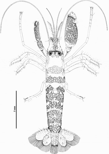
Alpheus cristulifrons Rathbun, Reference Rathbun1900, colour pattern of living adult female from Portobelo, Caribbean coast of Panama. Drawing by J. Jara.

Alpheus utriensis Ramos & Von Prahl, Reference Ramos and Von Prahl1989, colour pattern of living adult male from Las Perlas, Pacific coast of Panama. Drawing by J. Jara.

Alpheus utriensis Ramos & Von Prahl, Reference Ramos and Von Prahl1989, adult male from Las Perlas (USNM 1100674): (a) frontal region, dorsal view; (b) same, lateral view; (c) first segment of antennular peduncle, tooth on ventromesial carina, lateral view; (d) right third maxilliped, lateral view; (e) right (male major) cheliped, dorsolateral view; (f) same, lateral view; (g) same, chela, mesial view; (h) same, chela, lateral view; (i) same, plunger of dactylus, lateral view; (j) left (male minor) cheliped, mesial view; (k) same, lateral view; (l) same, carpus and chela, mesial view; (m) right second pereiopod, lateral view; (n) right third pereiopod, lateral view; (o) right fifth pereiopod, lateral view; (p) telson and right uropod, dorsal view. Scale bars: 1 mm.

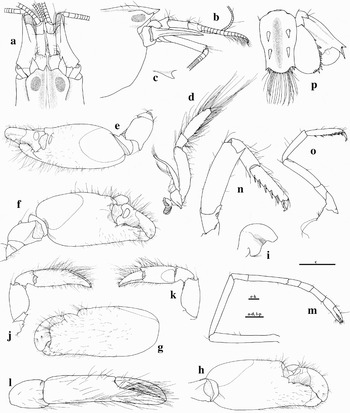
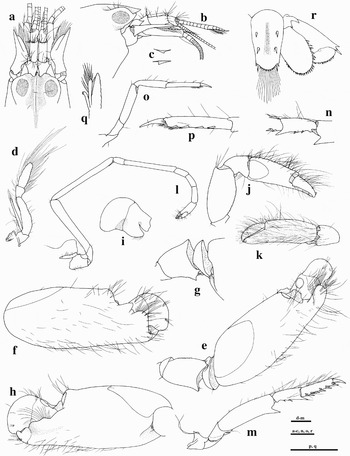
Alpheus xanthocarpus sp. nov., paratype: adult male from São Tomé (USNM 1100676): (a) frontal region, dorsal view; (b) same, lateral view; (c) first segment of antennular peduncle, right and left tooth on mesioventral carinae, lateral view; (d) right third maxilliped, lateral view; (e) left (male major) cheliped, dorsolateral view; (f) same, chela, mesial view; (g) same, merus and carpus, mesial view; (h) same, chela, lateral view; (i) plunger of dactylus, lateral view; (j) right (male minor) cheliped, lateral view; (k) same, chela, mesial view; (l) right second pereiopod, lateral view; (m) right third pereiopod, lateral view; (n) left third pereiopod, carpus, lateral view; (o) right fifth pereiopod, lateral view; (p) same, propodus and dactylus, ventromesial view; (q) endopod of right second pleopod, mesial view; (r) telson and right uropod, dorsal view. Scale bars: 1 mm.

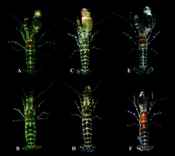
Colour patterns of species of the Alpheus cristulifrons Rathbun, Reference Rathbun1900 complex: Alpheus cristulifrons Rathbun, Reference Rathbun1900: (a) male and (b) female (USNM 1100670) from Bocas del Toro, Caribbean coast of Panama; Alpheus utriensis Ramos & Von Prahl, Reference Ramos and Von Prahl1989; (c) male and (d) female (USNM 1100674) from Las Perlas, Pacific coast of Panama; Alpheus xanthocarpus sp. nov.; (e) male (holotype, USNM 1100675) and (f) female (paratype, NHM 2006.613) from São Tomé.

Alpheus obeso-manus (not Dana, 1852)—Pocock, Reference Pocock and Ridley1890: 520.
Alpheus cristulifrons Rathbun, Reference Rathbun1900: 152;—Rathbun, Reference Rathbun1901: 106; Coutière, Reference Coutière1910: 485; Chace, Reference Chace and Mendez1956: 146; Fausto-Filho, Reference Fausto-Filho1970: 56; Chace, Reference Chace1972: 64; Coelho & Ramos, Reference Coelho and Ramos1972: 149; Pequegnat & Ray, Reference Pequegnat, Ray, Bright and Pequegnat1974: 246, figures 49c, 50; Ray, Reference Ray1974: 90, figures 70, 71; ? Fausto-Filho, Reference Fausto-Filho1974: 5; Abele, Reference Abele1976; Christoffersen, Reference Christoffersen1979: 308; Corredor et al., Reference Corredor, Criales, Palacio, Sánchez and Werding1979: 32; Christoffersen, Reference Christoffersen1980; Rodríguez, Reference Rodríguez1980: 143, figure 40l–n; Carvacho, Reference Carvacho1982: 18; Reed et al., Reference Reed, Gore, Scotto and Wilson1982: 768; Cubit & Williams, Reference Cubit and Williams1983: 24; Criales, Reference Criales1984: 313; Grajal & Laughlin, Reference Grajal and Laughlin1984: 224; Young, Reference Young1986: 109; Abele & Kim, Reference Abele and Kim1986: 198, 210–211, figures d, e; Rodríguez, Reference Rodríguez1986: 137; Knowlton & Mills, Reference Knowlton and Mills1992: 1; Knowlton et al., Reference Knowlton, Weigt, Solorzano, Mills and Bermingham1993: 1630; Martínez-Iglesias et al., Reference Martínez-Iglesias, Gómez, Carvacho and Ríos1993: 11; Salazar-Rosas, Reference Salazar-Rosas1995: 40, pl. 7; Martínez-Iglesias et al., Reference Martínez-Iglesias, Carvacho and Ríos1996: 33; Hernández Aguilera et al., Reference Hernández, Toral and Ruiz1996: 31; Martínez-Iglesias et al., Reference Martínez-Iglesias, Ríos and Carvacho1997: 404, figure 8; Christoffersen, Reference Christoffersen and Young1998: 375; Moreno-Forero et al., Reference Moreno-Forero, Navas and Solano1998: 231, table 1; Williams et al., Reference Williams, Knowlton, Weigt and Jara2001: 377; Souza, Reference Souza and da2001: 25, figure 5; McClure, Reference McClure and Hernández2005: 136 (not figure 10).
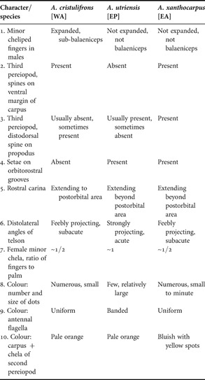
Major characters useful for separation of the three species of the Alpheus cristulifrons Rathbun, Reference Rathbun1900 species complex. EA, eastern Atlantic; EP, eastern Pacific; WA, western Atlantic.

Crangon cristulifrons—Schmitt, Reference Schmitt1924a: 65; Schmitt, Reference Schmitt1924b: 73; Schmitt, Reference Schmitt1935: 143; Schmitt, Reference Schmitt1936: 368; Schmitt, Reference Schmitt1939: 28.
Not Alpheus cristulifrons—Crosnier & Forest, Reference Crosnier and Forest1965: 606; Crosnier & Forest, Reference Crosnier and Forest1966: 260, figures 17, 18 (=A. xanthocarpus sp. nov.); Wicksten, Reference Wicksten1983: 44; Kim & Abele, Reference Kim and Abele1988: 44, figure 18; Villalobos-Hiriart et al., Reference Villalobos, Nates-Rodríguez, Cantu, Valle, Flores, Lira and Schmidtsdorf-Valencia1989: 19; Flores-Hernández, Reference Flores-Hernández1991: 99; Wicksten & Hendrickx, Reference Wicksten and Hendrickx1992: 4; Hendrickx, Reference Hendrickx1992: 8; Hendrickx, Reference Hendrickx, Salazar-Vallejo and González1993: 306; Hendrickx, Reference Hendrickx and Fischer1995: 432; Camacho, Reference Camacho1996: 72; Villalobos, Reference Villalobos2000: 45, figure 22; Wicksten & Hendricks, 2003: 63; McClure, Reference McClure and Hernández2005: 137, figure 10 (=A. utriensis Ramos & Von Prahl, Reference Ramos and Von Prahl1989).
MATERIAL EXAMINED
Brazil: Atol das Rocas: 2 males, 2 ovigerous females, 6 juveniles, MNRJ 17927, east of Laguna Interna, in calcareous algae, depth 1 m, coll. F.B. Pitombo and R. Barroso, 18 October 2000; 7 males, 7 ovigerous females, 8 juveniles, MNRJ 17912, Piscina Mapas, coll. P.S. Young et al., 1 January 2001; 15 males, 6 ovigerous females, 13 juveniles, MNRJ 17900, Station P8, Piscina das Rocas, among calcareous algae, coll. P.S. Young, P.C. Paiva and A.A. Aguiar, 7 October 2000; 1 male, 2 ovigerous females, MNRJ 17925, off Piscina das Tartarugas, in tide pools, coll. P.S. Young, P.C. Paiva and A.A. Aguiar, 17 October 2000; 7 males, 9 ovigerous females, 2 juveniles, MNRJ 17916, Piscina do Cemitério, in calcareous algae, coll. P.S. Young et al., 1 January 2001; 3 males, 2 ovigerous females, MNRJ 17895, LT 719, Canal da Barretão, coll. P.S. Young, P.C. Paiva and A.A. Aguiar, 10 October 2000; 1 male (dissected), 2 ovigerous females (1 dissected), MNRJ 20110, LT 702, between Piscina das Tartarugas and Piscina das Procas, in pools, among calcareous algae, coll. P.S. Young, P.C. Paiva and A.A. Aguiar, 28 October 2000; 7 males, 11 ovigerous females, 1 juvenile, MNRJ 17904, off Piscina das Tartarugas, in nodules of calcareous algae, coll. P.S. Young, P.C. Paiva and A.A. Aguiar, 25 October 2000; 7 males, 13 ovigerous females, MNRJ 17913, Piscina das Tartarugas, coll. P.S. Young et al., 30 December 2000; 1 male, 1 ovigerous female, MNRJ 17889, Piscina de Maré in front of Farol Velho, in calcareous algae, coll. F.B. Pitombo and R. Barroso, 21 December 2000; 1 juvenile (incomplete), MNRJ 17907, LT 711, in sponges and algae, coll. P.S. Young and P.C. Paiva, 16 October 2000; >50 specimens (males, females and juveniles), MNRJ 20111, Piscina das Rocas, in calcareous algae, coll. P.S. Young, P.C. Paiva and A.A. Aguiar, 26 October 2000 (one specimen dissected); 3 males, 1 ovigerous female, MNRJ 20112, Pedra de Tartaruga, north-east part of atoll, between Barretão and Barretinha, in calcareous algae, depth 10 m, coll. F.B. Pitombo and R. Barroso, 24 December 2000; 1 female, MNRJ 20113, near Barretão, in tide pools and nodules of calcareous algae, coll. P.S. Young, P.C. Paiva and A.A. Aguiar, 27 August 2000; 7 males, 8 ovigerous females, MNRJ 20114, Piscina das Rocas, in calcareous algae, coll. P.S. Young, P.C. Paiva and A.A. Aguiar, 26 October 2000. Fernando do Noronha: 1 ovigerous female, MNHN-Na 15722, coll. Rallier de Baly, 1913. Pernambuco: 1 ovigerous female, MNHN-Na 15658, Rio Formozo, coll. A.C. Branner Hartt, December 1875. French Antilles: 1 female, MNHN-Na 16240, Guadeloupe, Vieux Bourg, coll. F. Fasquel, 21 July 2002 (fcn 02-001). Belize: 1 female, OUMNH-ZC 2006-10-003, Carrie Bow Cay, Curlew Key, 15 m, in Xestospongia sp., coll. E. Tóth, 15 March 2006 (fcn 06-305) (corresponding male preserved in RNA-later). Honduras: 1 ovigerous female, OUMNH-ZC 2007-13-037, Bay Islands, Utila, southern shore, bay near Utila town, 16º05.458′N 086º54.533′W, from coral rubble, depth 1 m, coll. A. Anker and S. De Grave, 3 July 2007 (fcn H79). Panama: 1 male, USNM 1100669, Bocas del Toro, Isla Colón, Boca del Drago, from coral rocks, depth: 0.5–1 m, coll. A. Anker, 20 October 2005 (fcn 05-093, infested with protozoan parasite); 2 males, 1 ovigerous female, USNM 1100670, Bocas del Toro, Isla Colon, between Big Creek and Playa Bluff, from coral rocks, depth: 1–2 m, coll. A. Anker, 18 October 2005 (fcn 05-097); 1 juvenile (specimen preserved in RNA-later), Isla Grande, southern shore, from coral conglomerate, depth: 1 m, coll. A. Anker, 23 April 2006 (fcn 06-416); 1 male, 1 ovigerous female, USNM 1100671, Isla Grande, southern shore, from coral rubble, depth: 0.5–1 m, coll. A. Anker, 4 September 2006 (fcn 06-437). Costa Rica: 1 male, 1 female (immatures), USNM 1100672, Cahuita National Park, Puerto Vargas, from coral rocks, rubble and seagrass roots, depth: 0.5–1 m, coll. A. Anker, L. Harris and I. Wehrtmann, 26–27 November 2005 (fcn 05-094). Dominican Republic: 4 males, 3 ovigerous females, 1 female, MNHN-Na 16242, Bayahibe, from coral rocks, depth: 0.5–1 m, coll. A. Anker, 2–3 January 2005 (fcn 05-096).
Specimens examined at our request by Dr Sammy De Grave at the NHM: Brazil: Fernando do Noronha: 1 male, CL 6.0 mm, lectotype, NHM 1888.19, Fernando do Noronha, no other data; 7 specimens (males and females), paralectotypes, NHM 2006.619-627, same collection as for lectotype. Specimen examined at our request by Dr Rafael Lemaitre at the USNM: Brazil: Alagoas: 1 male, paralectotype (originally labelled ‘type’), USNM 25809, Maceió, coral reef.
DIAGNOSIS
Species of Alpheus crinitus group. Carapace depressed below orbital hoods. Rostrum short, triangular; rostral carina blunt, reaching posteriorly slightly beyond eyes. Orbital hoods unarmed, anterior margins produced as rounded lobes; orbitorostral grooves shallow, not abruptly delimited. Orbital hoods, orbitorostral grooves and rostral carina without setae. Pterygostomial angle rounded. Antennular peduncle with stylocerite not reaching distal margin of first segment; second segment longer than wide. Antennal basicerite lacking distoventral tooth; carpocerite reaching far beyond scaphocerite; scaphocerite with well developed blade and strong distolateral tooth. Third maxilliped with ultimate segment expanded, setose. Major cheliped stout; merus bearing subacute distal tooth on mesioventral margin, dorsal margin distally projecting; chela ovoid, smooth, rounded in cross-section, mesial face with strong rounded distal lobe; fingers about 1/3 length of palm; dactylus plunger short. Minor cheliped with ischium bearing blunt process on ventral margin; chela with palm strongly flattened on mesial side; fingers expanded, sub-balaeniceps in males, simple, not balaeniceps in females. Second pereiopod with second carpal segment almost three times as long as first. Third pereiopod with strong ventrolateral spine on ischium; both merus and carpus with strong distoventral tooth, ventral margin of carpus with spines; propodus with strong spines on ventral margin, occasionally with distodorsal spine; dactylus simple, conical. Fifth pereiopod without spine on ischium. Male second pleopod with robust appendix masculina, reaching far beyond appendix interna. Sixth abdominal somite with subacute dorsolateral projections on posterior margin. Uropod with exopod bearing large, subacute lateral tooth on diaeresis; lateral spine strong, reaching beyond exopod margin; exopod with row of spines on distal margin; endopod with row of spines on distolateral and distal margin. Telson medially depressed, with subacute posterolateral angles, two pairs of strong dorsal spines and two pairs of short posterolateral spines; distal margin rounded, with spines; anal tubercles well developed (Figures 1 & 2).
DESCRIPTION
Body stout, bulky, laterally not compressed, glabrous, depressed below orbital hoods, with dorsolateral grooves (Figure 1A); anterolateral region markedly inflated (Figure 1B & J). Rostrum subtriangular, about as long as wide, short, tip subacute not reaching half length of first segment of antennular peduncle (Figure 1B & H); rostral carina blunt, extending posteriorly to eye level or slightly beyond (Figure 1B & H). Orbital hoods inflated, unarmed, frontally closed, anterior margins produced anteriorly into rounded lobes on each side of rostrum (Figure 1B), sometimes reaching beyond rostrum (Figure 1H). Orbitorostral process developed as two short lobes (Figure 1J). Orbito-rostral grooves shallow, not abruptly delimited, extending slightly beyond eyes, gradually flattening. Orbital hoods, rostral carina and orbitorostral grooves without setae. Eyes completely covered in dorsal and lateral views (Figure 1B & C); eyestalks with large cornea, anterior margin without acute projections. Pterygostomial angle rounded, slightly protruding anteriorly (Figure 1C); cardiac notch well developed. Ocellar beak protruding between eyes, not visible in lateral view. Epistomial sclerite unarmed.
Antennular peduncles moderately slender; stylocerite short, with acute tip, falling short of distal margin of first segment (Figure 1B); ventromesial carina with subacute tooth as illustrated (Figure 1D); second segment longer than dorsally visible portion of first segment, about twice as long as wide; third segment shortest, about 1/3 length of second; lateral flagellum with groups of long aesthetascs, secondary ramus rudimentary (Figure 1C). Antenna with basicerite bearing small blunt lobe distoventrally, without acute tooth (Figure 1C); carpocerite slender, exceeding both scaphocerite and antennular peduncle (Figure 1B & C); scaphocerite with distolateral tooth reaching slightly beyond distal margin of antennular peduncle (Figure 1B & H); distolateral tooth strong, reaching far beyond anterior margin of blade, cleft between blade and tooth deep; lateral margin somewhat concave (Figure 1B & H).
Mouthparts typical for Alpheus; mandible with bisegmented palp and incisor process bearing eight or nine teeth. Third maxilliped robust (Figure 1E); coxa with lateral plate ear-shaped, blunt distally (Figure 1E); exopod not reaching penultimate segment, with long flexible setae on posterior margin; antepenultimate segment somewhat flattened, subtriangular in cross-section, ventral margin rugose; dorsal margin not projecting distally; penultimate segment cup-shaped, distally widening; ultimate segment compressed, broadened, unarmed distally, furnished with very long setae (Figure 1E).
Male major cheliped with ischium very short; merus very stout and short (Figure 2A), ventral surface flattened, dorsal margin distally projecting as strong subacute tooth, ventrolateral margin slightly rugose (Figure 2B), ventromesial margin with subacute distal tooth (Figure 2A); carpus short, cup-shaped (Figure 2A & B); chela large, inflated, rounded in cross-section, not compressed, smooth, mesial and ventral faces setose (Figure 2C & D); linea impressa well marked (Figure 2C); distomesial margin with large blunt lobe (Figure 2D); adhesive discs large (Figure 2C); fingers about three times as long as palm; pollex slightly shorter than dactylus, with curved tip (Figure 2E); dactylus with rounded tip, plunger short, truncated (Figure 2F); female major chela much smaller and more slender (Figure 2 N). Male minor cheliped with very short ischium; merus broad, robust, ventrolateral margin proximally serrated (Figure 2G); ventromesial margin distally without marked tooth; carpus cup-shaped; chela relatively broad; palm smooth, setose, particularly on mesial face, latter conspicuously flattened (Figure 2H); linea impressa well marked (Figure 2G); distomesial margin with blunt lobe (Figure 2G & H); adhesive discs small (Figure 2G); fingers shorter than palm; dactylus proximally expanded, with short row of sub-balaeniceps setae on lateral face (Figure 2I); cutting edges smooth; female minor cheliped with ischium and merus almost as in male, but carpus and chela more elongated (Figure 2O); chela fingers half as long as palm, distomesial margin of latter lacking blunt lobe (Figure 2O); dactylus without sub-balaeniceps setae (Figure 2P).
Second pereiopod elongate, slender (Figure 2J); ischium slightly shorter than merus; carpus with five segments having ratio approximately equal to 1:3:1:1:1.2 (Figure 2J); chela simple, fingers with tufts of setae (Figure 2K). Third and fourth pereiopods similar in size and shape; third pereiopod with ischium armed with strong ventrolateral spine (Figure 2L); merus more or less four times as long as wide, strongly compressed, ventral margin armed with strong acute distal tooth (Figure 2L); carpus more slender than merus, slightly more than half as long as merus, compressed, ventral margin with usually one or two spines and strong acute distal tooth (Figure 2L); propodus slightly longer than carpus, ventral margin armed with several spines or pairs of robust spines, including one pair of strong distoventral spines (Figure 2L); distodorsal margin sometimes with short spine above propodo-dactylar articulation; dactylus simple, subconical, curved, with acute tip (Figure 2L), about 1/3 length of propodus.
Abdominal somites with posteroventral margins broadly rounded to somewhat angular; sixth segment without articulated flap, distodorsal margin bluntly to subacutely projecting on each side of telson (Figure 1G); sternites unarmed. First pleopod with small endopod furnished with setae. Male second pleopod with appendix masculina robust, elongated, reaching well beyond appendix interna, but falling short of distal margin of endopod, furnished with numerous, long slender setae (Figure 1F); female second pleopod with appendix interna only. Uropod (Figure 1F) with protopod bearing two subacute teeth; exopod and uropod with numerous long, thick setae; exopodal diaeresis bearing one large subacute tooth near lateral spine, remaining portion more or less straight (Figure 1G); posterior margin furnished with row of spines (Figure 1G); lateral spine and adjacent lateral tooth strong, lateral spine almost reaching level of distal margin (Figure 1G); endopod with row of spines starting at lateral margin and stopping at distomesial margin (Figure 1G). Telson subrectangular, tapering posteriorly, less than half as long as wide at base, with lateral margins slightly convex (Figure 1G); dorsal surface with medio-longitudinal depression, numerous long, stiff setae and two pairs of strong spines inserted at some distance from lateral margin, first pair well anterior to half telson length, second pair posterior to half telson length (Figure 1G); posterior margin broad, convex, with subacute, non-projecting angles (Figure 1K) and two pairs of small posterolateral spines, mesial longer than lateral, margin between spines furnished with numerous plumose setae and row of small spines (Figure 1G & K); anal tubercles well developed. Gill/exopod formula typical for Alpheus: five pleurobranchs (P1–5); one arthrobranch (Mxp3); no pleurobranchs; two lobiform epipods (Mxp1–2); five mastigobranchs (=strap like epiopods) (Mxp3, P1–4); five setobranchs (P1–5); three exopods (Mxp1–3).
COLOUR IN LIFE
Carapace and abdomen dark brown to brown-greenish, speckled with numerous, small, rounded dots of pale greenish or yellowish colour, forming more or less clear transverse bands, five on abdominal somites and one on posterior portion of carapace; yellow-green chromatophores disposed along some of these bands and inside some dots; telson distally brown-grey, proximally brown with pale dots, dorsal spines whitish; uropods, antennal and antennular peduncles brown with pale dots and yellow-green chromatophores; antennal and mesial antennular flagella pale yellow, lateral antennular flagella with reddish bands; cheliped meri and carpi, as well as third to fifth pereiopods reticulated with brown, with pale dots and yellow chromatophores; second pereiopods proximally colourless, distal carpus and chela orange; major chela brown with numerous whitish and yellowish dots, proximal portion of palm sometimes entirely white, distal portion beige-white with some brown patches, some interconnecting; orange patch usually present proximal to dactylar articulation; dactylus white with pink tinge; minor chela brown with pale and yellow dots, distally increasingly whitish (Figures 3 & 7A, B). Similar colour descriptions were provided by Pequegnat & Ray (Reference Pequegnat, Ray, Bright and Pequegnat1974, with photograph), Christoffersen (Reference Christoffersen1979) and Rodríguez (Reference Rodríguez1986). However, Criales (Reference Criales1984) noted that the Santa Marta specimen associated with a crinoid (see below) was ‘dark brown’, matching the colour of its ‘host’.
SIZE
The largest Atol das Rocas specimens attain 7.0 mm CL and 18.6 mm TL; the largest examined Caribbean male is 5.3 mm CL, 17.1 mm TL, female 6.0 mm CL, 17.5 mm TL. The maximum TL of this species is probably around 20 mm (Rodriguez, 1986).
VARIATION
As pointed out by Christoffersen (Reference Christoffersen1979), this species is variable in the relative length of the scaphocerite (as long as to slightly longer than antennular peduncle, cf. Figure 1B & H), the development of the distomesial tooth on the merus of the chelipeds (usually well developed, but sometimes reduced or even absent), and the shape of the frontal lobes (cf. Figure 1B, H & I). Sexual dimorphism affects the major cheliped, mainly the size and shape of the chela (much larger and thicker in males), and the shape of the dactylus (cf. Figure 2D, N & 7A, B), as well as the minor cheliped, mainly the shape of the carpus and chela (much more slender in females), and the presence (males) or absence (females) of sub-balaeniceps setae on the dactylus (cf. Figure 2G & O).
ECOLOGY
Most specimens from Atol das Rocas were collected intertidally from nodules and cavities in calcareous algae; some specimens came from sponges. Most of the Caribbean specimens were collected on coral reefs, ‘reef edges near tide level’ (Chace, Reference Chace1972), from corals and coral rocks (mostly massive Porites but also branching corals incorrectly identified as Pocillopora), coral rubble and conglomerate rocks, at depths ranging from 1 to 5 m. The pair from Belize was found in a sponge, Xestospongia sp., collected at 15 m, however, seemed to occupy a superficial region of the sponge (E. Tóth, personal communication). Chace (Reference Chace and Mendez1956) also reported A. cristulifrons from sponges, Callyspongia sp. Nevertheless, A. cristulifrons is only an occasional sponge dweller that does not occupy the deeper canals. Other observed habitats include intertidal reefs of the sabellariid polychaete Phragmatopoma sp.; bryozoan colonies, Schizoporella sp. (Christoffersen, Reference Christoffersen1979); oculinid corals, Oculina sp. (Reed et al., Reference Reed, Gore, Scotto and Wilson1982); dead and living portions of various corals, Acropora, Porites, Siderastrea and Mussismilia (Christoffersen, Reference Christoffersen1979; Grajal & Laughlin, Reference Grajal and Laughlin1984; Young, Reference Young1986); and colonies of the zoanthid, Zoanthus sociatus (Ellis & Solander, 1786) (Schmitt, Reference Schmitt1936). The association of A. cristulifrons with the comasterid feather star, Nemaster grandis Clark, 1909 (Criales, Reference Criales1984), is questionable and needs confirmation, especially because the colour pattern of this specimen was noted as uniform dark brown (see above). Although A. cristulifrons is not considered a boring shrimp, it is capable of excavating small cavities or enlarging natural cavities using its major claw. Young (Reference Young1986) reported that A. cristulifrons excavates tunnels into the coral Mussismilia or into the coral's epibiontic growth consisting mainly of coralline algae, ascidians and bryozoans. As many other alpheids, A. cristulifrons lives in male/female pairs.
TYPE LOCALITY
Fernando do Noronha, Brazil.
DISTRIBUTION
Tropical and subtopical western Atlantic from Florida to southern Brazil. Specific localities include Florida: Dry Tortugas (Coutière, Reference Coutière1910), Indian River (Reed et al., Reference Reed, Gore, Scotto and Wilson1982); Texas: West Flower Garden Bank (Pequegnat & Ray, Reference Pequegnat, Ray, Bright and Pequegnat1974; Ray, Reference Ray1974); Mexico: Quintana Roo: Isla Mujeres, Bahía de Ascensión, Bahía de Espíritu Santo (Chace, Reference Chace1972; Salazar-Rosas, Reference Salazar-Rosas1995), Veracruz (Ray, Reference Ray1974; Hernández Aguilera et al., 1996), Campeche (Hernández Aguilera et al., 1996); Panama: Isla Grande, Bocas del Toro (present study), Colón (Cubit & Williams, Reference Cubit and Williams1983); Costa Rica: Cahuita (present study); Belize: Carrie Bow Cay (present study); Honduras: Utila (present study); Cuba: Batabano Gulf (Martínez-Iglesias et al., Reference Martínez-Iglesias, Gómez, Carvacho and Ríos1993, Reference Martínez-Iglesias, Ríos and Carvacho1997); Dominican Republic: Bayahibe (present study); Puerto Rico: Ponce, Culebra (Rathbun, Reference Rathbun1901), San Juan (Schmitt, Reference Schmitt1935); Colombia: Providencia, Santa Marta, Islas del Rosario (Schmitt, Reference Schmitt1939; Corredor et al., Reference Corredor, Criales, Palacio, Sánchez and Werding1979; Criales, Reference Criales1984); Venezuela: Sucre, Falcon (Rodríguez, Reference Rodríguez1980), Los Roques (Chace, Reference Chace and Mendez1956; Rodriguez, 1980, 1986; Grajal & Laughlin, Reference Grajal and Laughlin1984); British Virgin Islands: Guana, Virgin Gorda; Barbuda; St Christopher; Antigua; Guadeloupe; Dominica; Grenada: Carriacou; Tobago; (Chace, Reference Chace1972); Martinique (Carvacho, Reference Carvacho1982); Curaçao; Aruba; Bonaire; Klein Bonaire (Schmitt, Reference Schmitt1924a, Reference Schmitt1936); Barbados (Schmitt, Reference Schmitt1924b); Brazil: Fernando do Noronha (Pocock, Reference Pocock and Ridley1890), Atol das Rocas (present study), Rio Grande do Norte (Fausto-Filho, Reference Fausto-Filho1970; Coelho & Ramos, Reference Coelho and Ramos1972), Alagoas (Rathbun, Reference Rathbun1901; Coelho & Ramos, Reference Coelho and Ramos1972; Christoffersen, Reference Christoffersen1979; Souza, 2001), Pernambuco (present study), Paraíba (Young, Reference Young1986), Espiritu Santo, Rio de Janeiro (Christoffersen, Reference Christoffersen1979, Reference Christoffersen and Young1998).
REMARKS
Alpheus cristulifrons can be separated from both A. utriensis and A. xanthocarpus sp. nov. by several features, including the presence of sub-balaeniceps setae on the minor chela and much shorter rostral carina; specifically from A. utriensis by the presence of spines on the carpus of the third pereiopod, the longer fingers (relative to the palm) of the female minor chela, as well as the smaller and more numerous dots on the carapace and abdomen (Figure 5); and specifically from A. xanthocarpus sp. nov. by the absence of bright yellow spots on the second pereiopods (see Table 1).
The record of A. cristulifrons by Fausto-Filho (Reference Fausto-Filho1974) is probably erroneous. This author noted the colour pattern of his specimen from Fernando do Noronha as ‘light yellow’, a colour pattern more typical of A. simus Guérin-Méneville, 1856, which is also known from that area (Christoffersen, Reference Christoffersen and Young1998).
GENBANK NUMBER
COI 5′ EF092276 (06-304); COI 3′ AF309887, AF309888, AF308995 (98-143, 98-151).
Alpheus utriensis Ramos & Von Prahl, Reference Ramos and Von Prahl1989: 477, figures 1 & 2.
Alpheus cristulifrons (not sensu Rathbun, Reference Rathbun1900)—Wicksten, Reference Wicksten1983: 44; Kim & Abele, Reference Kim and Abele1988: 44, figure 18; Villalobos-Hiriart et al., 1989: 19; Flores-Hernández, Reference Flores-Hernández1991: 99; Wicksten & Hendrickx, Reference Wicksten and Hendrickx1992: 4; Hendrickx, Reference Hendrickx1992: 8; Hendrickx, Reference Hendrickx, Salazar-Vallejo and González1993: 306; Hendrickx, Reference Hendrickx and Fischer1995: 432; Camacho, Reference Camacho1996: 72; Villalobos, Reference Villalobos2000: 45, figure 22; Wicksten & Hendricks, 2003: 63; McClure, Reference McClure and Hernández2005: 136 (partial), figure 10 (not description).
MATERIAL EXAMINED
Panama: 1 male, 1 ovigerous female, USNM 1100673, Taboga Islands, Taboguilla, in coral rubble dredged from less than 30 m, coll. P. Barber et al., 30 September 2005 (fcn 05-090, 05-091); 1 ovigerous female, UP, Taboga Island, exposed coral and coral rubble flat, from crevices, coll. A. Anker, C. Hurt and J. Jara, 19 February 2007 (fcn 07-050); 1 male, 1 female, UP-Na, Taboga Island, exposed coral and coral rubble flat, from crevices, coll. A. Anker, C. Hurt and J. Jara, 21 February 2007 (fcn 07-064); 1 male, 1 ovigerous female, MNHN-Na 16243, Las Perlas Islands, Saboga, from coral rocks, depth at low tide: 1–2 m, coll. A. Anker, C. Hurt and J. Jara, 15 November 2005 (fcn 05-092); 1 male, 1 ovigerous female, USNM 1100674, Las Perlas Islands, off Contadora, from basaltic rock outcrops, near extreme low tide mark, coll. A. Anker, C. Hurt, J. Jara, E. Gómez, E. Tóth, 31 March 2006 (fcn 06-378, male partially dissected); 1 male, 1 female, OUMNH-ZC 2006-10-002, same collection data as previous specimens (fcn 06-381, 06-383); 1 male, 1 female, MNHN-Na 16390, Coiba Marine National Park, Coibita, rocky intertidal, from crevices in rocks, extreme low tide, coll. A. Anker, 22 March 2007 (fcn 07-170); 1 ovigerous female, UP, Coiba Marine National Park, Coibita, rocky intertidal, from crevices in rocks, extreme low tide, coll. I. Wehrtmann, 17 March 2006 (fcn 06-648). Colombia: 1 female (holotype) (AHF 842), Utría Sound, 5 August 1984; 1 male (allotype) (AHF 842a), same collection data as for holotype.
Specimens examined at our request by Dr Rafael Lemaitre at the USNM: Panama: 1 male, 1 female (USNM 237743), Las Perlas Islands, Chapera, Station 39, identified and reported as Alpheus cristulifrons by Kim & Abele (Reference Kim and Abele1988).
DIAGNOSIS
Species of Alpheus crinitus group. Carapace depressed below orbital hoods. Rostrum short, triangular; rostral carina blunt, reaching posteriorly to about half of carapace length. Orbital hoods unarmed, anterior margins produced as rounded lobes; orbitorostral grooves shallow, not abruptly delimited. Orbital hoods, orbitorostral grooves and rostral carina with setae. Pterygostomial angle rounded, slightly protruding anteriorly. Antennular peduncle with stylocerite not reaching distal margin of first segment; second segment longer than wide. Antenna with basicerite lacking distoventral tooth; carpocerite reaching far beyond scaphocerite; scaphocerite with well developed blade and strong distolateral tooth. Third maxilliped with ultimate segment expanded, setose. Major cheliped stout; merus bearing subacute distal tooth on mesioventral margin, dorsal margin distally projecting; chela ovoid, smooth, rounded in cross-section, mesial face with strong rounded distal lobe; fingers about 1/3 length of palm; dactylus plunger short. Minor cheliped with ischium bearing subacute projection on ventral margin; palm strongly flattened on ventral and mesial sides, sub-quadrangular in cross-section; fingers simple, not balaeniceps in both sexes. Second pereiopod with second carpal segment about three times as long as first. Third pereiopod with small ventrolateral spine on ischium; both merus and carpus with strong distoventral tooth, ventral margin of carpus unarmed; propodus with strong spines on ventral margin, occasionally with distodorsal spine; dactylus simple, conical. Fifth pereiopod without spine on ischium. Male second pleopod with robust appendix masculina, reaching far beyond appendix interna. Sixth abdominal somite with subacute dorsolateral projections on posterior margin. Uropod with exopod bearing large lateral tooth on diaeresis; lateral spine strong, reaching beyond exopod margin; exopod without spines in distal margin, endopod with row of spines on distolateral and distal margin. Telson medially depressed, with acutely projecting posterolateral angles, two pairs of strong dorsal spines and two pairs of short posterolateral spines; distal margin rounded, with spines; anal tubercles well developed (Figure 5).
DESCRIPTION
For a complete description of this species see Kim & Abele (Reference Kim and Abele1988, as A. cristulifrons) and Ramos & Von Prahl (Reference Ramos and Von Prahl1989). Kim & Abele (Reference Kim and Abele1988) apparently overlooked a short but stout spine inserted in a deep pit on the ischium of the third pereiopod (Figure 5 N). Furthermore, their description and illustrations do not contain some important details, e.g. the shape of the posterolateral angles of the telson (Figure 5P); the setae on the orbito-rostral region (Figure 5B); and the plunger of the cheliped dactylus (Figure 5I). Ramos & Von Prahl (Reference Ramos and Von Prahl1989) provided illustrations of the female major and minor chelipeds; however, these drawings may not be accurate and should be considered with some reservation (see above). The male second pleopod of A. utriensis (see Ramos & Von Prahl, Reference Ramos and Von Prahl1989, figure 1D) is very similar to that of A. cristulifrons and A. xanthocarpus sp. nov.
COLOUR IN LIFE
Carapace brown to brown-greenish, speckled with numerous relatively large, rounded dots of pale yellowish to greenish colour, forming two narrow transverse bands in middle of carapace and along posterior margin; abdomen dark or chocolate brown, speckled with rounded dots, some with yellow-green chromatophores, dots forming chains or transverse bands on abdominal somites (one per somite); telson and uropods mostly dark brown with greenish dots, dorsal spines white; antennal and antennular peduncles brown with whitish patches and dots and some yellow-green chromatophores; antennular and antennal flagella with conspicuous, alternating brown and pale-greenish bands; cheliped meri and carpi, as well as third to fifth pereiopods reticulated with brown, with pale dots and yellow chromatophores; second pereiopods proximally colourless, distal carpus and chela orange; major chela pale or chestnut-brown, speckled with numerous small white dots, most proximal portion of palm sometimes entirely white, distal portion pale orange with larger and smaller white patches and dots; dactylus white with pink tinge; minor chela brown with pale and yellow dots, distally paler (Figures 4 & 7C, D). Ramos & Von Prahl (Reference Ramos and Von Prahl1989) described the colour pattern of A. utriensis as ‘Eyes black; chelae translucent green with yellow finger tips; carapace and abdomen with intercalated transverse bands of dark green and light green, and numerous yellow chromatophores; pereiopods and antennal and antennular peduncles translucent with greenish spots; pleopods translucent green; eggs orange’.
SIZE
The largest male from Las Perlas is 8.6 mm CL and 24.8 mm TL; the largest female is 7.5 mm CL and 22 mm TL.
VARIATION
The size and shape of the major cheliped and proportions of the minor chela differ between males and females, in particular, the female major chela is smaller and less inflated compared to the male major chela (cf. Figure 7C & D). However, in A. utriensis, the sexual dimorphism appears to be generally less strong than in A. cristulifrons sensu stricto.
ECOLOGY
Most specimens were collected from crevices in coral rubble and coral pavement outcrops; some were found in basaltic rocks, where they dwelled mostly in pairs in relatively superficial crevices. Most probably, the shrimps are capable of boring small tunnels into rocks, or at least enlarging natural cavities of rocks into more spacious domiciles. Wicksten (Reference Wicksten1983) reported some specimens from sponges. Ramos & Von Prahl (Reference Ramos and Von Prahl1989) reported A. utriensis from hollow spines of the fan clam, Pinna rugosa Sowerby, 1835, and observed them to feed on algae growing on the clam's surface. The depth range appears to be from the lower intertidal down to about 35 m (Hendrickx & Hermoso-Salazar, Reference Hendrickx and Hermoso-Salazar2005).
TYPE LOCALITY
Ensenada Utría, Colombia.
DISTRIBUTION
Tropical and subtopical eastern Pacific, from central Gulf of California to Colombia. Specific localities include Mexico: Baja California Sur, Guerrero, Oaxaca (Wicksten, Reference Wicksten1983; Villalobos, Reference Villalobos2000), Sinaloa (Kim & Abele, Reference Kim and Abele1988), Nayarit (Camacho, Reference Camacho1996; Hendrickx & Hermoso-Salazar, Reference Hendrickx and Hermoso-Salazar2005); Costa Rica: Puntarenas; Panama: Las Perlas, Taboga, Punta Paitilla, Coiba (Kim & Abele, Reference Kim and Abele1988; present study); Colombia: Ensenada Utría (Ramos & Von Prahl, Reference Ramos and Von Prahl1989).
REMARKS
Alpheus utriensis differs from A. cristulifrons mainly by the absence of sub-balaeniceps setae on the fingers of the male minor chela; the posteriorly further extending rostral carina; the carpus of the third pereiopod lacking ventral spines; and the telson with distinctly protruding, acute posterolateral angles (cf. Table 1). The most conspicuous and diagnostic character of the colour pattern of A. utriensis is the presence of greenish-brown bands on the antennal and antennular flagella, absent in both A. cristulifrons and A. xanthocarpus sp. nov. (cf. Table 1; Figure 9). For further differences between A. utriensis and A. xanthocarpus sp. nov. see Table 1 (see also remarks under A. xanthocarpus sp. nov. below).
GENBANK NUMBER
COI 5′ EF092279, EF092280 (AA-AU-29, 06-378); COI 3′ AF309889, AF309890 (98-22, 98-25).
Alpheus cristulifrons (not sensu Rathbun, Reference Rathbun1900)—Crosnier & Forest, Reference Crosnier and Forest1965: 606; Crosnier & Forest, Reference Crosnier and Forest1966: 260, figures 17 & 18.
TYPE MATERIAL
São Tomé: Holotype: 1 male, USNM 1100675, Station 12, Ilha Santana, from rock crevices, 15–20 m, coll. N. Knowlton and F. Nunes, 9 February 2006 (fcn 06-207). Paratypes: 1 male, USNM 1100676, same collection data as for holotype (fcn 06-206), partially dissected; 1 male, MNHN-Na 16244, same collection data as for holotype (fcn 06-203); 1 ovigerous female, MNHN-Na 16245, same collecion data as for holotype (fcn 06-195); 1 ovigerous female, NHM 2006.613, same collection data as for holotype (fcn 06-208); 1 ovigerous female, USNM 1100677, Station 4, 2 km west of Lagoa Azul, near ship wreck, rocks with growth of coralline algae, from crevices of rocks and coralline algae, depth at low tide: 1–2 m, coll. A. Anker and N. Knowlton, 1 February 2006 (fcn 06-107); 1 male, RMNH D 51704, same collection data as previous paratype (fcn 06-108); 1 male, OUMNH-ZC 2006-10-001, Station 5, Lagoa Azul, shallow bay with rocky shore, from coralline algae and rocks, 1–3 m, coll. A. Anker and N. Knowlton, 3 February 2006 (fcn 06-138); 1 male, NHM 2006.614, Station 3, 200 m west of Lagoa Azul, rocky shore with coralline algae, from holes and under rocks, near extreme low tide mark, coll. A. Anker and N. Knowlton, 31 January 2006 (fcn 06-066).
DIAGNOSIS
Species of Alpheus crinitus group. Carapace depressed below orbital hoods. Rostrum short, triangular; rostral carina blunt, reaching posteriorly 1/2 to 3/4 of carapace length. Orbital hoods unarmed, anterior margins produced as rounded lobes; orbitorostral grooves shallow, not abruptly delimited. Orbital hoods, orbitorostral grooves and rostral carina with setae. Pterygostomial angle angular–rounded. Antennular peduncle with stylocerite not reaching distal margin of first segment; second segment longer than wide. Antenna with basicerite lacking distoventral tooth; carpocerite reaching far beyond scaphocerite; scaphocerite with well developed blade and strong distolateral tooth. Third maxilliped with ultimate segment expanded, setose. Major cheliped stout; merus bearing subacute distal tooth on mesioventral margin, dorsal margin distally projecting; chela ovoid, smooth, rounded in cross-section, mesial face with strong rounded distal lobe; fingers about 1/3 length of palm; dactylus plunger short. Minor cheliped with ischium bearing subacute projection on ventral margin; palm flattened on mesial side; fingers simple, not balaeniceps in both sexes. Second pereiopod with second carpal segment about three times as long as first. Third pereiopod with strong ventrolateral spine on ischium; both merus and carpus with strong distoventral tooth, ventral margin of carpus with spines; propodus with strong spines on ventral margin, usually with dorsodistal spine; dactylus simple, conical. Fifth pereiopod without spine on ischium. Male second pleopod with robust appendix masculina, reaching far beyond appendix interna. Sixth abdominal somite with subacute dorsolateral projections on posterior margin. Uropod with exopod bearing large lateral tooth on diaeresis; lateral spine strong, reaching beyond exopod margin; exopod with row of spines on distal margin; endopod with row of spines on distolateral and distal margin. Telson medially depressed, with subacute posterolateral angles, two pairs of strong dorsal spines and two pairs of short posterolateral spines; distal margin rounded, with spines; anal tubercles well developed (Figure 6).
DESCRIPTION
For an extensive description of this species see Crosnier & Forest (Reference Crosnier and Forest1966, as A. cristulifrons). Some of the important details omitted in Crosnier & Forest's description are the setae on the orbitorostral region (Figure 6B); the dactylus plunger of the major chela (Figure 6I); the tooth on the mesioventral carina of the first segment of the antennular peduncle (Figure 6C); and the male second pleopod (Figure 6Q).
COLOUR IN LIFE
Carapace dark or purple brown, sometimes greenish brown, speckled with numerous small to medium-sized, rounded dots of pale yellowish colour, some fused, forming one more or less distinct transverse band on middle of carapace, some dots with yellow-green chromatophores; abdomen dark or chocolate brown, speckled with rounded dots and spots, some with yellow-green chromatophores, some dots chained, forming diffuse, narrow, transverse bands on abdominal somites (one per somite); telson and uropods mostly dark brown with greenish dots and yellow chromatophores, dorsal spines white; antennal and antennular peduncles dark brown with greenish patches and dots and some yellow-green chromatophores; scaphocerite whitish with dark lateral margin, distolateral tooth brown with greenish dots; antennular and antennal flagella uniform pale grey; cheliped meri and carpi, as well as third to fifth pereiopods brown with white bands near article joints; second pereiopods bluish-grey, joints marked with bright yellow dots, most conspicuous on carpus and chela; major chela dark brown, proximally and distally with white patches; dactylus white with pink tinge; minor chela brown with whitish dots (Figure 7E & F).
SIZE
The largest male from São Tomé reaches 5.9 mm CL and 18.2 mm TL; the largest female is 6.8 mm CL and 20.3 mm TL.
VARIATION
The size and shape of the major chela and proportions of the minor chela differ between males and females, the major chela of females being smaller and less inflated (cf. Figure 7E & F).
ECOLOGY
Most specimens were collected from crevices in rock outcrops and occasionally from coral rubble. The shrimps may bore tunnels and cavities into superficial portions of rocks or enlarge natural cavities into more spacious domiciles. The depth range of this species reaches from the lower intertidal to at least 20 m (present study).
TYPE LOCALITY
São Tomé, Gulf of Guinea.
DISTRIBUTION
Tropical eastern Atlantic: Gulf of Guinea. Presently known only from the islands São Tomé and Principe (Crosnier & Forest, Reference Crosnier and Forest1966; present study).
REMARKS
Alpheus xanthocarpus sp. nov. differs from A. cristulifrons by the absence of sub-balaeniceps setae on the fingers of the male minor chela (Figure 6J & K) and the posteriorly further extending rostral carina (Figure 6A); from A. utriensis by the carpus of the third perieopod bearing spines on the ventral margin (Figure 6M) and the much less protruding posterolateral angles of the telson (Figure 6R) (see also Table 1). The most characteristic feature of the colour pattern of the new species is the presence of bright yellow spots on the merus, carpus and chela of the second pereiopod; these spots are absent in both A. cristulifrons and A. utriensis (Figure 7).
GENBANK NUMBER
COI 5′ EF092277, EF092278 (06-109, 06-107).
DISCUSSION
The three species of the A. cristulifrons species complex may be distinguished by several features of morphology and colour pattern (see Table 1) and occur in different oceanic provinces: eastern Pacific (A. utriensis), western Atlantic (A. cristulifrons) and eastern Atlantic (A. xanthocarpus sp. nov.), making a misidentification virtually impossible. The sub-balaeniceps condition of the male minor chela appears to be the main character separating A. cristulifrons from A. utriensis and A. xanthocarpus sp. nov. (Table 1). Three other morphological features may also help in the discrimination of the three species: (Reference Abele1) the presence/absence of setae on the orbital grooves; (Reference Abele and Kim2) the posterior extension of the rostral carina; and (Reference Anker3) the presence/absence of spines on the carpus of the third pereiopod (Table 1). In live shrimps, the banded antennular/antennal flagella and the yellow-spotted second pereiopods are the most obvious diagnostic features of A. utriensis and A. xanthocarpus sp. nov., respectively.
The barcode COI sequence from A. utriensis differs from those of A. cristulifrons and A. xanthocarpus sp. nov. by 20.2% and 20.4%, respectively, while COI from A. cristulifrons and A. xanthocarpus sp. nov. differ by 16.7%. These values suggest the following topology: A. utriensis (A. cristuliftons–A. xanthocarpus sp. nov.). Thus A. cristulifrons and A. utriensis do not represent a pair of transisthmian geminate species as previously assumed (Knowlton & Weigt, 1998). Therefore, it is not surprising that they are among the most genetically differentiated transisthmian pairs of Alpheus, with an estimated divergence time of over 13 million years ago (mya) (see Materials and Methods). The divergence values for the trans-Atlantic sister taxa A. cristulifrons and A. xanthocarpus sp. nov. are also relatively large compared to values obtained for other alpheid cryptic taxa from the western/eastern Atlantic and eastern Pacific, which averaged 6.3% in four comparisons (Williams et al., 2001); the estimated divergence time for this pair is about 11 mya.
The molecular data are corroborated by data from morphology and colour, with the two trans-Atlantic sister taxa sharing the greatest number of characters (cf. Table 1): A. cristulifrons and A. xanthocarpus sp. nov. are united by five characters: 2, 6, 7, 8 and 9; A. cristulifrons and A. utriensis are united only by the character 10; finally, A. utriensis and A. xanthocarpus sp. nov. share three characters: 1, 4 and 5. Without an outgroup, in this particular case a closely related taxon from the Indo-West Pacific (see below), it is difficult to polarize and code these characters; some of them may be either autapomorphies or plesiomophies without phylogenetic significance. Leaving out the possible autapomorphies of colour (9: banded antennae in A. utriensis; 10: yellow spots on P2 in A. xanthocarpus sp. nov.) and morphology (1: sub-balaeniceps chela of the male minor cheliped in A. cristulifrons), A. cristulifrons and A. xanthocarpus sp. nov. still appear to be closer to each other, sharing four characters (Reference Abele and Kim2, Reference Camacho6, 7 and Reference Chace and Mendez8), versus only two (4 and Reference Banner and Banner5) shared by A. utriensis and A. xanthocarpus sp. nov., and remarkably none shared by the two transisthmian taxa, A. cristulifrons and A. utriensis.
The three species of the A. cristulifrons complex belong to the mostly Indo-West Pacific A. crinitus species group, which presently includes over 35 species (A. Anker, personal observation). This group is defined mainly by the frontal margin of the carapace lacking orbital teeth and bearing a small or reduced rostrum (occasionally without rostrum); smooth and cylindrical, sometimes greatly swollen major chela; relatively small and simple minor chela; and very stout third and fourth pereiopods, with meri often armed with strong distoventral teeth (see also Banner & Banner, 1982). Most species of the A. crinitus groups are sponge-dwellers and remarkably similar in their general appearance to the sponge-dwelling species of Synalpheus Bate, 1888. Other species (including species of the A. cristulifrons complex) inhabit crevices or self-made galleries in hard substrates, such as corals and rocks. At least four species are known to felt filamentous algae (cyanobacteria) to fabricate peculiar sausage-like tubes under coral rocks or among living corals (Banner & Banner, 1982); this type of symbiosis appears to be unique among the Decapoda. These examples aim to illustrate the ecological diversification and the biological interest of the A. crinitus group.
The position of the A. cristulifrons complex within the A. crinitus group remains unknown. The possibly closest Indo-West Pacific relatives appear to be A. bucephalus Coutière, 1905 and A. bucephalus Nobili, 1905 (Crosnier & Forest, 1965), although the colour pattern of A. bucephalus as described in Banner & Banner (Reference Banner and Banner1981, 1982) and Miya (Reference Miya1984) is quite different from the patterns of the three species of the A. cristulifrons complex. In addition, the A. crinitus group may not be monophyletic in its present composition. For instance, A. ovalipes Coutière, 1905, a species currently assigned to the A. crinitus group, appears to have more affinities with other groups or lineages, while A. acutofemoratus Dana, 1852 and A. amirantei Coutiére, 1905, both currently assigned to the non-monophyletic A. sulcatus Kingsley, 1878 group, may actually be members of the A. crinitus group (cf. Banner & Banner, 1982; A. Anker, personal observation). Therefore, both morphological and genetic coherence of the A. crinitus group should be examined as part of a comprehensive (as many representative taxa as possible) and multidisciplinary (DNA, morphology and colour) phylogenetic assessment of the genus Alpheus.
ACKNOWLEDGEMENTS
The present study was supported by the John Dove Isaacs Professorship of Natural Philosophy and the Scripps Institution of Oceanography, La Jolla, USA (SIO), and is part of the Census of Marine Life/CReefs Project. We express our gratitude to the Fisheries Department (Direcção das Pescas) of the Democratic Republic of São Tomé and the Autoridad Nacional del Ambiente (ANAM) of the Republic of Panama for allowing us to collect and export scientific specimens. This work was done at the Smithsonian Tropical Research Institute (STRI) facilities at Naos, Panama City, Republic of Panama. Javier Jara (STRI) prepared the detailed line drawings of colour patterns of the transisthmian species, and also checked the Spanish spelling in some references. Rafael Lemaitre (USNM) and Sammy De Grave (OUMNH) kindly examined specimens at our request. Professor Tereza Cristina dos Santos Calado (Universidade Federal de Alagoas, Maceió, Brazil) and Sammy De Grave scanned or checked unaccessible references. Flavia Nunes (SIO) checked the Portuguese spelling in Brazilian references. Paolo Young (late of MNRJ) arranged the collection and loan of the Atol das Rocas specimens; Cristiana Serejo (MNRJ) provided MNRJ numbers.