Introduction
With the proportion of the European population over the age of 65 years estimated to increase from 19.1% in 1995 to over 20% by 2025 and over 42.2% by 2050,Reference Aebi, Gunzburg and Szpalski1 and with even higher estimates in Western Europe, there will be increased demand for services that treat degenerative conditions related to ageing. An increase in demand for services of this kind (in the area of spine surgery) has already been reported in the United States.Reference Weinstein2
Spine surgery in older people can provide improved mobility, relief from pain, increased levels of function, and a better quality of life for many. The incidence of different pathologies affecting the spine changes with increasing age, as does the nature of surgical intervention. The frequency and severity of co-morbidities as well as life expectancy (particularly in a palliative care setting) may all influence treatment decisions. Careful multidisciplinary assessment prior to consideration of surgery is frequently required.
The following article will provide an overview of spine surgery in older people, focusing specifically on the particular considerations and conditions that are prevalent in this age group.
Age-related changes in the spine
The degenerative process that affects the spine is complex, begins at an early age and is almost ubiquitous. Men are affected at an earlier age, and more frequently than women. Progression of the age-related change is relentless and affects all anatomical structures. The process appears to begin with the intervertebral disc.
Increasing age is associated with dehydration of the intervertebral disc. This dehydration is associated with progressive histological, biochemical and biomechanical change.Reference Boos, Weissbach, Rohrbach, Weiler, Spratt and Nerlich3 Histologically, the boundary between nucleus pulposus and annulus fibrosus becomes less distinct and collagen fibre organization less ordered. Biochemical changes include the loss of agrecan and water from the extracellular matrix and an increase in the proteases responsible for enzymatic degradation.Reference Roberts, Caterson, Menage, Evans, Jaffray and Eisenstein4 There is an increased keratin sulphate to chondroitin sulphate ratio. Histological and biochemical changes lead to mechanical changes in the function of the intervertebral disc. Loss of disc height leads to instability within the motion segment, which in turn leads to compensatory calcification, osteophyte formation, hypertrophy and buckling of the ligamentum flavum. Facet joint arthrosis develops. Annular tears and disc herniation may occur. Progressive loss of disc height in combination with these changes leads to narrowing of the neural exit foramen, increased loading of the arthritic facet joint and narrowing of the spinal canal. This narrowing (stenosis) can either be central, affect only the lateral recesses, or affect the neural exit foramen.
Within the spinal column altered biomechanics affect different regions in different ways. The support and relative stability provided by the thoracic rib cage causes this region to be relatively spared. In the more mobile cervical and lumbar regions degenerative change is more common. In the cervical spine, the lower levels (C5/6 and C6/7) are more frequently affected. Likewise the lower lumbar motion segments are more frequently affected. Instability can progress and degenerative spondylolisthesis may occur. Degenerative spondylolisthesis is most common between the 4th and 5th lumbar vertebra.
The primary aetiology for these age-related changes is unknown but a decrease in the nutrient supply to the disc may play an important role. The main pathway for nutrition to reach the intervertebral disc is via the vertebral body endplate. Permeability of the endplate to nutrients decreases with increasing age, thus decreasing nutrient supply to the disc.
Degenerative conditions affecting the lumbar spine – neurogenic claudication and radiculopathy
Neurogenic claudication and radiculopathy are the most common indications for spine surgery in older people.
Neurogenic claudication is a symptom complex caused by lumbar spinal stenosis.Reference Aebi, Arlet and Webb5 The symptom complex typically includes a reduction in walking distance due to bilateral leg pain, a feeling of heaviness, fatigue, aching, numbness and unsteadiness, all affecting the lower limbs. Symptoms are frequently reduced by rest and bending forward. Bending forward flexes the lumbar spine, reducing the lumbar lordosis and increasing the space available for the cauda equina within the spinal canal. Activities that involve flexion of the lumbar spine (e.g. walking uphill, upstairs, pushing a shopping trolley and cycling) are frequently found to be easier than less arduous tasks that extend the lumbar spine (increasing the lordosis).Reference Frymoyer6
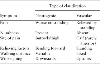
One of the challenges for the clinician is to effectively distinguish a neurological cause for the claudication from a vascular one (Table 1). The characteristics of the symptoms described above will help, and clinical examination with palpation of peripheral pulses as well as ankle-brachial pressure recording is also useful. Vascular claudication is relieved by standing, whereas neurogenic claudication may be made worse.Reference Nadeau, Arellano and Gurr7
Symptoms in neurogenic and vascular claudication

Radiculopathy is characterized by pain, paraesthesia, numbness and muscle weakness caused by injury to a specific spinal nerve or nerve root. Symptoms affect (radiate to) the dermatome and myotome of the nerve. The term ‘sciatica’ is frequently misused by patients and clinicians and is perhaps a more general term. Sciatica encompasses the various different lumbar radiculopathies.
Neurogenic claudication is caused by spinal canal stenosis. Facet joint hypertrophy, posterior bulging of the intervertebral disc and thickening of the ligamentum flavum combine to narrow the available space for the nerves of the cauda equina and cause the symptoms. This degenerative process may or may not be associated with a spondylolisthesis, which further narrows the canal and compounds the problem. In some cases a spondylolisthesis occurs without symptoms or it may compress only the neural exit foramen creating unilateral or bilateral radiculopathy. Different degrees of loss of disc height, exit foraminal narrowing and osteophytosis will determine the neurological structure first compressed and therefore the symptoms.
Investigations
Magnetic resonance scanning has become the gold standard for investigating these symptoms. Detailed images of the spinal canal in sagittal and transverse section can be used to assess the space available for neural structures. Its main disadvantage is that investigations are performed supine, which may cause a spondylolisthesis to reduce. Standing X-rays can be helpful in this regard. Alternatives include CT myelography.
Outcomes of surgery for spinal stenosis and spondylolisthesis
There are many different surgical techniques that have been described for treating compressive lesions including laminectomy, undercutting facetectomy, laminotomy, selective nerve root decompression and many others. Where there is instability (spondylolisthesis), decompression may be supplemented with fusion. Whichever technique is undertaken, treatment is aimed at removing the mechanical compression, and recovery of symptoms relies on recovery of the compressed neural structure. However, the outcome of surgery of this type is relatively predictable and dramatic improvements in leg pain, back pain,Reference Sanderson and Wood8 walking distance (and associated disability), and quality of life can be expected. Recent high-quality data from prospective studies in a large number of patients have shown the clinically significant benefits of spine surgery over conservative treatment and maintenance of these improvements in the medium term.Reference Weinstein, Lurie, Tosteson, Zhao, Blood, Tosteson, Birkmeyer, Herkowitz, Longley, Lenke, Emery and Hu9–Reference Matsuda, Shibata, Oki, Kawatani, Mashima and Oishi11 Patients undergoing surgery have shown significant improvements in pain, physical function and disability as assessed using validated instruments such as the Short Form 36 Health Survey (SF-36) and the Oswestry Disability Index (ODI).
Controversies and novel treatments
One of the main dilemmas that the spine surgeon faces when treating a degenerative spondylolisthesis is whether to fuse the spine to stabilize and prevent progression of the spondylolisthesis. This may be undertaken with or without instrumentation. Instrumentation is associated with higher fusion rates, and in younger patients with higher demands may theoretically help prevent progression of the slip. However, in an older age group, the spondylolisthesis rarely progresses and simple decompression avoids the complications associated with instrumented or non-instrumented fusion surgery.
Novel technologies including inter-spinous spacer devices have been marketed and are currently being evaluated. Typically they attempt to treat symptoms by flexing the spine at the symptomatic level, relieving symptoms in a similar but exaggerated way to bending forward. Encouraging early results have been reported but the indications for surgery of this type have still to be established.
Degenerative conditions affecting the cervical spine – radiculopathy and myelopathy
Cervical spondylosis is a widely used but non-specific term referring to the generalized degeneration of the cervical spine frequently seen in older people, although it can begin at an early age (over 30 years). Characteristic X-ray changes can be identified. Patients may present complaining of neck pain, or have signs and symptoms of radiculopathy or myelopathy. Neck pain caused by cervical spondylosis typically presents as episodic bouts of pain that resolve over days or weeks. Symptoms may have been exacerbated by increased activity, and may be associated with occipital headache. Treatment is largely conservative with analgesia. Assessment is aimed at identifying and excluding surgically treatable conditions.
Cervical radiculopathy is typically caused by a compressive lesion affecting a single nerve root in the cervical spine. Compression can be caused by osteophytes, herniated disc fragments, facet joint hypertrophy and loss of disc height. It presents with unilateral neck pain, and pain radiating into the upper limbs in the distribution of the affected nerve root. These symptoms are associated with decreased or altered sensation and lower motor neurone signs in a similar distribution. The most commonly affected levels are the C6 and C7 nerve roots. The natural history of radiculopathy is benign, with a significant proportion of cases resolving spontaneously or with conservative treatment. Its peak incidence is in middle age and this decreases with increasing age.
Radiculopathy affecting the C6 nerve root (exiting above the 6th cervical vertebra) will produce pain radiating to the radial side of the forearm and hand affecting the thumb and index fingers, decreased or altered sensation in a similar distribution, with weakness of biceps (supination) and wrist extension. The brachioradialis reflex may be decreased.
Radiculopathy affecting the C7 nerve root will produce pain radiating to the long (middle) finger, decreased sensation in the same area, weakness of wrist flexion, elbow extension and a decreased triceps reflex.
Cervical myelopathy can be defined as dysfunction of the spinal cord caused by compression of the cervical cord within the cervical spine. It is the most common type of spinal cord lesion in older people. The compression is commonly caused by osteophytes, ligamentum flavum hypertrophy and bulging or prolapse of the intervertebral disc.
Cervical myelopathy presents with upper motor neurone signs and symptoms in both upper and lower limbs. Initial presentation may be subtle, but detection and treatment is essential before irreparable cord damage occurs. Typically the myelopathy follows a slow, progressive course, deteriorating in a stepwise manner with stable periods and periods of rapid deterioration. It rarely resolves spontaneously. Symptoms include decreased co-ordination, loss of fine dexterity (e.g. buttoning a shirt, handwriting, manipulating small objects), balance and gait problems, and problems with bowel and bladder function. Balance and walking problems may lead to frequent trips, falls or bumping into things. There may be diffuse altered sensation and occasionally the sensation of shooting pains or ‘electric shocks’ radiating down the arms when moving the head in a particular position.
Associated (upper motor neurone) signs include: a wide based unsteady gait, upper and lower limb weakness, hyper-reflexia, intrinsic muscle wasting in the hand, positive Babinski and Hoffman signs and an inverted radial reflex.
Anterior cervical decompression and fusion (ACDF)
Anterior cervical decompression and fusion can be thought of as the ‘gold standard’ for cervical spine surgery. It frequently provides excellent results and has a low complication rate.Reference Emery, Bohlman, Bolesta and Jones10, Reference Matsuda, Shibata, Oki, Kawatani, Mashima and Oishi11 The anterior approach allows access to the cervical disc that can be removed along with osteophytes at the posterior aspect of the vertebral body. It allows removal of most lesions causing myelopathy or radiculopathy. Placement of anterior bone graft between the vertebral bodies in the excised disc space helps to decompress the exit foramen indirectly and facilitates fusion.
The procedure is associated with low rates of complications, and scars tend to heal in a cosmetically acceptable manner. Complications include pseudarthrosis (increased in smokers), hoarse voice and swallowing problems caused by retraction or injury to the recurrent laryngeal nerve (2–5%). Graft complications also include the graft loosening and migration. Fusion alters the mechanics of the cervical spine, increasing the lever arms of forces acting at adjacent levels and there is a significant rate of adjacent level degeneration.
Cervical disc replacement
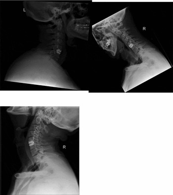
Cervical disc replacement is an emerging surgical technique offering an alternative to anterior cervical decompression and fusion (ACDF). It is aimed at treating the same pathologies through the same anterior approach, but preserving motion in the cervical spine by replacing the cervical disc with materials similar to those used in large joint arthroplasty (Figure 1). Initial results are encouraging and appear to be at least comparable to ACDF.Reference Murrey, Janssen, Delamarter, Goldstein, Zigler, Tay and Darden12
Cervical disc replacement (pictures courtesy of Mr J. Andrews)

Posterior surgery – foramenotomy/laminoplasty
Posterior surgery may also be used in the cervical spine, either to decompress a single nerve root (foramenotomy) or to decompress multiple levels. It has the advantage of being motion preserving, and when surgery is required at more than two levels it is the treatment of choice. However, its disadvantage is that the surgery can potentially destabilize the spine posteriorly, leading to a tendency towards developing a kyphosis. Additionally, in many cases, as cervical spondylosis develops, progressive loss of disc height anteriorly leads to a progressive loss of the normal cervical lordosis and may itself produce cervical kyphosis. To avoid this combination of risk factors, kyphosis is a relative contraindication to posterior surgery in spondylotic patients.
Spinal tumours
Metastatic disease is the most common form of skeletal tumour and the spine is the most common site for skeletal metastasis. Breast, lung, prostate, renal, thyroid and gastrointestinal malignancies are the most common sources of primary disease. With increasing age, metastatic disease becomes more common.
Primary spinal tumours occur but are much less common. Location within the vertebra and age are important prognostic indicators. Tumours that affect the vertebral body are more likely to be malignant and posterior tumours benign. Under the age of 21, most spinal tumours are benign; over 21, most are malignant.Reference Weinstein and McLain13 The thoracic spine is the most commonly affected region, and the cervical spine the least.
The role of spine surgery is dependent on the aetiology, stage and grade of the lesion. Curative resection is possible in a few casesReference Abe, Kobayashi, Murai, Suzuki, Chiba and Okuyama14 but is the least common indication for spinal surgery. The most common presenting complaint is pain, which can either be bony (mechanical) or occur as the result of neural compression. Muscle weakness may also occur and can be detected in up to 50% of cases of metastatic disease.Reference Constans, Divittiis and Donzelli15 Palliative resection for prevention of (or prevention of progression of) neurological impairment is a common reason for surgical intervention. Also, pain from bony destruction and mechanical instability may respond to surgical stabilization.
The grading system proposed by Tokuhashi et al. is useful in establishing indications for treatment and subsequent surgical approach.Reference Tokuhashi, Matsuzaki, Toriyama, Kawano and Ohsaka16
Surgical treatment
Surgical resection of tumour may be aimed at improving survival or palliating symptoms by decompressing neural structures. Alternatively, removal of healthy bone (corpectomy/laminectomy) to facilitate the decompression or a combination of both may be considered. This may be undertaken anteriorly or posteriorly or both and depends on the size and location of the lesion. If a curative resection is hoped for or survival is likely to extend beyond a year, intervertebral bony fusion should be undertaken to avoid instrumentation failure. If life expectancy is short and a palliative procedure is being considered, fusion and its associated morbidity may not be required.
Novel techniques – minimally invasive surgery and vertebroplasty
If palliative procedures either for mechanical pain or neurological symptoms are being considered, novel treatments can be considered. Minimally invasive stabilization may minimize soft tissue trauma, help reduce post-operative pain and facilitate a faster post-operative recovery. Fusion may not be required as the strength of the implants is likely to outlast the demands of the patient.
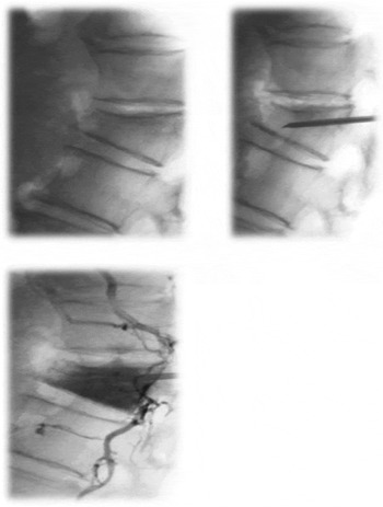
In many cases of malignant disease with associated multiple co-morbidities, patients are too unwell or are unwilling to consider major surgery. Surgery may also interrupt chemotherapy or radiotherapy regimens. When there is pain caused by instability that does not require decompression, vertebral body augmentation with high viscosity cement may be considered. Galibert et al. were the first to report the use of polymethylmethacrylate (PMMA) injection under fluoroscopic guidance to treat vertebral tumoursReference Galibert, Deramond, Rosat and Le Gars17 (Figure 2). PMMA is well known to orthopaedic surgeons and is used routinely to support ‘cemented’ joint replacements. Cement augmentation provides mechanical support at the site of the tumour and mechanical stabilization of the innervated eriostemon.Reference Bekoff, Maroney, Fenton and Matthis18 An exothermic reaction is generated as the cement sets with possible attenuation of the sensory nerve fibres, which may also improve pain. What effect this exothermic reaction will have on reducing viable tumour is as yet unknown. Biopsy is possible at the same time as treatment. The procedure is relatively quick, minimally invasive, associated with minimal blood loss, and has a lower complication rate than major spinal surgery.
Vertebroplasty

Encouraging results have been reported in multiple myeloma treated in this way.Reference Thielen and Kallmes19 Vertebroplasty has been used effectively to treat symptoms in pathological fractures regardless of cause.Reference Layton, Theilen and Kallmes20
Spinal trauma and osteoporosis
Spinal trauma can occur at any age. However, with increasing age, total bone mass and bone mineral density decrease, increasing the likelihood of insufficiency fractures. Vertebral compression fractures are the most common osteoporotic fractures, associated with increased morbidity and mortality and an impact on quality of life.Reference Dreinhöfer, Féron, Herrera, Hube, Johnell, Lidgren, Miles, Panarella, Simpson and Wallace21–Reference Kallmes, Comstock and Heagerty23 A significant proportion (25%) of these fractures remain, or become sufficiently symptomatic for patients to seek medical attention. Associated with the mechanically mediated pain there is a decrease in function and an increase in dependence on others. Life expectancy is also reduced with an increased mortality rate following fracture. Until recently the only treatments available were conservative and based on pain relief and rehabilitation.
Vertebroplasty for osteoporotic fractures
Vertebroplasty (described above) has also been proposed as a treatment for pathological fractures caused by osteoporosis. Although excellent results have been reported, more recent studies have reported conflicting outcomes. Two randomized studies with a sham control intervention seemed to show equal efficacy of percutaneous vertebroplasty and sham intervention in patients with osteoporotic vertebral fracture.Reference Buchbinder, Osborne, Ebeling, Wark, Mitchell, Wriedt, Graves, Staples and Murphy22, Reference Kallmes, Comstock and Heagerty23 However, both studies were limited by methodological issues.Reference Clark, Lyon and Burnes24 A positive bone scan has been reported as predicting symptomatic relief following vertebroplasty and studies reporting poor results did not use this imaging modality.Reference Maynard, Jensen and Kallmes25 A more recent randomized study comparing vertebroplasty against conservative care concluded in favour of vertebroplasty in those patients with ‘acute’ fractures.Reference Klazen, Lohle, de Vries, Jansen, Tielbeek, Blonk, Venmans, van Rooij, Schoemaker, Juttmann, Lo, Verhaar, Van Der Graaf, van Everdingen, Muller, Elgersma, Halkema, Fransen, Janssens, Buskens and Mali26 Treatment remains controversial.
Spinal deformity (degenerative scoliosis)
As instrumentation systems improve and the demands of older people increase, more major surgery is considered in this age group. Surgery for spinal deformity is considered major surgery in any age group and presents particular challenges when undertaken in patients with co-morbid disease.
Defined as a three-dimensional spinal deformity with a lateral curve of greater than 10 deg that occurs in a skeletally mature person, degenerative scoliosis has a mean age of presentation of 60 years. As well as deformity, patients frequently complain of symptoms of neurogenic claudication (secondary to spinal stenosis). However, as the spine is relatively inflexible, the symptoms of claudication typically do not resolve with sitting or bending forward. With severe curves, patients may complain of disabling severe back pain as a result of mechanical malalignment and a displaced centre of mass. Radicular leg pain is also common, occurring in association with exit foraminal stenosis on the concavity of the curve. It occurs (‘de novo’) in skeletally mature individuals rather than as a progression of an adolescent deformity. The prevalence of adult spinal deformity is increasing with the average age of the population, and is currently estimated at between 1.4 and 12% for curves measuring >10 deg. Men and women are equally affected.
Indications for surgery include severe persistent pain (stenotic, radicular or mechanical) that is resistant to conservative treatment, progressive unacceptable deformity and the risk of further progression. The goals of treatment are symptom relief achieved with a solid arthrodesis and a balanced spine over a level pelvis.
The most common surgical approach is posterior spinal fusion, but additional anterior procedures and osteotomies may be required to achieve sufficient correction of the scoliosis to achieve a balanced spine. Careful pre-operative assessment is essential and two stage procedures may be required.
Most surgical patients obtain significant pain relief and report that they would undergo the same procedure again for the same benefit. However, surgery for degenerative scoliosis is associated with a high complication rate. Pseudarthrosis (5–25%), infection (0.5–8%), neurological complications (0.5–5%) and pulmonary embolism (1–20%) are all more frequent in this population.
Conclusion
Spine surgery can provide improved mobility, relief from pain, increased levels of function, and a better quality of life for patients in many age groups. As the mean age of the population increases, demand for the surgical treatment of degenerative conditions is likely to increase. Also, as surgical techniques and instrumentation systems develop, successful treatment of previously untreatable conditions becomes more possible.
Conflicts of interest
The authors declared no conflicts of interest in relation to this article.