Introduction
Haemodynamically significant patent ductus arteriosus is commonly encountered in premature infants with an incidence inversely correlated with gestational age and it is associated with comorbidities and worse outcomes. Reference Noori, McCoy and Friedlich1–Reference Gentle, Travers, Clark, Carlo and Ambalavanan3 In many programmes, the advent of new devices that can be delivered through small catheters led to transcatheter patent ductus arteriosus closure replacing surgical ligation as the gold standard for definitive closure. Reference Sathanandam, Gutfinger and O’Brien4–Reference Mohammad Nijres, Hassan, Winsdor, Bischoff, McNamara and Aldoss8 While several devices are available and yield excellent outcomes, none are considered the “ideal” device due to limitations in the delivery process or catheter design. Reference Sathanandam, Gutfinger and O’Brien4–Reference Mohammad Nijres, Hassan, Winsdor, Bischoff, McNamara and Aldoss8 Traditionally, the catheter used to cross the tricuspid valve and ductus arteriosus to obtain an angiogram [typically a 4-Fr Angled Glide catheter (Terumo, Somerset, NJ)] differs from the one used for device deployment, often necessitating catheter and potentially wire exchanges. Reference Shibbani, Mohammad Nijres and McLennan7–Reference Heyden, El-Said, Moore, Guyon, Katheria and Ratnayaka9 In tiny infants, despite the 4-Fr Angled Glide catheter being soft and small, it is still relatively large, and haemodynamic instability can occur when crossing the tricuspid valve, likely due to regurgitation. Moreover, tricuspid valve injury with worsening regurgitation was reported with transcatheter patent ductus arteriosus closure in premature infants. Reference Sathanandam, Gutfinger and O’Brien4,Reference Dalby, Shibbani and Mercadante5,Reference Mohammad Nijres, Hassan, Winsdor, Bischoff, McNamara and Aldoss8,Reference Heyden, El-Said, Moore, Guyon, Katheria and Ratnayaka9 In this report, we describe our initial experience with zero-contrast transcatheter patent ductus arteriosus closure using only a single microcatheter of the KA micro plug device (Merit Medical, South Jordan, UT), eliminating the need for catheter or wire exchanges. This modification aims to simplify and shorten the procedure while reducing the risk of catheter-induced haemodynamic instability—particularly in infants weighing less than 1500 g, who are often unstable and have multiple comorbidities.
Material and methods
Study design and patient selection
This retrospective study focused on premature infants weighing less than 1500 g who underwent attempted transcatheter patent ductus arteriosus closure using a modified 2.9-Fr microcatheter and a KA micro plug device at Stead Family Children’s Hospital/University of Iowa between May 1, 2024, and July 22, 2024. The study was approved by the University of Iowa Institutional Review Board. Patients were excluded if their procedural weight exceeded 1500 g or if the closure was not attempted using this new technique. At our institution, the choice of device and technique for transcatheter patent ductus arteriosus closure is left to the operator’s discretion. The primary objective of this study was to describe the procedural technique and report both immediate and short-term outcomes.
Collected data
Demographic, procedural, and short-term outcome data were collected. The procedure was deemed successful if the new technique resulted in patent ductus arteriosus closure with no more than a trivial residual shunt and safe return of the patient to the neonatal intensive care unit. Major haemodynamic instability was defined as the need for chest compressions, cardioversion, or occurrence of catheter-induced hypotension or desaturation requiring equipment removal. Minor haemodynamic instability was characterised by the need for inotropic support or an increase in inspired oxygen concentration. Haemodynamic instability was considered catheter-related only if it occurred after crossing the tricuspid valve until the equipment was removed from the heart. Inotropic support initiated before transporting the patient to the cardiac catheterisation laboratory or as a result of anaesthesia induction was not classified as catheterisation-related instability.
As previously described, post-procedural iatrogenic aortic and left pulmonary artery stenoses were defined as a peak instantaneous pressure gradient greater than 20 mmHg measured by transthoracic echocardiography. Reference Dalby, Shibbani and Mercadante5,Reference Shibbani, Mohammad Nijres and McLennan7 More than mild tricuspid valve regurgitation was classified as a catheter-related complication unless it was similar in severity when compared to the pre-procedural echocardiogram. Acute and short-term follow-up data were collected, including mortality, time to extubation, time to discharge, cardiac function, residual shunt, tricuspid valve function, left pulmonary artery stenosis, and aortic arch obstruction.
Statistical analysis
Statistical analysis was conducted using the SPSS Statistics package [ver. 28.0.11(14)]. Categorical data are reported as counts and percentages, while continuous data are presented as medians and ranges.
Device description
The KA micro plug consists of three identical soft nitinol discs, and it is available in four sizes: 3, 4, 5, and 6 mm. All devices come in an unconstrained length of 2.5 mm and can be delivered through a straight 2.9-Fr 125 cm in length microcatheter, included in the package. The package also contains two short Y-connectors (Fig. 1).
Image shows the manufacturer provides a straight microcatheter and the KA micro plug device connected to the delivery wire.

Procedure description and microcatheter modification
After securing the patient on the cardiac catheterisation table, an intraoperative transthoracic echocardiogram is performed to confirm the presence of the patent ductus arteriosus and measure its smallest diameter, which is used to select the appropriate KA micro plug device. Additionally, cardiac function, tricuspid valve function, presence of pericardial effusion, and flow velocity in the aortic arch and left pulmonary artery are assessed. The device chosen is 1.5–2 mm larger than the narrowest dimension of the ductus arteriosus.
The microcatheter included with the KA micro plug device is manually shaped using a heat gun to create a curved distal tip with an angle of 90–110 degrees (Fig. 2). The device is then prepared and de-aired, and a 0.014” Balance Middle Weight (BMW) guide wire (Abbott, Santa Clara, CA) is selected.
Image shows the tip of the microcatheter being modified using a heat gun to create a distal curve.

Venous access is obtained in the common femoral vein using a 4-Fr sheath. The access wire is then exchanged for the BMW wire. The shaped microcatheter is advanced inside the sheath over the BMW wire. Under fluoroscopic guidance, the combination of the BMW wire and microcatheter is used to cross the tricuspid valve and the patent ductus arteriosus in an antegrade fashion. The wire is removed, and the KA device is inserted inside the microcatheter and advanced toward the tip. The microcatheter is then pulled inside the patent ductus arteriosus and retracted over the delivery wire, allowing the entire device to be deployed inside the patent ductus arteriosus. The previously inserted oral temperature probe, nasogastric tube, or orogastric tube are used as a landmark of the patent ductus arteriosus ampulla (Moving Clip 1). A transthoracic echocardiogram is performed to assess device positioning, evaluate the flow in the left pulmonary artery and descending aorta, and quantify the degree of any residual shunt. If the echocardiogram findings are reassuring, the device is released, and additional echocardiogram images are obtained to assess the degree of tricuspid valve regurgitation, flow in the left pulmonary artery and descending aorta, device position, residual shunt, and presence of pericardial effusion (see Fig. 3). The sheath is then removed, and manual pressure is applied until hemostasis is achieved.
Image shows the KA micro plug device inside the PDA (white arrow) with no residual shunt. PDA, patent ductus arteriosus.

Results
During the study period, five patients [two males (40%) and three females (60%)] underwent attempted transcatheter patent ductus arteriosus closure using the modified microcatheter technique with KA micro plug devices. The procedure was successful, achieving complete immediate patent ductus arteriosus closure in all patients. The median birth weight, gestational age, procedural weight, and age at the time of the procedure were 740 g (530–1080), 24.6 weeks (23.3–26.7), 1055 g (730–1330), and 22 days (16–46), respectively (Table 1).
Summary of demographic data

HFJV = high-frequency jet ventilator.
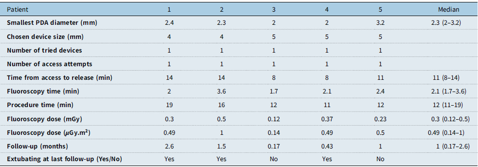
The median times from obtaining access to releasing the device, procedural duration, and fluoroscopy duration were 11 minutes (8–14), 12 minutes (11–19), and 2.1 minutes (1.7–3.6), respectively. The median radiation doses, expressed in air kerma and dose area product, were 0.3 mGy (0.12–0.5) and 0.49 µGy·m2 (0.14–1), respectively (Table 2). None of the patients experienced minor or major haemodynamic instability. Only one device was attempted in all patients. Notably, for patients #3 and #4, the ideal device size was 4 mm; however, due to unavailability at the time of the procedure, a 5 mm device was used. Post-procedure, none of the patients exhibited iatrogenic worsening of tricuspid valve regurgitation, left pulmonary artery stenosis, or aortic stenosis. At a median follow-up of 1 month (0.17–2.6), there were no concerning echocardiogram findings and three patients were extubated.
Summary of procedure and short-term follow-up data

PDA = patent ductus arteriosus.
Discussion
We report our early experience in closing the patent ductus arteriosus in small premature infants using a new technique with a modified microcatheter and zero-contrast use. The procedure was successful in all five patients in whom this new technique was attempted, with immediate complete closure observed in each case. No catheter-related complications or haemodynamic instability were encountered. This study supports the accumulating evidence of the safety and effectiveness of transcatheter patent ductus arteriosus in tiny premature infants. Reference Sathanandam, Gutfinger and O’Brien4–Reference Parkerson, Sathanandam and Molloy14
In contrast to previous reports from our group and others, only one device was attempted in all cases, which can be attributed to the operators’ increased familiarity with selecting the appropriate device size and assessing the adequacy of closure based solely on echocardiogram measurements. Reference Mohammad Nijres, Khallaf and Rahde Bischoff10–Reference Johnson, Sathanandam, Naik and Philip12
Traditionally, a 4-Fr Angled Glide catheter is used to cross the tricuspid valve and the patent ductus arteriosus in an antegrade fashion. After obtaining an aortic/ductal angiogram, the Angled Glide catheter must be exchanged for a stiffer 4-Fr delivery catheter (TorqVue LP catheter) if a Piccolo device is intended for use. Reference Sathanandam, Gutfinger and O’Brien4,Reference Shibbani, Mohammad Nijres and McLennan7 Alternatively, if the KA micro plug device is to be used, the microcatheter for the KA micro plug needs to be cut and inserted inside the Angled Glide catheter. Reference Mohammad Nijres, Hassan, Winsdor, Bischoff, McNamara and Aldoss8,Reference Heyden, El-Said, Moore, Guyon, Katheria and Ratnayaka9 The use of the 4-Fr catheter can lead to tricuspid valve injury or haemodynamic instability when crossing the tricuspid valve, with the risk likely increased when utilising the stiffer TorqVue LP delivery catheter. Reference Sathanandam, Gutfinger and O’Brien4,Reference Dalby, Shibbani and Mercadante5,Reference Mohammad Nijres, Hassan, Winsdor, Bischoff, McNamara and Aldoss8,Reference Heyden, El-Said, Moore, Guyon, Katheria and Ratnayaka9
In comparison to traditional methods of transcatheter patent ductus arteriosus closure using Piccolo and KA micro plug devices, Reference Sathanandam, Gutfinger and O’Brien4,Reference Shibbani, Mohammad Nijres and McLennan7–Reference Mohammad Nijres, Khallaf and Rahde Bischoff10 this new technique offers several advantages and simplifies the procedure in several ways. First, it eliminates the need for catheter exchange, as the same microcatheter used to cross the patent ductus arteriosus is also utilised for device deployment. This is crucial for mitigating the risk of haemodynamic instability and minimising the potential for catheter-induced tricuspid valve injury or regurgitation, which can be attributed to the softness and smaller size (approximately 1 mm in diameter) of the microcatheter. Notably, none of the patients in this series developed any catheter-related minor or major haemodynamic instability or worsened tricuspid valve regurgitation. Second, this technique shortens the procedure time by eliminating some steps, such as the need for angiograms and catheter exchanges. Reference Sathanandam, Gutfinger and O’Brien4,Reference Shibbani, Mohammad Nijres and McLennan7–Reference Mohammad Nijres, Khallaf and Rahde Bischoff10 Additionally, the device is selected and de-aired, the microcatheter is manually shaped, and the guide wire is pulled from the package before obtaining access contributing to further reduction in the procedure time. Third, since contrast is not required, this technique eliminates the risk of contrast-related kidney and thyroid dysfunction and further decreases radiation exposure. However, this new technique has a few drawbacks. First, an angiogram cannot be obtained through the microcatheter, necessitating the operators to be well-acquainted with device selection and capable of assessing the appropriate position via the echocardiogram. Second, the straight tip of the microcatheter must be manually shaped to create a favourable curve for crossing the tricuspid valve with the guide wire. Third, the excessive length of the microcatheter (125 cm) may be cumbersome to control in such small infants. On the other hand, using the classic technique for deploying the KA device can also be uncomfortable, as it requires the operator to simultaneously manage two catheters (the cut microcatheter within the Angled Glid catheter). Reference Mohammad Nijres, Hassan, Winsdor, Bischoff, McNamara and Aldoss8,Reference Heyden, El-Said, Moore, Guyon, Katheria and Ratnayaka9 The authors recommend that the manufacturer provide a company-made, curved, short microcatheter, ideally 40–50 cm in length.
The authors advise against cutting the microcatheter, as this is only appropriate when using a 4-Fr Angled Glide catheter. Reference Mohammad Nijres, Hassan, Winsdor, Bischoff, McNamara and Aldoss8,Reference Heyden, El-Said, Moore, Guyon, Katheria and Ratnayaka9 If the microcatheter is cut and the interventionalist intends to use the front segment, Reference Heyden, El-Said, Moore, Guyon, Katheria and Ratnayaka9 it will be very difficult to backload the device without the hub; frontloading is only feasible when the Angled Glide catheter is used and is across the patent ductus arteriosus, as this requires the microcatheter to be outside the body. Conversely, if the back segment of the cut microcatheter is utilised, Reference Mohammad Nijres, Hassan, Winsdor, Bischoff, McNamara and Aldoss8 this allows for device backloading through the hub and de-airing of the microcatheter. However, the end of the cut microcatheter may not be well visualised under fluoroscopy, as the discarded portion is the only segment containing a radiopaque marker close to the catheter tip. Reference Mohammad Nijres, Hassan, Winsdor, Bischoff, McNamara and Aldoss8 Additionally, the tip of the cut catheter could be sharp and pose a risk of trauma, especially when visibility is compromised.
The KA micro plug device is soft and short (2.5 mm in length), making it relatively straightforward to deploy the device entirely intraductally without encroaching on neighbouring vessels. Reference Guyon, Duster and Katheria6,Reference Mohammad Nijres, Hassan, Winsdor, Bischoff, McNamara and Aldoss8,Reference Heyden, El-Said, Moore, Guyon, Katheria and Ratnayaka9 For this reason, the authors select the device solely based on the smallest ductal diameter, regardless of the length and ampulla sizes. Reference Mohammad Nijres, Hassan, Winsdor, Bischoff, McNamara and Aldoss8–Reference Mohammad Nijres, Khallaf and Rahde Bischoff10 Notably, none of the patients in this series, including the two who received oversized devices (5 mm used instead of the 4 mm), experienced immediate or at follow-up aortic or left pulmonary artery obstruction.
A couple of technical tricks and observations are worth discussing. First, the authors recommend exchanging the access wire for the guide wire, which can be accomplished using the dilator of the sheath. This facilitates the advancement of the microcatheter inside the sheath. The microcatheter is soft and cannot be introduced into the sheath without the aid of the wire. Furthermore, attempting to insert the microcatheter through the sheath without the wire could result in losing the handcrafted curve. Second, the authors recommend using a soft wire, such as the BMW wire. Stiff wires can compromise the handcrafted curve of the microcatheter and complicate crossing the tricuspid valve. Additionally, a softer wire is, at least theoretically, less likely to distort the anatomy and induce haemodynamic instability.
It is important to note that transcatheter patent ductus arteriosus closure with exclusive echocardiogram guidance may be feasible using this device. However, in our setting, we found that the quality of echocardiogram images was suboptimal for guiding catheter and wire manipulation. This poor acoustic window is likely due to the use of a high-frequency jet ventilator as the preferred mode of ventilation for premature infants in our institution. Reference Shibbani, Mohammad Nijres and McLennan7 Furthermore, relying on the echocardiogram would necessitate frequent probe position adjustments, likely prolonging procedure time with a reported median procedure time of 40 minutes. Reference Shibbani, Mohammad Nijres and McLennan7,Reference Mohammad Nijres, Hassan, Winsdor, Bischoff, McNamara and Aldoss8,Reference Baruteau, Méot and Benbrik13–Reference Georgiev, Tanase and Eicken15
To minimise procedure duration, we utilised fluoroscopy to guide catheter and wire manipulation, as well as device deployment. Our median procedure time was 12 minutes (11–19) and the procedure was completed within 12 minutes in the last three patients. Notably, the radiation doses—Air Kerma of 0.3 mGy (0.12–0.5) and Dose Area Product of 0.49 µGy·m2 (0.14–1)—were considerably lower (more than 10 times lower) than previously reported levels. Reference Dalby, Shibbani and Mercadante5,Reference Heyden, El-Said, Moore, Guyon, Katheria and Ratnayaka9,Reference Sathanandam, Justino, Waller, Radtke and Qureshi16,Reference Philip, Tailor and Johnson17 This reduction is attributed not only to shorter radiation exposure times and the exclusion of cine angiograms but also to practising the “As Low As Reasonably Achievable” principle.
Limitations
This study is limited by its very small sample size and retrospective design. Further researches on a larger scale are needed to validate the findings of this pilot study and to compare the safety of this method with other available devices and techniques. Despite the promising short-term outcomes, the long-term effects and the superiority of transcatheter patent ductus arteriosus closure over other management options (including conservative, medical, and surgical approaches) have yet to be established, preferably through randomised controlled studies.
Conclusion
Manual modification of the microcatheter for the KA micro plug simplifies the patent ductus arteriosus closure procedure and reduces the procedure time and radiation exposure in infants weighing less than 1500g without compromising efficacy and safety. Further studies are necessary to refine the technique and confirm our findings. The authors encourage the manufacturer to develop a factory-made curved short microcatheter.
Supplementary material
The supplementary material for this article can be found at https://doi.org/10.1017/S104795112500006X.
Authors contribution
BMN developed the study concept/design, analysed and interpreted the data, and drafted the article.
TP collected the data and critically revised the article.
Competing interests
The authors declare that there is no conflict of interest.
Research involving human participants
All procedures performed in studies involving human participants were in accordance with the ethical standards of the institutional and/or national research committee and with the 1964 Helsinki Declaration and its later amendments or comparable ethical standards.
Ethical approval
The Institutional Review Board at the University of Iowa approved the study.
Informed consent
The Institutional Review Board at the University of Iowa determined that obtaining informed consent was not needed for this study.