Kawasaki disease was first described by Tomisaku Kawasaki in 1967.1 There are reports, however, of adults with undiagnosed coronary arterial lesions caused by presumed Kawasaki disease occurring before 1967.2 An understanding of the clinical course of this population should be useful, not only for their diagnosis and treatment, but also for the future follow-up of young patients with coronary arterial lesions caused by Kawasaki disease. We have now encountered 6 adults with coronary arterial lesions presumed to be the consequence of Kawasaki disease. In this report, we describe their clinical features.
Patients and methods
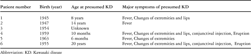
From 1980 to 2003, 6 male patients were referred to our hospital, with dates of birth ranging from 1947 to 1963. Of the 6 patients, 4 had several major symptoms raising the suspicion that the coronary arterial lesions were the sequels of prior acute Kawasaki disease (Table 1). In 1 patient, there was a history of unknown fever, and coronary arterial lesions were known to be present. Based on the coronary angiographic appearances, the coronary arterial lesions in the sixth patient were typical for Kawasaki disease. The period of follow-up ranged from 2 to 19 years, with a median of 3 years. We analyzed the clinical features and clinical courses of all the patients.
Table 1. History of presumed Kawasaki disease.

All patients underwent a 12-lead electrocardiogram, cross-sectional echocardiography, a 24-hour Holter electrocardiogram, and cardiac catheterization and coronary angiography. The age at the time of the coronary angiograms ranged from 35 to 55 years. We measured left ventricular ejection fraction and left ventricular end-diastolic volume by left ventriculography. Treadmill testing and radioisotope myocardial imaging were performed to detect myocardial ischemia in all patients. Electron beam computed tomography was done in four patients, and electrophysiologic studies were done in two patients.
Results
Onset and age at diagnosis of the coronary arterial lesions presumed to be the consequence of Kawasaki disease
At the time of discovery of the coronary arterial lesions, the ages of the patients ranged from 26 to 48 years, with a mean of 34 years. The presenting features were: chest pain in 2, general malaise in 1, recognition of electrocardiographic abnormalities in 2, and a history of unknown fever in one. Symptomatic myocardial infarction had occurred in one, while 2 patients had no symptoms.
The first patient had developed chest pain on effort at the age of 41 years. The second patient had an acute myocardial infarction when 26 years old. The third patient, aged 32, complained of general malaise and was diagnosed as having an ischaemic cardiomyopathy after a previous myocardial infarction. He experienced palpitations and transient unconsciousness on two or three occasions after that time. At the age of 48 years, he experienced presyncope, with a cold sweat and an oppressive sensation in his chest immediately after getting up to the toilet at midnight. The fourth patient was noted to have an abnormal Q wave in lead III, and a QS pattern in leads V1 and V2, on an electrocardiogram done when he visited the hospital because of headache at the age of 34 years. When he had a common cold at the age of 43 years, he visited another hospital because of fever, and an electrocardiogram then revealed a wide QRS tachycardia at a rate of 198 beats per minute. The fifth patient was diagnosed as having an abnormal electrocardiogram during a routine medical at the age of 35 years old. The sixth patient had a history of unknown fever, with the major symptoms of Kawasaki disease, when he was aged 20. His mother had recorded the symptoms of the acute episode at that time, but he had not undergone cardiac examination, because the diagnosis of Kawasaki disease was not suspected. He visited our hospital at the age of 48 years. Although he had been asymptomatic, a calcified giant aneurysm of the left anterior ascending artery was suspected on the basis of cross-sectional echocardiography, and was confirmed by electron beam computed tomography (Fig. 1).

Figure 1. Calcification of giant aneurysms as seen in our sixth patient. The upper panel shows calcification of the right coronary artery. The lower panel shows a giant aneurysm with calcification at the proximal portion of the left anterior descending artery.
Risk factors for atherosclerosis
Of the patients, 5 smoked, with the duration of smoking ranging from 4 to 20 years. Obesity with a body mass index greater than 25 was present in three. Hyperlipidemia and hypertension were found in two. None of the patients was diabetic.
Electrocardiogram at rest
The resting electrocardiogram was abnormal in five patients, and normal in the sixth. An abnormal Q wave was present in three, a QS pattern in the chest leads in one, and poor R wave development in the chest leads in five (Fig. 2). Abnormalities of the ST-T segments and T waves in V5 and V6 were observed in one.

Figure 2. The 12-lead electrocardiogram from our third patient. Note the flat T waves in leads V5 and V6, and an abnormal Q wave in leads III and aVF.
Coronary angiography
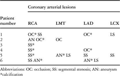
Among the 6 patients, 5 had multi-vessel disease (Table 2). Three vessel disease was found in three, and two vessel disease in two. The other patient had single vessel disease. Giant calcified aneurysms in the proximal portion of the major branches were present in five patients. Localized stenosis, with a giant calcified aneurysm, was detected in two. Segmental stenosis was found in five patients (Fig. 3), with 3 patients showing complete occlusion. Coronary arterial calcification was found in all patients.
Table 2. Coronary arterial lesions in coronary angiograms.


Figure 3. The coronary angiograms from our sixth patient. The left panel shows segmental stenosis of the right coronary artery, while the right panel shows the left coronary angiogram, which reveals a giant aneurysm with calcification of the left anterior descending artery.
Left ventricular ejection fraction and ventricular arrhythmia
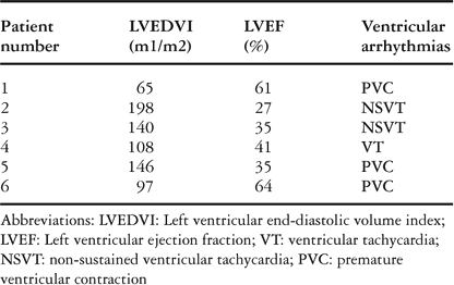
Left ventricular end-diastolic volumes, left ventricular ejection fractions, and ventricular arrhythmias are shown in Table 3. In 3 patients, the left ventricular ejection fraction was less than 40%. The left ventricular end-diastolic volume was increased in 3 patients. Sustained ventricular tachycardia was found in one, and non-sustained ventricular tachycardia in 2 patients. In these 2 patients, rapid ventricular tachycardia with syncope was induced during electrophysiologic studies. Although catheter ablation was attempted in one, it was not effective. A defibrillator was implanted in these 2 patients (Table 4).
Table 3. Left ventricular function and arrhythmias.

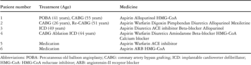
Table 4. Treatment.

Radioisotope myocardial imaging and treadmill test
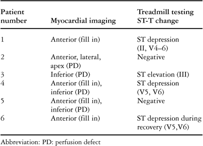
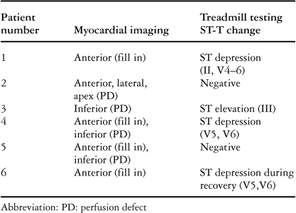
Exercise myocardial imaging results are shown in Table 5. Complete perfusion defects were discovered in 4 patients, indicating irreversible myocardial damage. Perfusion defects that filled in the late phase were discovered in 3 patients, indicating viability of the myocardium. ST-T changes during treadmill testing were detected in 3 patients.
Table 5. Myocardial imaging and treadmill testing.
Coronary arterial revascularization
This was performed in 3 patients (Table 4). The first patient underwent percutaneous transluminal balloon angioplasty at the age of 41 years old, followed by coronary arterial bypass grafting when he was 55 year old. When 26 years old, the second patient had saphenous vein grafts placed to the left anterior descending artery and the right coronary artery. The saphenous vein graft to the right coronary artery was occluded 11 years after the operation. He underwent recoronary arterial bypass grafting at the age of 51 years. The left internal thoracic artery was anastomosed to the first diagonal branch, and a saphenous graft was placed to the inferior interventricular artery. Although the grafts were patent, the left ventricular ejection fraction was severely decreased, and he was found to have deteriorated to the third class of the categorization established by the New York Heart Association. Bone marrow transplantation was tried because of severe left ventricular dysfunction at the age of 55 years. This proved unsuccessful. In the fourth patient, grafts were placed to the left anterior descending and inferior interventricular arteries when he was aged 44 years.
Medications
All 6 patients were given aspirin, and 3 received warfarin for antithrombosis. Diuretics, and beta-blockade were administered to 2. In 3 patients, allopurinol and HMG-CoA were used, whilst one patient each received amiodarone, mexiletine, digoxin, pimobendane, a calcium blocker, and a blocker of angiotensin-II receptors, respectively, as shown in Table 4.
Discussion
It is almost certain that, at least in Japan, there are aymptomatic adults with coronary arterial lesions caused by Kawasaki disease occurring before and after 1967 who remain undiagnosed. The diagnosis of Kawasaki disease is based on the major characteristic clinical features noticed in its acute phase.1 These acute symptoms, however, are self-limiting, and all features do not occur in every patient. Furthermore, the severity of the symptoms varies, and atypical Kawasaki disease may also cause coronary arterial lesions. Consequently, it has not been unusual to miss the diagnosis of both acute Kawasaki disease, and the subsequent development of coronary arterial lesions. Because the parents do not always remember the characteristic symptoms of the acute illness, the diagnosis of Kawasaki disease may remain unsuspected for many years. Knowledge of the characteristic features of coronary arterial lesions in this population, therefore, should prove useful for the eventual diagnosis of Kawasaki disease.
One typical feature is a calcified giant aneurysm, usually involving the proximal portion of the major branches of the coronary arteries, and not including small vessels at peripheral arteries in most of cases.3 An aneurysm at the bifurcation of the main stem of the left coronary artery is particularly characteristic. Further, affected segments and almost normal segments may coexist in the same patient, simply because the degree of involvement of the affected coronary arterial wall due to acute vasculitis can vary markedly.4 In the setting of atherosclerosis, the coronary arteries are diffusely affected, with the disease involving not only the major branches, but also the peripheral coronary arteries. In the coronary arterial lesions produced secondary to Kawasaki disease, calcification is usually limited to the site of the large aneurysms seen during the acute phase of the disease. Furthermore, calcification is detected in the outer side of the coronary aneurysms, because the calcification is the consequence of thickening of the intimal layer of the coronary arterial wall late after the initial destruction of the wall due to acute vasculitis. On the other hand, calcification seen in the setting of atherosclerosis is not always localized. Much more frequently, it is diffuse and widely spread, often being seen in vessels of normal diameter.5 In our opinion, therefore, the distribution of the coronary arterial lesions, and the nature of the coronary arterial calcification, serves to distinguish the two populations. Atherosclerosis, in addition, would be expected to be found not involving only the coronary arteries, but also other major arteries, such as the carotid arteries and the aorta. These calcified aneurysms can be easily detected by computerized tomography,6 making this technique a useful screening test for presence of calcified coronary arterial aneurysms.
Another typical lesion is segmental stenosis, which is often found in patients with a history of Kawasaki disease.3 It implies the development of several new small vessels, which represent recanalization after initial coronary arterial occlusion. Such collateral vessels are often well developed in the lesions produced by Kawasaki disease, forming after the initial occlusion of the major vessels. The presence of multi-vessel disease was a typical finding in most of our patients who developed cardiac events as adults. Such multi-vessel disease is more likely to lead to cardiac events, particularly those which result in left ventricular dysfunction.
Our 4 patients had had characteristic coronary arterial lesions, and a low left ventricular ejection fraction, from their thirties to forties. Patients with severe coronary artery lesions caused by presumed Kawasaki disease are likely to have cardiac events earlier in life than the normal Japanese population. Another 2 patients became symptomatic by developing ventricular tachycardia in their forties.7 Left ventricular dysfunction without symptomatic myocardial infarction can exist unrecognized for many years in such patients. Ventricular tachycardia developing with ageing after previous myocardial infarction is usually a late complication in patients with left ventricular dysfunction due to the coronary arterial lesions produced by Kawasaki disease.8
In the past, some patients had been diagnosed after cardiac events such as acute cardiac infarction or sudden death.9–14 The ideal goal in this population, of course, is to prevent a fatal cardiac event. To detect patients with coronary arterial lesions due to previous Kawasaki disease, it is important to include an electrocardiogram in annual health examinations. Not only abnormal Q waves and a QS pattern, but also low voltage R waves in the chest leads and flat T waves should arouse suspicion. If coronary arterial lesions are suspected, electron beam computed tomography and multislice spiral computed tomography should be used to screen patients with a history suggestive of Kawasaki disease. Calcification of the coronary arteries at a young age is characteristic in this population. Furthermore, it is probable that the population previously affected by Kawasaki disease is the one which develops symptoms in early adulthood, and includes patients with multi-vessel disease or left ventricular dysfunction. The cardiac events often occur in association with other risk factors for atherosclerosis, such as smoking and obesity,11, 12 risk factors which ideally should be avoided from a young age. Long-term and careful follow-up is essential for this population, even if they are asymptomatic. Treatment becomes more essential as they age.
In conclusion, there is a population of adults with undiagnosed coronary arterial lesions caused by previous Kawasaki disease. Coronary angiography in such patients is characterized by segmental stenosis, and presence of giant calcified aneurysms at the proximal portions of the major branches. Patients with multi-vessel disease or left ventricular dysfunction caused by undiagnosed Kawasaki disease can develop serious cardiac events as they age, becoming symptomatic many years after the initial attack of Kawaski disease in childhood.
Acknowledgements
We thank Professor Peter Olley for his kind help with the English language.