Introduction
Vidius (1509–1569) identified the vidian nerve and canal in the floor of the sphenoid sinus. The vidian nerve, through its autonomic fibres, is thought to have a role in rhinitis, epiphora, ‘crocodile tears’, Sluder syndrome, cranial and cluster headaches, and corneal ulceration.Reference Osawa, Rhoton, Seker, Shimizu, Fujii and Kassam1
Desmoplastic fibroma is a rare, benign neoplasm of bone characterised by aggressive local infiltration of fibroblasts or myofibroblasts and mature collagen, which typically occurs in young patients.Reference Moorjani and Stockton2, Reference Wippold, White, Jamroz, Haughey and Forsen3
To our knowledge, the presented patient represents the first reported case of a child with a desmoplastic fibroma of the vidian canal, compressing the vidian nerve.
Case report
History and examination
In November 2009, a 12-year-old girl with several years' history of right-sided facial pain was referred to the rhinology clinic of Guy's and St Thomas' National Health Service Foundation Trust (London, UK). Her symptoms were felt most intensely around the right ear and temporomandibular joint. She had been seen extensively in pain clinics, as well as in psychotherapeutic clinics, and had received unsuccessful conservative treatment. The patient suffered from daily pain, causing numerous school absences, and also reported several episodes of right-sided epistaxis and nasal congestion.
Imaging
In March 2008, the patient had undergone computed tomography (CT) and magnetic resonance imaging (MRI) scans at another centre, which had demonstrated a small lesion in the right sphenoid bone. However, this had not been thought to be related to the underlying symptoms.
In November 2009, repeated MRI and CT scans showed an expansile mass measuring 9 × 8 × 10 mm, which involved the right vidian canal and indented the floor of the right sphenoid sinus medial to the carotid artery (Figure 1).
Axial magnetic resonance imaging scan showing the mass in the right vidian canal (arrow). AF = anterior foot; R = right; L = left; PH = posterior head

Operation
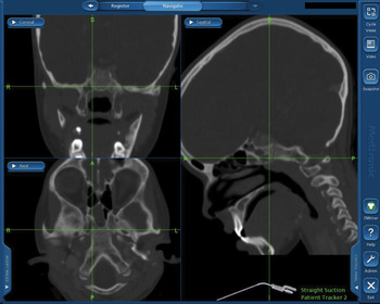
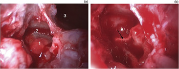
In December 2009, the patient underwent a complete endoscopic surgical resection, aided by the FusionTM ENT navigation system (Medtronic Navigation, Louisville, CO, USA) (Figures 2 and 3). A transnasal, trans-septal, trans-sphenoidal route was utilised, via the right nostril. A biopsy of the lesion was sent for histopathological examination.
Screengrab showing coronal, sagittal and axial computed tomography imaging scans of the right vidian canal, as presented using the FusionTM ENT navigation system.

(a) Pre-operative endoscopic image showing the lesion compressing the nerve (1), the right vidian canal (2) and the right sphenoid sinus (3). (b) Post-operative endoscopic image after resection of the lesion, showing the freed vidian nerve (arrow).

The patient was discharged the day after surgery, and did not report any post-operative complications.
Follow up
By post-operative day seven, the patient's symptoms had resolved.
At a second post-operative follow-up appointment, four months post-operatively, no further facial pain was reported.
Histopathological findings
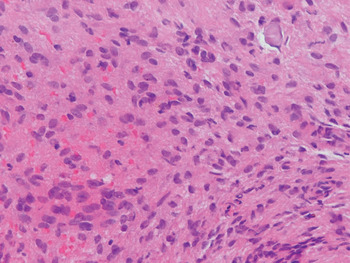
Upon histopathological analysis, the lesion consisted of a moderately cellular proliferation of cytologically bland spindle cells, some with the appearance of myofibroblasts. These were separated by collagen, and focally by a myxoid matrix. Small foci of calcification were present within the lesion. There was no evidence of mitosis or nuclear atypia. A subset of the spindle cells stained positive with smooth muscle actin and desmin stains, confirming myofibroblastic differentiation; however, these cells were negative for glial, neural and epithelial marker staining. The lesion appeared to be closely associated with the bone, but there was no evidence of osteoclastic or osteoblastic activity.
The features were those of a fibrous lesion, and were similar to what one would expect for fibromatosis of soft tissue. The lesion was therefore diagnosed as a desmoplastic fibroma (Figure 4).
Photomicrograph showing cytologically bland spindle cells with extravasated red blood cells and a collagenous background. (H&E; ×20)

Discussion
The vidian nerve carries parasympathetic fibres from the greater superficial petrosal nerve, sympathetic fibres from the deep petrosal nerve, and the pericarotid nerve.Reference Osawa, Rhoton, Seker, Shimizu, Fujii and Kassam1, Reference Ginsberg, De Monte and Gillenwater4 It is located in the pterygoid canal along with the vidian artery. Microanatomically, the pterygoid canal is located approximately 5 mm posterior and lateral to the sphenopalatine foramen. The canal is approximately 1 cm long and has a funnel-like mouth at the posterior aspect of the pterygopalatine fossa. It is separated from the foramen rotundum by a distinct vertical osseous ridge forming the lateral aspect of the mouth of the canal in the pterygopalatine fossa.Reference Konno5–Reference Cheong, Kim, Bak, Kim, Oh and Park7
Desmoplastic fibromas are extremely rare tumours. They are slowly progressive, with well differentiated cells that produce collagen. The mandible is the most common site, accounting for approximately 40 per cent of cases involving bony sites, followed by the femur and pelvis. This benign tumour is characterised by aggressive local infiltration. It occurs most frequently in the first three decades, and is found equally in males and females.Reference Moorjani and Stockton2
The radiological appearance of desmoplastic fibroma is nonspecific. Diagnosis is only established by histological examination.Reference Kendi, Erakar, Saglik, Yildiz and Erekul8 Computed tomography and MRI are useful to delineate the lesion and its relationship to adjacent structures. On MRI, desmoplastic fibromas display a heterogeneous appearance; unenhanced T1-weighted images show nonspecific low signal intensity, while T2-weighted images show intermediate to high signal intensity with areas of low intensity. The presence of such low intensity regions on T2-weighted images is helpful in determining the diagnosis.Reference Moorjani and Stockton2
Histopathologically, these tumours display uniform fields of fibroblasts amid abundant collagen, and resemble locally aggressive desmoid tumours of the abdominal wall. Hypercellularity, nuclear pleomorphism, mitotic activity and any traces of odontogenic epithelium are absent, differentiating the lesion from fibrosarcoma and ameloblastoma. Although these lesions do not have a capsule, the margins tend to be well defined, explaining the compact appearance on cross-sectional imaging.Reference Wippold, White, Jamroz, Haughey and Forsen3
Treatment of desmoplastic fibroma consists of wide excision of the lesion. With adequate excision, this tumour is usually curable. Radiotherapy has been reported as an effective treatment method. However, in young patients the detrimental effects of radiation on growth and the increased risk of malignancy make this an unappealing treatment option in most instances.Reference Kendi, Erakar, Saglik, Yildiz and Erekul8
• Desmoplastic fibromas have a nonspecific radiological appearance; diagnosis is only established on histopathological examination
• Treatment consists of wide excision of the lesion; if excision is adequate, the lesion is usually curable
• In the presented case, earlier diagnosis and resection would have avoided delays in resolving the child's disabling pain, emphasising the importance of early referral of unusual cases to tertiary centres
Our patient's lesion was accessible endoscopically, enabling macroscopic clearance with minimal peri-operative morbidity. However, diagnosis and resection of this rare lesion at an earlier stage would have avoided delays in resolving the child's disabling pain. This emphasises the importance of early referral of unusual cases to tertiary centres.