Implications
Following the first successful cloning of a mammal from an adult somatic cell (‘Dolly the sheep’), it has taken approximately 10 years to produce the world’s first cloned swamp buffalo. This accomplishment represents the outcome of a long-term programme of research and development in buffalo and cattle reproduction, combined with extensive on-farm experience. The technique may play a role in the conservation of the valuable Thai swamp buffalo.
Introduction
Nuclear transfer of adult somatic cells from farm animals is the most efficient technique for obtaining large numbers of genetically identical animals (Kato et al., Reference Kato, Tani, Sotomaru, Kurokawa, Kato, Doguchi, Yasue and Tsunoda1998), and could greatly enhance current agricultural production (Lu et al., Reference Lu, Jiang, Li, Zhang, Sun, Luo, Wei and Shi2011). Approximately once per year, since the birth of Dolly, a viable cloned sheep derived from adult mammalian cells (Wilmut et al., Reference Wilmut, Schnieke, McWhir, Kind and Campbell1997), has been produced successfully, along with several other cloned mammals (Lu et al., Reference Lu, Jiang, Li, Zhang, Sun, Luo, Wei and Shi2011). The production of cloned buffalo (Bubalus bubalis) is the latest addition to these successes in the application of the nuclear transfer technique. Lu et al., (Reference Lu, Jiang, Li, Zhang, Sun, Luo, Wei and Shi2011) noted that cloning of buffalo presents a greater challenge than other livestock. This may be attributable to their low fertility, as evidenced by delayed puberty, silent oestrous, long postpartum ovarian inactivity and lower oocyte recovery number from ovaries in comparison with cattle (Shi et al., Reference Shi, Lu, Wei, Cui, Yang, Wei and Liu2007). Our first reports on swamp buffalo cloning showed that foetal fibroblast could be reprogrammed following transfer into recipient cytoplasts to produce cloned embryos up to blastocyst stage (Parnpai et al., Reference Parnpai, Tasripoo and Kamonpatana1999). Moreover, foetal and granulose cells exhibited similar abilities (Parnpai et al., Reference Parnpai, Tasripoo and Kamonpatana2000). Subsequently, our team succeeded in producing the first cloned Brangus cattle calf born on 6 March 2000, followed by a second that did not survive delivery. A third cloned American Brahman calf was born on 3 April 2001 (Kamonpatana, Reference Kamonpatana2003).
Cloning of buffalo presents a number of challenges, both logistic as well as technical. First, access to swamp buffaloes (typically owned by remotely located small farmers) is time-consuming. Moreover, the challenge of preparing animals for implantation is compounded by irregular oestrous with weak oestrous symptoms (Kamonpatana, Reference Kamonpatana2003); this hampers detection of the correct date for transfer of cloned embryos. As a result, work on cloning of buffaloes has made significant progress, mainly at the laboratory level (e.g. Parnpai et al., Reference Parnpai, Tasripoo and Kamonpatana2001, Reference Parnpai, Tasripoo and Kamonpatana2002 and Reference Parnpai and Tasripoo2003). Kamonpatana (Reference Kamonpatana2003) also highlighted key preimplantation differences between cattle and buffalo that reduce the cloning success rate in buffalo relative to comparable species (Parnpai et al., Reference Parnpai, Tasripoo and Kamonpatana2002). Thus, specialized techniques for swamp buffalo must be developed to overcome these challenges and increase the success rate.
A significant component in determining the success of somatic cell nuclear transfer (SCNT) is the source of the nuclear donor cells (Powell et al., Reference Powell, Talbot, Well, Kerr, Pursel and Wall2004). Skin cells offer the advantages of easy accessibility and non-invasiveness, without sex or age limitations (Kubota et al., Reference Kubota, Yamakuchi, Todoroki, Mizoshita, Tabara, Barber and Yang2000). Moreover, skin cells offer an additional attraction as the phenotype of the animal is known (Forsberg et al., Reference Forsberg, Strelchenko, Augenstein, Bethauser, Childs, Eilertsen, Enos, Forsythe, Golueke, Koppang, Lange, Lesmeister, Mallon, Mell, Misica, Pace, Pfister-Genskow, Voelker, Watt and Bishop2002). Finally, ear fibroblasts can be obtained without risk to the donor animal’s health and may be more convenient and practical to harvest and sub-culture than other cells (Lu et al., Reference Lu, Shi, Wei, Yang and Wei2005; Hosseini et al., Reference Hosseini, Moulavi, Foruzanfar, Hajian, Abedi, Rezazade-Valojerdi, Parivar, Shahverdi and Nasr-Esfahani2008). Moreover, Tasripoo et al. (Reference Tasripoo, Srisakwattana, Sophon, Nualchuen and Usawang2007) showed that there was no difference in blastocyst formation among buffalo donor skin fibroblasts of a 6-month old calf, a 2-year old and a 4-year old buffalo. Furthermore, Saikhun et al. (Reference Saikhun, Kitiyanant, Songtaveesin, Pavasuthipaisit and Kitiyanant2004) found no significant difference in the development potential of embryos reconstructed with foetal, calf and adult fibroblast cells.
Shi et al. (Reference Shi, Lu, Wei, Cui, Yang, Wei and Liu2007) reported successful cloning of swamp buffalo by SCNT of foetal fibroblast and granulose cells. Of the three female cloned calves produced, only one survived. Subsequently, Singla et al. (Reference Singla, Singh, Shah, George, Chauhan, Manik and Palta2010), using handmade cloning, produced the world’s first two cloned dairy buffalo (Murrah) derived from the embryonic stem cells. However, despite a number of attempts using SCNT, no successful cases have thus far been reported on live-cloned swamp buffalo derived from adult ear fibroblasts. Our previous study (Parnpai et al., Reference Parnpai, Tasripoo and Kamonpatana2000) found that foetal fibroblasts and granulosa cells demonstrated a similar capacity for reprogramming following transfer into recipient cytoplasts to produce cloned embryos up to the blastocyst stage.
This paper describes the first successful application of SCNT in producing live-cloned swamp buffalo derived from adult ear skin fibroblasts. The technique carries significant implications for breeding and conservation of rare species, particularly owing to the greater practicality of using adult ear fibroblast relative to foetal fibroblast. In this study, a prized Thai swamp buffalo with high genetic potential was selected as the cell donor. It is hoped that as a result of this proof of concept, SCNT may be further developed as a technique to conserve rare and endangered breeds.
Material and methods
Chemicals
Unless otherwise stated, chemicals were purchased from Sigma Aldrich Corp. (St. Louise, MO, USA).
Production of adult SCNT embryos
The procedure for producing embryos by SCNT followed the method of Parnpai et al. (Reference Parnpai, Tasripoo and Kamonpatana1999, Reference Parnpai, Tasripoo and Kamonpatana2001 and 2002) with some modification as described below.
Preparation of donor cells
A male swamp buffalo (5 years old) with specific colour markings was selected as the cell donor. Tissue samples (0.25 cm2) were collected by ear biopsy and stored in normal saline. The samples were washed several times in phosphate buffered saline (PBS), then manually cut into small pieces, and treated with CaCl2 and MgCl2-free PBS containing trypsin and ethylene diamine tetraacetic acid (EDTA) for 5 min, before washing with MEM+10% (v/v) foetal bovine serum (FBS; Gibco, Invitrogen, Grand Island, NY, USA). The tissue pieces were then placed in a 60-mm culture Petri dish (Nunc, TC dish, Vents nunclon, Roskilde, Denmark), covered with a glass slide and cultured for 7 days with 5 ml of Modified Eagle’s Medium (MEM; Sigma) plus 10% FBS+0.06 g/ml of penicillin G+0.10 g/ml of streptomycin and amphotericin B. The medium was replaced every 3 days. The tissue was cultured under a humidified atmosphere of 5% CO2 in air at 37°C. After 7 days, fibroblast cells had begun to grow out of these explants; the explants were then removed and the fibroblast outgrowths harvested by washing twice with Ca2+ and Mg2+-free PBS and then trypsinized with trypsin/EDTA. The fibroblast cells were counted and seeded in a 25 cm2 tissue culture flask. When the cells reached 90% confluence, at the third cell culture passage, they were collected by trypsinization and frozen in αMEM supplemented with 10% FBS and 10% dimethyl sulfoxide (v/v), and stored in liquid nitrogen. Frozen-thawed fibroblasts were cultured for five passages in MEM+10% FBS and used as donor cells. They were isolated from culture flasks supporting a fully confluent cell monolayer for 2 days in MEM+2 mM l-glutamine+0.5% FBS (v/v) before transfer. The cell monolayer was trypsinized and the cells washed by centrifugation in MEM. Cells (diameter=14–16 µm) with smooth membrane surfaces were selected for nuclear transfer.
In vitro maturation of oocytes
Buffalo ovaries were obtained from an abattoir and transported to our laboratory in 0.9% normal saline plus antibiotics at 25 to 30°C. Within 3 h of collection, the oocytes were aspirated from follicles with a diameter of 2 to 6 mm through an 18-gauge needle attached to a 10 ml disposable syringe. The oocytes were washed with TCM199+10% FBS (Gibco, Invitrogen). A group of 10 cumulus–oocyte complexes with compact cumulus cells were placed in a 50 µl drop under mineral oil in a 60×15 mm plastic dish (Nunc) of the medium TCM199 supplemented with 10% buffalo follicular fluid (v/v), 50 IU/ml human chorionic gonadotrophin (Chorulon Intervet-International BV., Boxmeer, the Netherlands), 0.02 AU/ml FSH and 1 µg/ml oestradiol-17β (E2). The cells were then cultured for 19 to 20 h at 38.5°C in a humidified atmosphere of 5% CO2.
The oocytes were freed from the cumulus by initial treatment with 0.2% hyaluronidase in Dulbecco’s phosphate buffered saline, supplemented with 1 mg/ml polyvinylpyrrolidone (K-30) with gentle pipetting. The oocytes were washed in TCM199 supplemented with 10% FBS and 12.5 mM Hepes. Oocytes with a visible first polar body were selected and transferred into drops (5 µl each) of 12.5 mM Hepes in TCM199 with 10% FBS, which had been previously placed under mineral oil.
Enucleation
The oocytes were washed five times with Hepes-buffered TCM199+20% FBS and enucleated by micromanipulation; each oocyte was then placed in a small drop of TCM199+10% FBS (v/v), 12.5 mM Hepes and 5 µg/ml of cytochalasin B. Oocytes with extruded first polar body (metaphase II arrest) were selected for enucleation. The zona pellucida above the first polar body was cut with a glass needle, and a small volume of adjacent cytoplasm removed, where possible, together with the first polar body (Supplementary Figure S1). Complete enucleation was confirmed by staining with Hoechst 33342 (Supplementary Figure S1).
Nuclear transfer, fusion and activation
A single donor cell was transferred into the perivitelline space of enucleated oocytes from 50-µl culture drops of TCM199+12.5 mM Hepes and 10% FBS (v/v) (Supplementary Figure S2). The oocyte–fibroblast complexes were then placed individually between electrodes and induced to fuse with 2 DC pulses of 30 V for 15 µs each by electro cell fusion (BTX Electro Cell Manipulator 200, San Diego, CA, USA) in Zimmerman fusion medium (Zimmermann and Vienken, Reference Zimmermann and Vienken1982; Parnpai and Tasripoo, Reference Parnpai and Tasripoo2003). The electrical pulse simultaneously induced initial oocyte activation (Kubota et al., Reference Kubota, Yamakuchi, Todoroki, Mizoshita, Tabara, Barber and Yang2000; Supplementary Figure S2). The nuclear transferred embryos were then washed seven times with TCM199+12.5 mM Hepes+20% FBS and incubated for 45 min at 38.5°C in a humidified atmosphere of 5% CO2. Fusion was then confirmed by microscopic examination. All the fused embryos were further activated by culturing with 50-µl drops of TCM199+12.5 mM Hepes+10% FBS+5 µM calcium ionophore A23187 for 5 min. The fused embryos/reconstructed embryos were washed five times and then cultured in SOFaa+10% FBS (v/v)+10 µg/ml cycloheximide+1.25 µg/ml cytochalasin D at 38.5°C in a humidified atmosphere of 5% CO2 for 5 h.
In vitro culture of nuclear transfer embryos
The nuclear transfer embryos were cultured in 50-µl drops of SOFaa+1% FBS, overlaid with mineral oil, and incubated at 38.5°C in a humidified atmosphere of 5% CO2, 5%O2 and 90% N2 for 48 h. Cleavage rates were recorded and the eight-cell stage embryos were selected and cocultured with buffalo oviductal epithelial cells of SOF+5% FBS at 38.5°C in a humidified atmosphere of 5% CO2. The medium was replaced every 2 days. The embryos were cultured for 6 days before transfer.
Recipients and embryo transfer
Recipient animals
The recipient animals were a group of 10 female swamp buffaloes, owned by farmers, in Saraburi province, located about 100 km from Bangkok. The animals were raised by grazing in natural pasture and paddy fields during the day, and fed with rice straw according to availability. Oestrous cycles occurred naturally without any specific programme for detection.
Embryo transfer, pregnancy monitoring and diagnosis of pregnancy
All candidate recipient animals were subjected to fertility diagnosis by plasma progesterone radioimmunoassay (Kamonpatana et al., Reference Kamonpatana, Chantaraprateep and Ngramsuriyaroj1981) to determine the non-return to oestrous recipients and to predict whether the transferable embryos would be conceived (Supplementary Material S1). They were also assayed for progesterone profiles in plasma every 10 days. The oestrous cycles of candidate recipients were checked by their respective plasma progesterone profiles. Before embryo transfer, the presence of corpus luteum in the ovaries of the recipient animals was checked by rectum palpation. Day 6 blastocyst in SOF+FBS 10%, were transferred into the recipient animal on 6 days after detection of oestrous (day 0=day of oestrous). On day 6, one to three embryos were deposited into the uterine horn non-surgically, ipsilateral to the ovary bearing a corpus luteum by a trans-cervical method. Following embryo transfer, pregnancy status was determined by monitoring blood progesterone levels at 10-day intervals, continuing until 3 months of pregnancy. Pregnancy and calving details were recorded.
Microsatellite analysis
A microsatellite analysis of genomic DNA from the cloned buffalo calf, the donor fibroblast cells, the surrogate mother and one random buffalo sample was performed with 12 primer sets: ITSLT006, ITSLT033, CSSN022, CSSM038, CSSM043, CSSM057, CSSM029, CSSM019, CSSM041, ETH121, BMC1013 and BM1818 to confirm the genetic identity of the offspring. The DNA of the donor cell was prepared from trypsinized ear skin fibroblast cells, but other samples were prepared from whole blood. The genomic DNA was extracted using a commercial kit (QiAamp® DNA blood Mini kit) according to the manufacturer’s instructions, and isolated DNA was then assessed for quantity and quality using 2% agarose gel electrophoresis.
The PCR primers for microsatellite markers were labelled with fluorescent dye (FAM, Yakima Yellow, ATTO550 and ATTO565; Schubbert and Rittler, Reference Schubbert and Rittler2012) synthesized by Microsynth AG, Switzerland. PCR amplification was carried out by denaturing at 95°C for 5 min, and 35 subsequent cycles at 95°C for 30 s, annealing for 56 to 60°C for 30 s, extension at 72°C for 30 s and a final extension at 72°C for 10 min. The products were separated by 2% agarose gel and stained with ethidium bromide to assess their quality and quantity. All microsatellite loci were analysed by detection on vertical electrophoresis units using 7% denaturing polyacrylamide. After electrophoresis at 60 V for 16 h, the gel was stained with EDTA (10 mg/ml) for 50 min, and DNA fragments were visualized by UV transilluminator, PCR product size was confirmed by Gene Mapper V3.5 software performed on ABI3130 automated sequencer (Applied Biosystem, Foster City, CA, USA).
Results
To the best of the authors’ knowledge, this cloned calf is the first swamp buffalo derived from adult ear skin fibroblast cells. The development of reconstructed embryos from frozen-thawed adult fibroblasts is shown in Table 1.
Development potential of swamp buffalo-cloned embryos using freeze-thawed ear skin fibroblast cells

Embryo transfer to recipients
The fresh embryos, 19 morula (Supplementary Figure S3) and 39 viable blastocysts (Supplementary Figure S3) were transferred (30 transfer attempts) into 12 recipient buffaloes (Table 2).
Pregnancy, calving and incidence of late embryo mortality (LEM) and foetal mortality (FM) of recipients after transferred fresh swamp buffalo-cloned embryo

Pregnancy diagnosis and calf birth after nuclear transfer
The percentage of pregnancy by day 45 following fresh cloned embryo transfer was 83.3% (Table 2). The pregnancy rate remained high (>80%) from day 45 to 60, but dropped to 50% at day 90, remaining at this level until day 120. By day 150, half of these losses occurred, at day 240, the pregnancy rate had fallen to 8.3% (one remaining recipient) where it remained until calving. Detection of aborted foetus was not determined. In October 2011, after a full-term pregnancy (326 days), the single remaining recipient naturally delivered a cloned swamp buffalo (Supplementary Figure S4).
Parturition proceeded naturally without any assistance. The calf weighed 40 kg at birth and was developmentally normal. The calf possessed similar phenotypes and phenotypical identity characteristics to the donor (Supplementary Figure S5). The results of microsatellite analyses shown in Table 3 confirmed that the cloned calf was genetically identical to the somatic cell donor. The calf has since been raised on the farm under a good feeding regime, and began grazing at about the age of 2 months. The cloned calf has shown normal physiological and behavioural parameters, and is currently ageing normally (Supplementary Figure S6).
Microsatellite analysis of nuclear transfer donor cells, calf blood, recipient blood and a non-related swamp buffalo

Genetic analysis of cloned calf
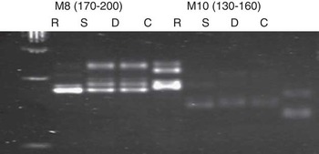
A comparison of the microsatellite alleles of genomic DNA from the cloned calf derived from the donor cell line from ear fibroblasts was made with 12 microsatellite markers. Parentage analysis showed that the cloned calf DNA was genetically identical to that of the donor adult skin fibroblasts, with a 100% genetic identity to the donor cell and a clear difference from the surrogate mother DNA (Table 3, Figure 1) at all 12 microsatellite markers.
Representative of microsatellite analyses of donor cell, cloned calf, non-related recipient using M10 and M8 markers in genomic DNA. R=recipient; S=cloned calf; D=donor cell; C=non-related swamp buffalo.

Discussion
This study has described the first known successful application of SCNT derived from adult ear skin fibroblast cells to produce a live-cloned offspring of swamp buffalo. The cloned calf displayed the unique phenotype of the donor karyoplasts, and microsatellite analysis with 12 markers confirmed that the cloned calf genotype was also identical to that of the donor animal. The blastocyst and hatched blastocyst rates in this study were similar to those reported in our previous studies (Parnpai et al., Reference Parnpai, Tasripoo and Kamonpatana2002; Tasripoo et al., Reference Tasripoo, Srisakwattana, Sophon, Nualchuen and Usawang2007). Our previous report on the average number of nuclei in cloned swamp buffalo blastocyst derived from foetal fibroblast at 7 days was 80.6±11.6 (Parnpai et al., Reference Parnpai, Tasripoo and Kamonpatana1999). The natural delivery of the cloned calf following a normal gestation period (326 days) was within the range observed in normal breeding or natural insemination (325–332 days), as reported by Virakul (Reference Virakul1987). However, differences in gestation period have previously been observed by Shi et al. (Reference Shi, Lu, Wei, Cui, Yang, Wei and Liu2007) in cloned Chinese swamp buffalo, in which gestation periods for cloned pregnancies were found to be longer than for natural pregnancies. In the current experiment, birth weight of the cloned calf was at the higher end of the normal range (mean value, 26.68; range 15 to 41 kg), as established by Thevarnanoharan et al. (Reference Thevarnanoharan, Vandepitte, Mohiuddin and Chantalakhana2001). This is significant in light of previous studies on cattle, which found a 47% rate of large offspring syndrome in calves derived from the skin, ear or liver donor cells (Heyman et al., Reference Heyman, Chavatte-Palmer, LeBourhis, Camous, Vignon and Renard2002). At present, at 2 years and 4 months of age (Supplementary Figure S6), the cloned calf exhibits normal growth and development (Supplementary Figure S7; unpublished data) and appears healthy. Plasma progesterone concentration can be used in the selection of non-pregnant buffaloes. Plasma progesterone levels on day 10 offer an important indicator, with progesterone levels elevated in pregnant animals, while remaining low in non-pregnant animals (Kamonpatana et al., Reference Kamonpatana, Chantaraprateep and Ngramsuriyaroj1981). The progesterone test is particularly useful with buffalo as oestrous may be difficult to determine (Dobson and Kamonpatana, Reference Dobson and Kamonpatana1986).
In this study, the 29% pregnancy rate at day 60 was higher than that reported by Saikhun et al. (Reference Saikhun, Kitiyanant, Songtaveesin, Pavasuthipaisit and Kitiyanant2004), who performed SCNT in synchronized buffaloes. In animals derived from cloning or in vitro culture procedures, a high incidence of placental abnormalities is found during both early and late gestation (Hill et al., Reference Hill, Burghardt, Jones, Long, Looney, Shin, Spencer, Thompson, Winger and Westhusin2000). Buffaloes that were pregnant on day 30 but not on day 45 or day 90 were considered to have undergone embryonic mortality or foetal mortality (FM), respectively (Vecchio et al., Reference Vecchio, Di Palo, Zicaralli, Grassi, Cammarano, D’Occhio and Campanile2007). In nuclear transfer, first trimester losses of more than 50% are common for nuclear transfer pregnancy in cattle, sheep and goats (Hill et al., Reference Hill, Burghardt, Jones, Long, Looney, Shin, Spencer, Thompson, Winger and Westhusin2000). This study recorded FM of ∼50%, whereas the proportion of late gestation losses between day 90 and calving reached 91.7%.
In bovine SCNT pregnancies, foetal losses continue to occur sporadically throughout the second and third trimesters, affecting 25% to 75% of pregnancies from day 90 onwards, depending on the donor genotype and cell line (Chavatte-Palmer et al., Reference Chavatte-Palmer, Camous, Jammes, Le Cleac’h, Guillomot and Lee2012).
The causes of abortion remain unclear, although incomplete genetic reprogramming or abnormal gene expression of nuclear transfer embryos may contribute to high abortion rates after embryo transfer (Saikhun et al., Reference Saikhun, Kitiyanant, Songtaveesin, Pavasuthipaisit and Kitiyanant2004). Incidence of loss between day 90 of gestation and calving was 43.7% for bovine adult somatic clones (Heyman et al., Reference Heyman, Chavatte-Palmer, LeBourhis, Camous, Vignon and Renard2002), which was much lower than those encountered in the current study. Serum starvation may often result in reduced cell survival and increased DNA fragmentation, which in turn cause subsequent high embryonic loss after nuclear transfer (Shi et al., Reference Shi, Lu, Wei, Cui, Yang, Wei and Liu2007). In bovine nuclear transfer, Hill et al. (Reference Hill, Burghardt, Jones, Long, Looney, Shin, Spencer, Thompson, Winger and Westhusin2000) recorded 50% to 60% loss of cloned pregnancies by day 100; these rates are comparable to the loss rates encountered in the current study.
It is generally accepted that oocyte quality has an impact on early embryonic survival, establishment and maintenance of pregnancy, foetal development and even adult disease (Hossein et al., Reference Hossein, Jeong, Park, Kim, Lee, Ko, Hyuk, Hoon, Kim, Hyun, Shin and Hwang2009). Buffalo ovaries are often obtained from a slaughterhouse and the genetic background of the oocytes is unknown. The frozen-thawed buffalo adult fibroblast cell was successfully used for cloning in this study. Our results demonstrate that the frozen buffalo ear skin fibroblast cells could support the development of matured oocytes into the embryos, and the subsequent establishment of pregnancy to full term. It has been reported that factors such as age, type and treatment of donor cells, as well as embryo culture methods influence survival rates in the first trimester, and neonatal viability at term (Hill et al., Reference Hill, Burghardt, Jones, Long, Looney, Shin, Spencer, Thompson, Winger and Westhusin2000).
Failure to reprogramme the donor genome is thought to be a main reason for the low efficiency of cloning (Rodriguez-Osorio et al., Reference Rodriguez-Osorio, Urrego, Cibelli, Eilertsen and Memili2012). The efficiency of swamp buffalo cloning in the present study is about 1.7% (1 calf from 58 embryos). However, adult SCNT protocols were set up in this study. Several factors may account for the success of this adult SCNT. First, the quality of oocytes obtained was excellent. Second, recipient oestrous cycles were closely synchronized with the stage of reconstructed embryos. Third, implantation of two or three embryos into the reproductive tract of the recipient animal could improve success rates (Hufana-Duran et al., Reference Hufana-Duran, Pedro, Salazar, Venturina, Duran, Takahashi, Kanai and Cruz2008). In water buffaloes, a higher success rate is achieved in recipients receiving embryos following natural oestrous, compared with recipients with synchronized oestrous (Hufana-Duran et al., Reference Hufana-Duran, Pedro, Venturina, Hufana, Salazar, Duran and Cruz2004). Fourth, regular oestrous cycle assessment and pregnancy diagnosis by progesterone monitoring may also have contributed to the success of this adult SCNT (Supplementary Material S1). Furthermore, the cloned embryos were transferred in December. During this period (November to January, cool season) reproductive performance (ovarian activity) is elevated (Promdireg et al., Reference Promdireg, Presicce, De Rensis, Singlor and Techakumphu2008) and conception rates are the highest (Chaikhun et al., Reference Chaikhun, Hengtrakunsin, De Rensis, Techakumphu and Suadsong2012). The birth of the cloned calf occurred during the peak calving period of swamp buffalo (August to October, rainy season). Aside from these technical considerations, close cooperation between farmers and the research team is also an important supporting factor for a successful pregnancy. The current application of SCNT offers a promising new avenue for reproductive technologies to breed and conserve, and offers convenience in that animal transport is no longer required. However, freezing of donor cells will be required for large-scale cloning. In this study, limitations to the efficiency of SCNT are indicated by the low success ratio. The outcome of this experiment represents the world’s third live-cloned buffalo produced by nuclear transfer, the world’s second buffalo produced by nuclear transfer and the world’s first cloned swamp buffalo derived from adult somatic cell (based on published research articles). While acknowledging the challenges and low efficiency presented by current methods, this study demonstrates that pregnancy can be established via this route, although survival to term remains low for in vitro and in vivo embryos. The study improves our understanding of buffalo reproduction and the factors influencing success rates for adult SCNT. Further refinement of the SCNT technique carries potential for genetic conservation of superior buffalo Landraces.
Acknowledgements
The authors are grateful to The Use of Nuclear Technology to Improve Artificial Insemination in Dairy Cattle and Swamp Buffalo Project; a scientific Thai-Netherlands Co-operative Project; and Research and Development Centre for Livestock Production Technology, Faculty of Veterinary Science, Chulalongkorn University, Thailand, for financial support and provision of research facilities. We would like to thank all colleagues at the Centre for their support and dedication to the Centre’s artificial breeding (IVM/IVF/IVC/Embryo transfer/cloning) of buffalo and cattle. Special thanks to Professor Maneewan Kamonpatana, the Centre’s founder, for her invaluable directing. Mr Anucha Noiwong was responsible for providing the donor buffalo ear specimen, whereas Woua Thong Farm made an invaluable contribution in maintaining the recipient animals and rearing the cloned calf. Finally, the authors would like to thank Mr Napat Ketpratum and DVM Somsak Srisongchet for medical assistance and Mrs Supitchaya Chetasing for her assistance in microsatellite analysis.