INTRODUCTION
Legumes are able to thrive in nitrogen-impoverished soils through their symbiotic interactions with soil-borne rhizobia bacteria, which infect legume roots, suppress plant defence reactions and finally create specialist nodules within which they fix atmospheric nitrogen (N2). Fixed N is supplied to the plant as ammonium, thus reducing the plant's need for additional fertilizer N. The resulting high protein foliage and seeds are crucial for agricultural sustainability, providing feeds for animals (as grazing or as silage) and grains directly for human consumption.
Nitrogen in drinking water is a significant public health issue in Europe and, because of these concerns, agro-ecosystems in Europe are being subjected to increasingly rigorous policies focused on reducing diffuse environmental pollution into waterways, often resulting in regulations to reduce nitrogenous fertilizer use. In this context, the potential role that legumes can play in facilitating reductions in nitrogenous fertilizer use while sustaining adequate levels of agricultural productivity and economic viability is important. However, legumes can also leave an adverse environmental footprint. Field research in southern Australia has suggested that N leakage from annual legumes contributes to stream and ground water pollution (Ridley et al. Reference Ridley, Mele and Beverly2003) and, in Europe, Doyle & Topp (Reference Doyle and Topp2004) found that more nitrate is leached into waterways from pure stands of perennial legumes than from fertilized grass swards. The contribution that the senescence and subsequent decay of legume root systems, with their high-nitrogen content (particularly in nodules), makes to rhizo-deposition of N and subsequent N leaching is currently unknown. However, root senescence in Trifolium repens ranged from 0·29 to 0·73 over 21 days under field conditions (Watson et al. Reference Watson, Ross, Bagnaresi, Minotta, Roffi, Atkinson, Black and Hooker2000), which is likely to be ecologically significant. In an agricultural context, Trifolium pratense L. (red clover) is of particular interest, since it is grown in both low-input organic and conventional farms as a component of mixed grass-clover swards or in monocultures, and is often harvested for silage (Abberton & Marshall Reference Abberton and Marshall2005). The more extensive use of clover has significant potential to facilitate the reduction of nitrogenous fertilizer application, but N-leaching from these systems remains a risk. In order to design breeding programmes that will improve the environmental footprint of clover, it is crucial to understand the contribution that senescence of root systems and nodules makes to N loss and the genetic control of changes in mortality.
The morphology of white and red clover differs markedly: white clover is stoloniferous and produces roots at each node, while red clover has a single, large tap root. In clovers, relevant studies have previously focused on the mobilization of N reserves during re-growth following defoliation (Corre et al. Reference Corre, Bouchart, Ourry and Boucaud1996), determining root and nodule turnover (Watson et al. Reference Watson, Ross, Bagnaresi, Minotta, Roffi, Atkinson, Black and Hooker2000) and relating their growth and persistence in the field to their underground morphologies (Butler et al. Reference Butler, Greenwood and Soper1959; Watson et al. Reference Watson, Ross, Bagnaresi, Minotta, Roffi, Atkinson, Black and Hooker2000). Repeated defoliation (every 11 days) of both white and red clover resulted in cyclic decay of old roots and nodules, followed by a renewal of root growth with production of new nodules. Both root and nodule turnovers were more extensive in white clover than in red clover (Butler et al. Reference Butler, Greenwood and Soper1959). In contrast, shading allowed little new growth of roots or nodules in white clover and none in red clover. A recent study carried out in sand culture described a time course of events for senescence and N release from red clover roots following permanent excision of the shoot (Bingham & Rees Reference Bingham and Rees2008). Under these conditions, red clover roots remained viable (as defined by positive turgor pressure and cellular respiration) for more than 20 days and significant nitrogen loss was only observed once viability started to decline, 34 days after excision.
The morphology of both determinate and indeterminate root nodules have been described in detail (Puppo et al. Reference Puppo, Groten, Bastian, Carzaniga, Soussi, Lucas, De Felipe, Harrison, Vanacker and Foyer2005) and is relatively well understood, but there are few markers of root and nodule health or senescence and this has hampered the development of a better understanding of both temporal changes in state and the mechanisms through which they occur. Root nodules have distinct patterns of growth, depending on the legume species. The present study focuses on indeterminate nodules, which are characteristic of clovers and their close relatives, including the model species Medicago truncatula. Indeterminate nodules grow throughout their lifetime and comprise clear zones: an actively growing meristem, an infection zone, a central nitrogen fixing region and, in mature nodules, a senescent zone at the basal end adjacent to the root. During natural ageing, the senescing zone gradually extends until it eventually engulfs the apical meristem and the nodule degenerates. This natural process occurs more rapidly under stressful events, such as the application of nitrate, drought or reduced carbon supply (for example, by shading or defoliation), thus changing patterns of nodule senescence. The onset of nodule senescence is known to involve wide-ranging changes in primary metabolism, reactive oxygen species and antioxidants. Key enzymes involved in carbon flux can provide useful indicators of tissue health and functioning. Glyceraldehyde-3-phosphate dehydrogenase (GAPDH) expression is often used as a reference control in studies of biological systems and continued nodule sucrose synthase activity is crucial for continued nitrogen fixation in legume nodules (Gordon et al. Reference Gordon, Minchin, James and Komina1999). Gene expression changes have been analysed in M. truncatula nodules following simple defoliation (Curioni et al. Reference Curioni, Reidy, Flura, Vogeli-Lange, Nosberger and Hartwig2000). Two transcripts were preferentially up-regulated: methylenetetrahydrofolate dehydrogenase1 (MTD1) was identical to an expressed sequence tag (EST) clone of M. truncatula following infection with Phytophthora medicaginis and MTD2 was highly homologous to an Arabidopsis thaliana gene coding for an RNA binding-like protein. More recently, studies have focused on gene expression during natural ageing of indeterminate nodules and in specific regions of dissected healthy, mature nodules (Van de Velde et al. Reference Van de Velde, Guerra, De Keyser, De Rycke, Rombauts, Maunoury, Mergaert, Kondorosi, Holsters and Goormachtig2006). This global transcriptomic approach of dissected healthy nodules of M. truncatula has elucidated some molecular changes from carbon sink to nutrient source in naturally ageing tissues of nodules and highlighted similarities between nodule and leaf senescence (Van de Velde et al. Reference Van de Velde, Guerra, De Keyser, De Rycke, Rombauts, Maunoury, Mergaert, Kondorosi, Holsters and Goormachtig2006). Cysteine proteases are involved in aspects of plant development and leaf senescence and are expressed in senescing zones of mature nodules of T. repens (Asp et al. Reference Asp, Bowra, Borg and Holm2004) and in senescing nodules of Astragalus sinicus (Naito et al. Reference Naito, Fujie, Usami, Murooka and Yamada2000).
In temperate agricultural systems, red clover is subjected to periods of temporary stress, such as harvest (defoliation) followed by re-growth under favourable conditions, or prolonged stress, when harvest may be followed by re-growth under the low light and low temperatures of autumn and winter months. At these times, senescent tissues release N into the soil. In this context, the present study employed temporary stress and an accentuated prolonged stress (inhibiting re-growth) to examine the temporal relationship of root system senescence and N leaching from red clover and associated molecular changes using the combined approaches of analytical chemistry, biochemistry and molecular biology. This permits a description of the process and timing of N leaching and natural bacterial and fungal colonization in stressed roots and nodules. The aim was also to identify and analyse differentially expressed plant genes in order to permit the development of molecular markers for use in red clover breeding programmes.
MATERIALS AND METHODS
Plant material and nodulation
Single plants of a diploid, outbreeding red clover cultivar, Milvus, were established in sterilized clay granules (Agsorb 8/16, Oil-Dri Ltd, UK) in 1 litre pots in a controlled environment: 12 h photoperiod; 400 μmol/m/s; 20°C in the light period and 15°C in the dark; 0·6 vapour pressure deficit (74% relative humidity at 20°C). Seedlings (Expt 1) and clonal cuttings from three randomly selected plants (Expt 2) were all watered daily throughout the experiments with N-free nutrients (Ryle et al. Reference Ryle, Powell and Gordon1978) and inoculated with sterile-grown Rhizobium leguminosarum bv. trifolii. The surface of the growth medium was covered with black polystyrene granules and pot positions were randomized. Plants of similar sizes (5·27±0·32 g dry wt) were used for the experiments: plants were 8 weeks old for Expts 1a and 1b and 16 weeks old for Expt 2.
Experimental treatments
Experiments 1a, 1b and 2 were designed to study root senescence and N loss under stress involving defoliation at 30 mm above the growing point, followed either by shoot re-growth (temporary stress, defoliation: D) or by shading each plant with heavy black polythene to give 0 μmol/m/s (prolonged stress, defoliation and shade: DS). Control plants (C) were neither defoliated nor shaded. Plant position and order of harvest was fully randomized. Means and standard error of the mean are presented. Data were analysed using analysis of variance (ANOVA; Expt 1a) or restricted maximum likelihood (REML) analysis of repeated measures (Expt 1b) in GenStat® for Windows™ 11th edition (Payne et al. 2008).
Experiment 1: senescence and N loss following temporary or prolonged stress in seed-grown plants
Seed-grown plants (8 weeks old at start of treatment) were left intact (C), defoliated (D), or defoliated and shaded (DS).
Experiment 1a: A total of 30 plants (three plants per treatment) were sacrificially harvested, for shoot and root fresh weights and root tissues for biochemical and molecular analyses. Three plants were harvested at day 0, followed by three plants per treatment every 7 days for 21 days. A further 36 plants were subjected to DS and at each weekly harvest 12 equivalent DS plants were returned to light for 14 days before final assessment; recovering plants were protected from desiccation by Cryovac® bags.
Experiment 1b: A total of 24×8 week-old plants (six plants per treatment and plant-free control pots) were used. Every 7 days for 28 days the first 30 ml leachate following watering with 100 ml N-free nutrient solution (Ryle et al. Reference Ryle, Powell and Gordon1978) was collected from plants into sterile tubes, frozen and stored at −20°C until analysis for total N, total oxidized nitrogen (TON) and amino acids. Samples for quantitative analysis of amino acids were acidified with 2 m HCl (2·5 ml/100 ml). Data were analysed after subtraction of data for leachate from plant-free control pots. The results from C and D plants were not significantly different so only data from C and DS plants are presented.
Experiment 2: prolonged stress in clonal plants over 10 days
A total of 33 clonal cuttings were taken from three plants. After 8 weeks, these clonal cuttings were transplanted to individual pots and grown for a further 8 weeks. Nine plants were left intact (C) and 24 plants were subjected to DS treatment (three plants per harvest, one of each genotype). Clonal cuttings reduced the genetic variability observed in seed-grown plants. Nodules (200 mg) and roots stripped of visible nodules. About 1 g of roots were harvested and the visible nodules stripped (about 200 mg) from 3×C plants at 1, 5 and 10 days (data combined from regularly spaced harvests for control values at day 0) and from DS plants at 1, 2, 3, 4, 5, 8, 9 and 10 days.
Growth analysis and tissue harvest
Root:shoot ratio was calculated on a fresh weight basis. Stems, leaves, pedicel, peduncles, flowers, embryos and intact root systems (1 g), nodules alone (100 or 200 mg) and stripped roots (500 mg or 1 g) were harvested for analysis. Stripped roots had all visible nodules removed by hand. All tissues were flash-frozen and stored at −80°C.
Histochemical staining and biochemical analyses
Evans Blue stain (0·25 g/100 ml) was used to differentiate visually living and dead root tissues in either fresh tissues or in fixed tissues cold-embedded in methacrylate resin (Baskin et al. Reference Baskin, Busby, Fowke, Sammut and Gubler1992). Tissues were immersed in 0·5 ml stain at 20°C for 20 min, washed with water (×8) before embedding for cutting or examining fresh under an Olympus binocular or BH-2 microscope. Images were captured using a Canon Powershot Pro1 digital camera (low magnification) or an Optronics digital camera coupled with Magnafire™ software (high magnification). All image modifications were performed with Adobe Photoshop Elements 7.0 software.
Root death index (RDI) was measured by extracting and quantifying the stain retained by root systems (Baker & Mock Reference Baker and Mock1994). Roots (3×50 mg fresh weight) were immersed in Evans Blue stain and washed as described above, blotted dry and stored at −20°C. Tissue was ground in 0·5 ml 1 g/100 ml sodium dodecyl sulphate (SDS), made up to 1 ml with ultra pure water, incubated at room temperature for 20 min and centrifuged at 8000 g for 5 min. Absorbance was read at 600 nm and RDI presented as a proportion: OD600 nm living roots/OD600 nm equivalent dead roots killed by boiling in water for 20 min.
Root catalase activity (Doulis et al. Reference Doulis, Debian, Kingston-Smith and Foyer1997) and soluble protein concentration (Bradford Reference Bradford1976) were determined on three replicates of 500 mg root samples. Soluble protein (Bradford Reference Bradford1976), sucrose synthase and GAPDH activities were measured in 200 mg nodules homogenized in 1 ml extraction buffer (Gordon et al. Reference Gordon, Minchin, James and Komina1999). The supernatant (50 μl) was assayed for sucrose synthase activity in the sucrose cleavage direction and GAPDH in the glycolysis direction. For sucrose synthase, the increase in absorbance at 340 nm (A 340) was measured spectrophotometrically at 30°C at pH 7·0 (Gordon et al. Reference Gordon, Minchin, James and Komina1999). For GAPDH, production of D-glyceraldehyde-3-phosphate was coupled with the oxidation of nicotinamide adenine dinucleotide plus hydrogen (NADH) in the presence of 3-phosphoglyceric phosphokinase. The decrease in A 340 was measured at 25°C at pH 7·6. All measurements were expressed as a proportion of control at day 0.
Analysis of leachate samples
Leachate samples were analysed for ammonia and amino acids by ninhydrin reagent (Winters et al. Reference Winters, Lloyd, Jones and Merry2002). For total N, organic N was oxidized to nitrate by potassium persulphate in a ultra-violet digester. For both total N and TON, the nitrate was then reduced to nitrite by hydrazine and the nitrite diazotized with sulphanilamide and coupled with N-1-napthylethylenediamine dihydrochloride to form an azo dye with an absorbance maximum at 550 nm. Analyses for total nitrogen (N) and TON were performed on a Bran+Luebbe AutoAnalyzer 3 continuous flow analyser (Bran+Luebbe GmbH).
Molecular analyses
RNA isolation and cDNA synthesis
Total RNA was extracted (Ougham & Davies Reference Ougham and Davies1990), DNAse treated using DNA Free™ kit (Ambion Ltd, UK) and purified using RNeasy® MinElute™ Cleanup kit (Qiagen, Germany). Double-stranded cDNA was synthesized using oligo dT primers and AMV Reverse transcriptase kit (Promega Corporation, USA) and purified using QIAquick PCR purification kit (Qiagen, Germany). Procedures using kits were performed as described in the manufacturers' protocols.
cDNA–amplified fragment length polymorphism (AFLP) gels
RNA extracted from nodules was used for AFLP analyses (Bert et al. Reference Bert, Charmet, Sourdille, Hayward and Balfourier1999) using cDNA as a template. Pre-amplified cDNA was amplified selectively using EcoRI and MseI primer pairs E40, 5′-GACTGCGTACCAATTCAGC-3′, E42, 5′-GACTGCGTACCAATTCAGT-3′; with M36, 5′-GATGAGTCCTGAGTAAACC-3′; M50, 5′-GATGAGTCCTGAGTAACAT-3′ and M53, 5′-GATGAGTCCTGAGTAACCG-3′. Polymerase chain reactions (PCRs) were performed using a 9600 Perkin Elmer thermocycler. Gels were run and visualized using silver nitrate staining (Tixier et al. Reference Tixier, Sourdille, Roder, Leroy and Bernard1997). Polymorphisms were noted and candidate sequences were excised, amplified and sequenced on an ABI-3100 Genetic Analyser (Applied Biosystems, Warrington, UK).
Confirmation and analysis of candidate gene expression
Gene fragments were isolated and purified (QiaexII agarose gel extraction kit, Qiagen) before sequencing. Sequence analysis was carried out using Vector NTI version 9.1 and the National Centre for Biotechnological Information (NCBI) website. Sequences were assembled and analysed using basic local alignment search tool (BLAST) and fast DNA and protein sequence alignment software package (FASTA). DNA sequences were aligned using ClustalW. Gene structure was analysed using multiple alignment construction and analysis workbench (MACAW) and Softberry (Mount KIsco, New York, USA; http://linux1.softberry.com/berry.phtml).
Expression patterns of candidate genes were confirmed by cDNA Southern hybridization. cDNA was amplified using the same primers as used for the cDNA–AFLP gels and the resulting gels also Southern blotted. Filters were probed non-radioactively with DIG Easy Hyb DIG-High Prime and detected using Anti-Digoxigenin-AP, Fab fragments (Roche Applied Science, Penzberg, Germany).
Isolation of Tp-cp8
Tp-cp8 complete coding sequence was isolated from a red clover bacterial artificial chromosome (BAC) library created from cultivar Milvus using primers designed for T. repens (Tr-cp1 forward, 5′-TGGATTGGAGGAAGAAAGG-3′), (Tr-cp1 reverse, 5′-AAATTGGTTGATTTGCTACAGC-3′] (Asp et al. Reference Asp, Bowra, Borg and Holm2004).
cDNA-PCR
cDNA-PCR was used to determine the expression pattern of four selected genes Tp-cp8, Tp-gapdh1, Tp-nsr1 and Tp-nse1. cDNA template concentration was normalized for red clover actin expression over 30, 35 and 40 cycles (×3). PCR procedure used primer sequences: actin SS2 forward (fwd), 5′-TGGGATGACATGGAAAAGATCTGGCA-3′; actin AS1 reverse (rev), 5′-AGATTGGCACAGTGTGACTCACACCATC-3′; Tr-cp1 fwd and rev as above; Tp-gapdh1 fwd, 5′-ACCACTCATTCATCAACC-3′; Tp-gapdh1 rev, 5′-TTGGGAAAGAGTAAGAGGCC-3′; Tp-nsr1 fwd, 5′ATATGCTGGCTCCACTTTCTCC-3′; Tp-nsr1 rev, 5′TGAAATGCAACGTTATAGCC-3′; Tp-nse1 fwd, 5′GAATTCAGCATCTACACAACA-3′; Tp-nse1 rev, 5′ACTAACAAGGCCTAGTTATCAA-3′ and FastStart Taq DNA Polymerase (Roche Applied Science, Penzberg, Germany) kit: denaturation at 95°C for 4 min, followed by 35 cycles denaturation at 95°C for 30 s, annealing at either 55 or 50°C for 30 s, elongation at 72°C for 45 s and final extension at 72°C for 4 min. Images were scanned in Multimage™ Light Cabinet (Alpha Inotek Corporation) and captured using AlphaImager™ 1200.
Detection of 16S/18S rDNA in nodules
Genomic DNA was extracted from 100 mg nodules using a Nucleon Phytopure DNA extraction kit (Amersham Life Bioscience). Hypervariable regions of the 16S or 18S rDNA regions were amplified by PCR using eubacterial primers 101F-GC clamp 5′-CGC CCG CCG CGC CCC GCG CCC GTC CCG CCG CCC CCG CCC GTG GCG GAC GGG TGA GTA A-3′ and 518R 5′-GGT ATT ACC GCG GCT GCT GG-3′ (Liu et al. Reference Liu, Marsh, Cheng and Forney1997) and fungal universal primers NS2 and fung5-C (Van Elsas et al. Reference Van Elsas, Duarte, Keijzer-Wolters and Smit2000). PCR products were separated using Ingeny PhorU DGGE system (GRI, UK) (Muyzer et al. Reference Muyzer, Dewaal and Uitterlinden1993). Individual bands were excised and incubated in 30 μl SUPW for 24 h at 4°C. Fragments were re-amplified and sequenced using GenomeLab™ DTCS Quick Start Kit and CEQ 8000 Genetic Analysis System (Beckman-Coulter). Forward and reverse sequences were aligned and checked for similarity with sequences deposited in the Genbank databases using BLAST (Altschul et al. Reference Altschul, Madden, Schaffer, Zhang, Zhang, Miller and Lipman1997).
RESULTS
Roots and nodules in healthy control seedlings and plants
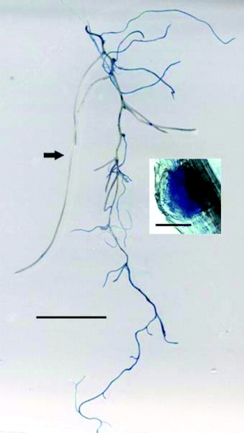
Living and dead tissues, which were differentiated visually in developing root systems using Evans Blue mordant stain, showed a changing pattern from germination to maturity. Few secondary roots of germinating seedlings retained the stain, but this increased with plant age. In older plants, the primary tap root remained white, whereas secondary, tertiary and fine roots retained stain to varying degrees. Some apparently healthy, unstained fine roots arose directly from roots which were heavily stained (Fig. 1). Transverse and longitudinal stained sections of such roots indicated localized death of the outer epidermal and cortical cells; the inner functioning vascular system and associated meristematic cells were unstained.
Root system stained with Evans Blue to visualize dead or dying cells. Living, white roots (arrowed) generated from Evans Blue stained, apparently dead roots. Scale: line=10 mm. Insert shows Evans Blue stained, apparently dead, immature nodule. Scale: line=0·5 mm.

Nodules also stained differentially with Evans Blue, indicating a mosaic of nodule survival in healthy plants. Nodules were at various stages of development from immature, small (<1 mm diameter) white nodules to mature, large (1–8 mm long) nitrogen-fixing nodules with single or multiple meristems; the latter nodules were pink in colour and firm in texture. In healthy root systems, 0·03 of nodules retained stain, including some immature nodules developing within the root (Fig. 1). In mature nodules, a dark brown region indicated a senescence zone at the basal end, which differentially retained Evans Blue stain but left N-fixing and meristematic zones unstained.
A novel, semi-quantitative method using Evans Blue mordant stain was adapted to estimate RDI in actively growing, healthy root systems. The RDI increased with plant age, increasing from 0·044± 0·0007 in roots of 7 day-old germinating seedlings to 0·231±0·0001 at 21 days and 0·346±0·0038 in 56 day old plants.
Experiments 1a and 1b: temporary or prolonged stress in seed-grown plants
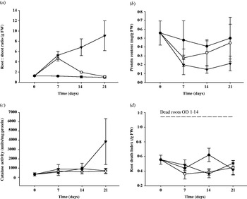
Root:shoot ratio differed significantly (P<0·001) according to imposed stress level and time (Fig. 2a). In control plants, root:shoot ratio remained almost constant at 0·50±0·071 throughout the experiment. In plants which were either defoliated or defoliated and shaded (treatments D and DS), root:shoot ratio increased 6·5-fold at defoliation: treatment D returned to the control level by 21 days, while treatment DS prevented recovery.
Root:shoot ratio, root protein content, catalase activity and RDI of control seed-grown plants compared to defoliated or defoliated and shaded plants over 21 days. (a) Root:shoot ratio, (b) protein content, (c) root catalase activity and (d) RDI. Control (C) (●); defoliated (D) (○); defoliated and shaded (DS) (▲). RDI for dead roots was 1·14. Vertical lines represent s.e.m. FW, fresh weight.

There were no significant differences between root protein content, catalase activity or RDI between treatments (Fig. 2b–d). Protein content of both D and DS plants declined following defoliation, recovering by 14 days in D plants, remaining low in DS plants (Fig. 2b). Catalase activity increased in roots of DS plants 21 days after defoliation, but there was considerable variation between plants (Fig. 2c). In contrast, RDI was similar for all treatments throughout the time course (Fig. 2d).
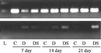
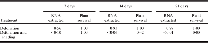
RNA concentrations extracted from roots of D and DS plants harvested at 7, 14 and 21 days after defoliation were expressed as a proportion of the RNA concentration in control roots (Table 1). Expression of actin and Tp-cp8 was detected even after 21 days DS (Fig. 3); all plants failed to survive 21 days DS, while nearly half the plants subjected to 14 days DS stress survived (Table 1).
Actin and Tp-cp8 expression in whole roots following stress over 21 days. cDNA–PCR analysis of whole senescing roots from healthy control plants (C), plants subjected to temporary stress (defoliated only, D) and prolonged stress (defoliated and shaded, DS) at 7, 14 and 21 days. PCR products of actin (upper band; 350 bp) and Tp-cp8 (lower band; 820 bp) were compared to 1 kb ladder (L).

RNA extracted from senescing roots and plant survival following defoliation or defoliation and shading. All values relative to control plants

Plant survival expressed as proportion of control plants (12 plants per treatment); RNA concentration expressed as proportion extracted from control roots (98–124 μg RNA/g fresh weight).
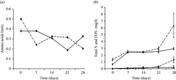
Experiment 1b: Amino acid, total N and TON concentrations in leachate were similar in controls and defoliated but not shaded plants (treatment D), therefore data for treatment D are not presented here. There was no significant difference in the concentration of amino acids between C and DS treatments (Fig. 4a); TON samples were below the limits of quantification until 28 days of stress (Fig. 4b) and could not be statistically analysed. There was no significant difference in total N (Fig. 4b) over time; however, total N increased by 214% and TON increased by 433% in DS treatment as compared with controls by 28 days. Sand alone, watered with nitrogen-free nutrient solution, contained low levels of total N (1·41±0·13 mg/l), TON (0·26±0·05 mg/l) and total amino acids (2·68±0·61×10−3 mM).
Analysis of leachate collected from control plants and plants defoliated and shaded for 28 days. Leachate samples analysed for (a) amino acids and (b) total N (▲) and TON (▪). Solid lines represent controls and dotted lines DS treatment. Data were based on 10 replicates per time point. Vertical lines represent s.e.m.

Experiment 2: prolonged stress in clonal plants
Biochemical analyses
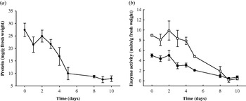
Nodules harvested from control plants after 1, 5 and 10 days from imposition of DS were pink and healthy. In contrast, nodules lost turgor and became dark brown or green during DS-induced nodule senescence. Protein concentration decreased to 28·8% of that in healthy nodules by 10 days DS (Fig. 5a). Sucrose synthase and GAPDH activities mirrored the decline in protein content and were expressed as units of activity/g nodule fresh weight; both decreased to 5–15% of control levels after 10 days DS (Fig. 5b).
Protein content and enzyme activities in nodules of clonal plants post-defoliation and shading. (a) Protein content of nodules, (b) sucrose synthase (●) and GAPDH (○) activities. Data were based on three replicate samples at each time point. Controls were taken at 1, 5 and 10 days and since there was no significant difference between replicate control samples over time, they were combined to give control values at day 0. Vertical lines represent s.e.m.

Molecular analysis
Nodules yielded more total RNA than stripped roots: 2160 μg/g compared with 112 μg/g in control tissues and 480 μg/g compared with 23 μg/g in senescing tissues.
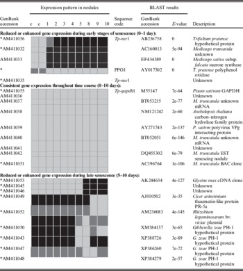
cDNA–AFLP analysis was used to identify differentially expressed sequences in healthy and senescing nodules by analysis of >500 cDNA amplification products and the expression of selected sequences was confirmed by cDNA Southern blots or cDNA-PCR (Table 2). While the majority of sequences were expressed in control and senescing tissues, three patterns of gene expression profile were identified: (a) consistent expression throughout the time course (0–10 days), (b) altered expression during early senescence (0–1 days) and (c) altered expression during late senescence (5–10 days).
cDNA–AFLP clones with differential or consistent expression in senescing nodules
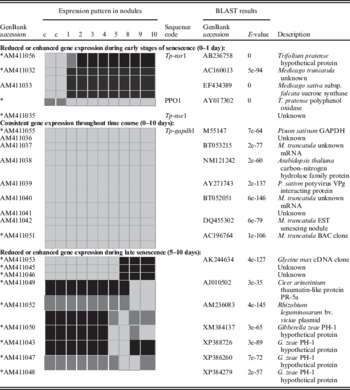
c control samples; 1–10 days post defoliation;
* cDNA–AFLP expression pattern confirmed by cDNA Southern blots; strong expression (light grey); weak expression (dark grey); no expression (black).
Various sequences from plant, bacteria and fungi were detected and identified (Table 2). At least seven plant sequences (including Tp-gapdh1) with consistent expression were identified. Five differentially expressed plant sequences (including sucrose synthase, polyphenol oxidase (PPO1), Tp-nse1 and Tp-nsr1) were detected during early nodule senescence (0–1 day), while in late senescence (5–10 days) at least five out of the nine differentially expressed genes were of fungal or bacterial origin. The functions of these fungal genes are unknown but all sequences have similarity to Gibberella zeae (anamorph Fusarium graminearum). Expression of three unknown plant genes decreased at this late stage, while a Rhizobium gene and a putative plant pathogenesis-related gene (which had a similarity of 0·93 to a thaumatin-like gene from Cicer arietanum PR-5a; Hanselle et al. Reference Hanselle, Ichinoseb and Barz2001), both increased.
Analyses of Tp-gapdh1, Tp-nsr1, Tp-nse1 and Tr-cp8 expression
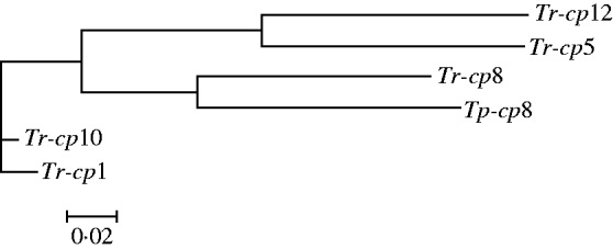
Tp-gapdh1, Tp-nsr1 and Tp-nse1 were identified from cDNA–AFLP analyses of nodules (Table 2). Tp-gapdh1 had a similarity of 0·90 to Pisum sativum chloroplast GAPDH; Tp-nsr1 was identical to Tp-211, a restriction fragment length polymorphism (RFLP) marker for red clover (Isobe et al. Reference Isobe, Klimenko, Ivashuta, Gau and Kozlov2003), which has a putative aspartyl protease domain; Tp-nse1 is an unknown gene. Expression patterns of these three genes were compared with the expression of a putative cysteine protease gene (Tp-cp8) homologous to Tr-cp8, which is expressed specifically in the senescing zone of nodules of white clover (Asp et al. Reference Asp, Bowra, Borg and Holm2004). Tp-cp8 was isolated from a red clover BAC library at the Institute of Biological, Environmental and Rural Sciences (IBERS), Aberystwyth (Winters et al. Reference Winters, Heywood, Farrar, Donnison, Thomas and Webb2009). Analysis by Softberry shows two exons. The predicted amino acid sequence of Tp-cp8 (AM409328) had a similarity of 0·95 to Tr-cp8 AAP32196, a similarity of 0·88 to Tr-cp1AAP32194 and Tr-cp10 AAP32197, and a similarity of 0·85 to Tr-cp5 AAP32195 and Tr-cp12 AAP32198 (Fig. 6).
Dendrogram of relationship of the predicted amino acid sequence of putative cysteine protease gene of T. pratense with amino acid sequences of cysteine protease family of T. repens. Tr-cp1 (GenBank accession no. AAP32194), Tr-cp5 (GenBank accession no. AAP32195), Tr-cp8 (GenBank accession no. AAP32196), Tr-cp10 (GenBank accession no. AAP32197), Tr-cp12 (GenBank accession no. AAP32198) and gene sequence Tp-cp8 (GenBank accession no. AM409328). The figure was generated in VectorNTI and formatted in GeneDoc.

Expression patterns of all four genes were analysed in cDNA from stems, leaves, pedicel, peduncles, flowers, embryos and root systems (data not shown): Tp-nsr1 was expressed in all tissues except mature leaves but both Tp-gapdh1 and Tp-nse1 were preferentially expressed in roots, with low expression in flower peduncles. Tp-cp8 was root-specific.
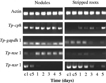
Expression patterns of Tp-cp8, Tp-gapdh1, Tp-nsr1 and Tp-nse1 were compared with actin in nodules and stripped roots of healthy and senescing root systems (Fig. 7). The four genes exhibited different expression patterns, but since gene expression patterns were consistent between days 5 and 10, only data for days 1–5 are shown. Both Tp-cp8 and Tp-gapdh1 were expressed consistently in healthy and senescing nodules, with no change in expression levels over 5 days. By contrast, Tp-nse1 expression increased and Tp-nsr1 decreased in senescing nodules. All four genes were expressed in stripped roots (Fig. 7): Tp-cp8 and Tp-nse1 expression was consistent in healthy and senescing stripped roots, while Tp-gapdh1 and Tp-nsr1 expression declined after 3 days' stress.
Actin, Tp-cp8, Tp-gapdh1, Tp-nse1 and Tp-nsr1 expression in nodules and stripped roots following stress over 5 days. cDNA–PCR analysis of nodules and stripped roots from control (c1 and c5) and defoliated and shaded plants (1–5). Controls were harvested at 1 day and 5 day; experimental plants were harvested at daily intervals over 5 days.

Detection of bacteria in control and senescing nodules
Rhizobium and Arthobacter species were associated with both control and senescing nodules throughout the time course. Three bacterial sequences were identified: two sequences (EF043446 and EF043447) had similarities of 0·98 and 0·94, respectively, to 16S rRNA from an unspecified Arthobacter species (AB242633) and one sequence (EF043448) has a similarity of 0·94 to 16S rRNA from the natural symbiont R. leguminosarum strain ICMP 14642 (Weir et al. Reference Weir, Turner, Silvester, Park and Young2004). No fungal 18S rDNA sequences were detected within nodules.
DISCUSSION
Red clover is subjected to regular cycles of defoliation and regrowth, and its persistence over winter is made more difficult by poor growing conditions of low light and low temperatures. In spring, slightly more N leaches into waterways from pure stands of white and red clovers than from grass (200 kg N/ha; Scholefield et al. Reference Scholefield, Halling, Tuori, Isolahti, Soelter, Stone, Wilkins and Paul2002), but there are few studies on the effect of management on root senescence and N release. Over winter, white clover persistence in pastures depends on the utilization of carbohydrates stored as starch in stolons: defoliation results in rapid, extensive starch remobilization from older stolons and prevents starch accumulation in young, developing stolons (Gallagher et al. Reference Gallagher, Volenec, Turner and Pollock1997). In red clover, carbohydrate reserves reside in roots, which can survive 20 days even after excision, and appreciable N loss from these excised roots has been detected only when cell membrane integrity has failed (Bingham & Rees Reference Bingham and Rees2008). The ability of red clover to buffer its root system against such fluctuations in carbohydrate supply, thereby conserving non-structural carbohydrate reserves, could thus be critical for its survival of regular grazing by ruminants (Bingham & Rees Reference Bingham and Rees2008).
The data presented in the present paper on whole red clover plants are consistent with the 600–800% increase in root longevity observed in excised roots of red clover (Bingham & Rees Reference Bingham and Rees2008), compared with tomato (Brouquisse et al. Reference Brouquisse, Gaudillere and Raymond1998) and maize (Devaux et al. Reference Devaux, Baldet, Joubes, Dieuaide-Noubhani, Just, Chevalier and Raymond2003). Root and cell viability can be estimated by the take-up of stains that reflect cell membrane integrity, such as Evans Blue (Baker & Mock Reference Baker and Mock1994) or by respiration rate (Bingham & Stevenson Reference Bingham and Stevenson1993; Bingham & Rees Reference Bingham and Rees2008). In the present study, red clover plants were grown under semi-sterile conditions where respiration rates could reflect bacterial or fungal contamination; therefore Evans Blue exclusion stain was utilized both to allow a visual determination of individual cell and tissue viability and also to provide a semi-quantitative indication of cell survival. The RDI (Fig. 2) indicated little change in average root viability over 21 days under temporary stress (defoliation and shoot re-growth, which simulated grazing (D)) or prolonged stress (defoliation and shading (DS)).
Over the 21 or 28 days of the present study, most measured parameters remained relatively constant both in control (C) and D plants. Only the relative concentration of extracted RNA from roots varied (Table 1). The DS treatment accentuated the root senescence response: nodule sucrose synthase and GAPDH activities decayed gradually and remained low, nodule protein was lost, possibly by N remobilization by proteases such as Tp-cp8, but RDI remained constant. However, after 21 days' prolonged stress, catalase activity increased and RNA was difficult to extract from the senescing roots, although Tp-cp8 could still be detected. Tp-cp8 has high homology with nodule-specific Tr-cp8, which is well-characterized in white clover and is involved in nitrogen remobilization (Asp et al. Reference Asp, Bowra, Borg and Holm2004). Assuming that Tp-cp8, like Tr-cp8, is nodule-specific and that no expression occurs in nodule-free roots, this continued or increased Tp-cp8 expression in stressed roots might be linked to senescence of residual immature nodules.
The concentration of amino acids in the leachate varied between pots, with no significant difference between C and DS: qualitative analysis indicated that serine was a main component, as has been reported previously in white clover root exudates (Lesuffleur et al. Reference Lesuffleur, Paynel, Bataille, LE Deunff and Cliquet2007). By day 28, both TON and total N increased in leachate from DS as compared to C.
In the present study, increased N loss (total N and TON) between 21 and 28 days indicated cell breakdown and coincided with the failure of plants to recover. Root longevity in red clover has been attributed to slow depletion of soluble sugars and starch reserves (Bingham & Rees Reference Bingham and Rees2008), suggesting an adaptive process to the natural cycles of defoliation occurring in clover swards. After a single defoliation, N2 fixation in clover declined by more than 80% within 3 h and by nearly 100% by 24 h (Gordon et al. Reference Gordon, Kessler and Minchin1990). However, in the present study, expression of many plant genes remains constant: this is probably due to the mixture of meristematic and senescing cells that exist in healthy indeterminate nodules and roots.
Molecular analyses of nodules indicated increased expression during late senescence of a thaumatin-like pathogenesis-related protein found in Cicer arinetinum, possibly in response to fungal or bacterial challenge. The plants were maintained under semi-sterile conditions and inoculated with a pure sterile culture of Rhizobium, but there was evidence of aerial contamination by fungi and bacteria, which could affect nodule decay and leachate N. Transcripts of fungal genes with similarity to G. zeae, the sexual stage of pathogenic fungus F. graminearum, were detected. Fusarium species are known to cause root rot in legumes (Dar et al. Reference Dar, Zargar and Beigh1997). However, the exact identity and source of these fungal genes is unclear; universal 16S rDNA fungal primers failed to detect any fungal sequences. In addition to R. leguminosarum, natural colonization of nodules by bacteria with genus-level similarity to Arthobacter species was detected. Arthobacter species are denitrifying bacteria found widely in the environment and are responsible for many breakdown processes occurring in soil and may be involved in N leaching from senescing nodules.
Potential genetic markers
There are currently few potential molecular markers available for nodule senescence. Two transcripts, MTD1 and MTD2, from M. truncatula were preferentially and specifically up-regulated in nodules following simple defoliation (Curioni et al. Reference Curioni, Reidy, Flura, Vogeli-Lange, Nosberger and Hartwig2000). Two more potential markers, nodulin 22 (down-regulated) and DD15 (up-regulated), were identified from soybean, using mRNA differential display (Alesandrini et al. Reference Alesandrini, Frendo, Puppo and Herouart2003); DD15 may be involved in remobilization of reserves during nodule senescence and is viewed as offering a potential marker. The two red clover genes identified in the present paper, Tp-nsr1 and Tp-nse1, have potential as molecular markers for nodule senescence. Tp-nsr1 is identical to an existing marker from a red clover RFLP linkage map and encodes a hypothetical protein with an aspartyl protease domain (Isobe et al. Reference Isobe, Klimenko, Ivashuta, Gau and Kozlov2003). Tp-nse1 has no known function and is clearly up-regulated in senescing nodules. A single nucleotide polymorphism (SNP) identified in Tp-nse1 has now been mapped to linkage group 5 in an F1 mapping population derived from a cross between two genotypes of cultivars Britta and Milvus (Leif Skøt, personal communication). The identification of this molecular marker is an important outcome of the present study and it will be incorporated into future red clover molecular breeding strategies.
The present studies of temporal changes in biochemistry, gene expression and N loss demonstrate clearly that it is possible to use molecular markers alongside traditional selection methods to identify clover genotypes with an increased ability to survive prolonged stress. Moreover, the ability to develop clover varieties with altered patterns of root and nodule senescence and thus select for timing of nitrogen leaching from root systems is enhanced significantly.
We thank Lynda Eakins for technical help, Andrew Bristow for nitrogen analyses and Kirsten Skøt for sequencing services. This work was supported by core funding from Biotechnology and Biological Sciences Research Council (BBSRC), UK; LE received a BBSRC vacation bursary. The work was done at IBERS, Aberystwyth University (formerly Institute of Grassland and Environmental Research), Aberystwyth, Ceredigion, UK and Department of Environmental and Geographical Sciences, Manchester Metropolitan University, Manchester, UK.