Introduction
Acute otitis externa is a common and painful condition that affects all age groups, and is most often caused by Pseudomonas aeruginosa or Staphylococcus aureus infections.Reference Halpern, Palmer and Seidlin 1 Topical agents are effective as primary treatment in uncomplicated otitis externa and preferable to systemic antibiotics if the disease is limited to the ear canal.Reference Kaushik, Malik and Saeed 2 Otological tampons or ‘otowicks’ are recommended in the treatment of otitis externa where there is significant oedema of the ear canal occluding the passage of topical treatment to the diseased area.Reference Rosenfeld, Brown, Cannon, Dolor, Ganiats and Hannley 3
The otowick is inserted under direct vision, and antibacterial ear drops are then applied onto the otowick once it is in the ear canal. When soaked, the otowick expands to fit the shape of the canal, which facilitates prolonged close contact between administered ear drops and the ear canal skin. This also applies pressure within the canal, thus reducing swelling. The recommended regimen of topical ear drops is two to four drops, two to four times daily.Reference Rakover, Smuskovitz, Colodner, Keness and Rosen 4 , 5 There are no separate recommendations if an otowick is used. In light of the theoretical risk of bacterial colonisation, current recommendations are to replace the ear wick every 2–3 days,Reference Pond, McCarty and O'Leary 6 although the otowick may fall out as the oedema decreases.
As the ear canal is 2.5 cmReference Faddis, Clark and Ohlemiller 7 and otowicks are not made to this length, there are circumstances where an otowick is removed but infection remains beyond the direct contact area. This causes a delay in symptom resolution and repeated follow-up appointments which can be labour intensive. The otowicks used in this experiment were tubular with a central lumen, which should allow drops to pass through. However, it is not clear how well drops penetrate through to reach the infected deeper ear canal or whether a colonised otowick can itself transmit infection within the ear canal during the treatment period.
This in vitro study aimed to: (1) investigate the permeability of otowicks to commonly used topical ear drops and ascertain the optimum regimen; (2) explore the ability of infected otowicks to pass infection through to the deeper canal; and (3) determine if otowicks can be safely left in the ear canal for longer than 3 days.
Materials and methods
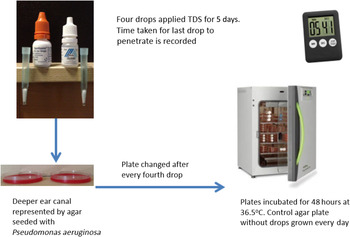
In part one of the study, sterile 15 mm × 9 mm tubular otowicks were inserted into two mock ear canals fabricated from plastic pipettes with narrowed ends. These were held vertically over agar plates. Two commonly used antibacterial ear drops – Gentisone HC™ (gentamicin 0.3 per cent and hydrocortisone acetate 1 per cent) and Ciloxan™ (ciprofloxacin hydrochloride 0.3 per cent) – were applied to the wicks at a rate of four drops, three times a day. The agar plates were seeded with P aeruginosa colonies and represented an infected deeper ear canal (Figure 1).
In vitro experiment with narrowed plastic pipettes representing the oedematous ear canal. Infected deeper ear canal is represented by bacterially seeded agar plates. TDS = three times per day

The time taken for drops to penetrate through the otowick was observed and recorded. To limit subjectivity, penetration was evidenced by the confirmed bactericidal effect on the agar plates. Each administration involved a newly seeded agar plate, which was incubated for 48 hours at body temperature; however, the two otowicks were fixed in the mock ear canal for 5 days. This was to determine if penetration and antimicrobial effects on colonised agar diminished with a used otowick.
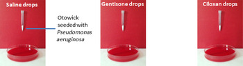
In a second model, otowicks seeded with P aeruginosa were inserted into three plastic pipettes and treated with sterile saline or antibacterial drops. These were held vertically over sterile agar, which represented an uninfected deeper ear canal. The drops penetrated the colonised otowick to land onto sterile agar, which was incubated at body temperature for 48 hours to ascertain bacterial penetration (Figure 2).
Colonised otowicks held over sterile agar representing an uninfected deeper ear canal.

Both experiments took place in the hospital microbiology laboratory. Preliminary bacterial counts were determined by a senior microbiology technician.
Results
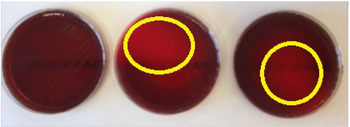
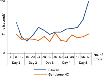
In part one of the study, it took six drops before penetration was observed. This occurred after 50 seconds for Gentisone HC and 105 seconds for Ciloxan. As the first four drops did not penetrate through the otowick, the first agar plate demonstrated an overgrowth of bacteria (Figure 3).
Agar plates from day 1 showing bacterial growth following: 1–4 (left), 5–8 (middle) and 9–12 (right) antibacterial drops (lighter shade demonstrates antibacterial activity).

Thereafter, the otowick treated with Ciloxan demonstrated a visible digress of penetration time (Figure 4) compared with Gentisone HC. Regardless, the otowicks stayed structurally intact in both models and still functioned as a form of treatment delivery.
Time taken for antibacterial drops to penetrate an otowick over 5 days. No. = number

In the second model, when sterile saline drops were applied to bacterially contaminated otowicks, the penetrating drops displayed bacterial growth on sterile agar plates, indicating that pseudomonas could penetrate the otowick material. However, when bacterially contaminated otowicks were treated with either Gentisone HC or Ciloxan, the penetrating drops showed no bacterial growth on the corresponding agar plate. This was despite the repeated application of bacterial cells on proceeding days.
Discussion
Uncomplicated otitis externa with a patent ear canal is simple to treat. Topical treatment allows contact of the affected area with a high concentration of medication, without causing systemic side effects. In the presence of ongoing ear canal oedema, removing otowicks every 2–3 days can be labour intensive. However, this in vitro experiment demonstrates that an otowick has the potential to still transmit treatment at 5 days, which may be useful if encountering a delay in patient review. The experiment also demonstrates that bacteria have the potential to spread from a colonised otowick, but this can be prevented by the continuous application of antibacterial ear drops, which will treat the contaminated wick and penetrate it to deliver treatment on the other side. If the otowick is not continually treated and left for a number of weeks, then there is a risk of systemic infection.Reference Burke, Gatton and Melville 8 It was observed that once the tubular otowick expanded, the lumen self-occluded, which suggested that it provided little advantage over a solid otowick.
The experiment demonstrates that otowicks themselves need to be primed with a minimum of six drops before starting a regimen because the initial dose is fully absorbed by the wick material. If a patient were to follow the British National Formulary guidelines of 2 drops 2–3 times per day, this would result in no antibiotic penetrating through for 24 hours, and thus delay treatment to the deeper canal. It is also important to recognise that only 40 per cent of patients who self-medicate do so appropriately during the first 3 days, often tending to undermedicate,Reference Rosenfeld, Brown, Cannon, Dolor, Ganiats and Hannley 3 which can cause further delays in symptom resolution.
-
• A 15 mm otowick should be primed by six antibacterial drops
-
• Removing an otowick in otitis externa every 2 days can be labour intensive
-
• Otowicks still transmit antibacterial drops at 5 days
-
• Bacteria can penetrate an otowick; however, this can be limited by the continuous application of topical treatment
-
• A tubular otowick may not provide any benefit over a solid wick
Treatment guidelines recommend that the head is kept tilted for 3–5 minutes, which is in agreement with the findings of this study, as there may be delayed penetration with ongoing treatment.
Limitations and recommendations
Otitis externa can be treated with a range of topical medications; it is difficult to be sure how permeability would be affected by other antibacterial drops. There is potential to investigate this further and to determine if solid wicks alter penetration significantly. As an in vitro experiment, the authors recognise the limitation of subjectivity and acknowledge that the otowick material may demonstrate different behaviour whilst in the ear canal; however, this is not easily reproducible with the benefits of observation.
We would encourage ENT departments to review their own otitis externa practice, as hospital care is often delivered acutely by a junior doctor or a non-specialist. With no separate guidelines for topical treatment in the presence of an otowick, we recommend that the treatment regimen begins with initial priming. We suggest that there is a low risk for leaving an otowick for 5 days, provided that the patient is compliant with treatment.
Acknowledgement
Many thanks to Nick Hurd for help in preparing the agar slides and establishing experimental controls.