Successful development of emotional functioning requires that children learn to express their feelings to others and accurately attend to, recognize, and interpret the emotions expressed by others (Wismer Fries & Pollak, Reference Wismer Fries, Pollak, Coch, Fisher and Dawson2009). Conversely, difficulties in socioemotional processing may lead to inappropriate social behaviors, as it is the case with individuals who develop antisocial personality disorder. Antisocial behavior committed by youths is a significant and particular clinical and societal concern (Van Goozen, Fairchild, & Harold, Reference Van Goozen, Fairchild and Harold2008). Children and adolescents with callous–unemotional (CU) traits are described as lacking guilt and empathy and a callous use of others for personal gain (Frick & Viding, Reference Frick and Viding2009; Lahey & Waldman, Reference Lahey, Waldman, Lahey, Moffitt and Caspi2003). These individuals are thought to lack emotional responsiveness to distress cues and may show both reactive and proactive aggression (de Wied, Gispen-de Wied, & van Boxtel, Reference de Wied, Gispen-de Wied and van Boxtel2010; Frick & White, Reference Frick and White2008). CU traits designate a subgroup of antisocial youth, who show a severe, violent, and difficult to treat pattern of antisocial behavior similar to the conceptualization of psychopathy made with adults (Cleckley, Reference Cleckley1976; Frick & Viding, Reference Frick and Viding2009; Hare, Glass, & Newman, Reference Hare, Glass and Newman2006). CU traits are considered to be the most important factor for identifying individuals with psychopathic tendencies despite cultural differences (Cooke & Michie, Reference Cooke and Michie1999). A large body of evidence has demonstrated that children with conduct disorder who display CU traits are distinct from children with conduct disorder who do not display CU traits in many ways, including clinical, neuropsychological, and genetic aspects (Frick & White, Reference Frick and White2008; Loney, Butler, Lima, Counts, & Eckel, Reference Loney, Butler, Lima, Counts and Eckel2006; Lynam et al., Reference Lynam, Charnigo, Moffitt, Raine, Loeber and Stouthamer-Loeber2009; Taylor, Loney, Bobadilla, Iacono, & McGue, Reference Taylor, Loney, Bobadilla, Iacono and McGue2003; Viding, Blair, Moffitt, & Plomin, Reference Viding, Blair, Moffitt and Plomin2005).
Empathy, the ability to share, appreciate, and respond adaptively to the emotional and affective states of others, is believed to act as a proxy in promoting prosocial behavior and inhibiting aggression (Decety & Jackson, Reference Decety and Jackson2004; Decety & Meyer, Reference Decety and Meyer2008). Conversely, the lack of empathy is thought to have a facilitating influence on offensive behavior (Eisenberg & Aalsma, Reference Eisenberg and Aalsma2005; Zahn-Waxler & Radke-Yarrow, Reference Zahn-Waxler and Radke-Yarrow1990). That is, if a person vicariously experiences the distress that they have caused to others because of their aggression, they will be less likely to continue to hurt others and more likely to help them. In contrast, lack of concern is an important risk factor for antisocial behavior problems such as conduct disorder (Lahey & Waldman, Reference Lahey, Waldman, Lahey, Moffitt and Caspi2003). The propensity for aggressive behavior is thought to reflect a blunted empathic response to the suffering of others (Blair & Blair, Reference Blair, Blair, Decety and Ickes2009), and it may be a consequence of a failure to be aroused by others' distress (Raine, Reference Raine, Stoff, Breiling and Maser1997). It has also been suggested that aggressive behavior can arise from abnormal processing of affective information, resulting in a deficiency in experiencing fear, empathy, and guilt, that, in normally developing individuals, inhibits acting out violent impulses (Davidson, Putnam & Larson, Reference Davidson, Putnam and Larson2000).
There is evidence that youth with CU traits are impaired in socioemotional communicative skills, which increases their risk of offending others (Richell et al., Reference Richell, Mitchell, Newman, Leonard, Baron-Cohen and Blair2003). Inconsistent findings have been reported, however. For instance, no specific impairment in the ability to infer others' mental states was found in offenders with psychopathy (Dolan & Fullam, Reference Dolan and Fullam2004).
Although researchers have speculated that offenders' callousness and lack of empathy may lead to the inclination toward instrumental and violent homicide (Woodworth & Waschbusch, Reference Woodworth and Waschbusch2008), little empirical research has investigated the neural processes underpinning socioemotional processing in children and adolescent offenders toward a victims in distress. The perception others' pain constitutes an ecologically valid paradigm to investigate the neural and cognitive mechanisms involved in empathy (Jackson, Meltzoff, & Decety, Reference Jackson, Meltzoff and Decety2005). Pain evolved as a protective function not only by warning a person suffering that something is awry but also by impelling expressive behaviors that attract the attention of others.
A handful of brain event-related potential (ERP) studies related to pain empathy demonstrated that frontal N120 and central–parietal late-positive potentials (LPPs) reflect an early affective arousal and a late cognitive evaluation component, respectively (Decety, Yang, & Cheng, Reference Decety, Yang and Cheng2010; Fan & Han, Reference Fan and Han2008; Han, Fan, & Mao, Reference Han, Fan and Mao2008). It is worth mentioning that the frontal N120 can be associated with a negativity bias, and that the LPPs reflects reappraisal of unpleasant stimuli along with enhanced attentional processes (e.g., Carretie, Hinojosa, Martin-Loeches, Merçado, & Tapia, Reference Carretie, Hinojosa, Martin-Loeches, Mercado and Tapia2004; Dennis & Hajcak, Reference Dennis and Hajcak2009; Hajcak & Nieuwenhuis, Reference Hajcak and Nieuwenhuis2006; Olofsson, Nordin, & Sequeira, Reference Olofsson, Nordin, Sequeira and Polich2008; Palomba, Angrilli, & Mini, Reference Palomba, Angrilli and Mini1997). In addition, it has been shown that the ~10 Hz (mu rhythm) suppression over the somatosensory cortex, indicative of sensorimotor resonance, can be modulated by pain empathy (Cheng, Yang, Lin, Lee, & Decety, Reference Cheng, Yang, Lin, Lee and Decety2008; Yang, Decety, Lee, Chen, & Cheng, Reference Yang, Decety, Lee, Chen and Cheng2009). Furthermore, a number of neuroimaging studies have demonstrated that the same neural circuit involved in the experience of physical pain is also involved in the perception or the imagination of another individual in a pain (for meta-analyses, see Jackson, Rainville, & Decety, Reference Jackson, Rainville and Decety2006; Lamm, Decety, & Singer, Reference Lamm, Decety and Singer2011). This neural network includes regions coding the affective–motivational and the sensory discriminative components of processing of pain in the observer (e.g., Akitsuki & Decety, Reference Akitsuki and Decety2009; Benuzzi, Liu, Duzzi, Nichelli, & Porro, Reference Benuzzi, Lui, Duzzi, Nichelli and Porro2008; Cheng, Chen, Lin, Chou, & Decety, Reference Cheng, Chen, Lin, Chou and Decety2010; Cheng et al., Reference Cheng, Lin, Liu, Hsu, Lim and Hung2007; Decety, Michalska, & Akitsuki, Reference Decety, Michalska and Akitsuki2008; Lamm, Batson, & Decety, Reference Lamm, Batson and Decety2007), namely, the supplementary motor area, somatosensory cortex, anterior cingulate cortex, periaqueductal gray, and anterior insula. Such a neural response to the pain of others has also been documented in school-age children who show a stronger activation of the insula and somatosensory cortex than adults (Decety & Michalska, Reference Decety and Michalska2010). Recently, a functional magnetic resonance imaging (fMRI) study compared the neurohemodynamic response elicited by empathy-eliciting stimuli in youth with aggressive conduct disorder and control adolescents. Results indicated that regions of the pain matrix were activated in both groups, but a specific activation of the amygdala and ventral striatum was detected in the former group, leading to the intriguing hypothesis that these individuals may find the perception of pain in others enjoyable and rewarding (Decety, Michalska, Akitsuki, & Lahey, Reference Decety, Michalska, Akitsuki and Lahey2009).
Empathy dysfunction in adults with psychopathy has been suggested to result from the reduced sensitivity to others' distress cues despite the appropriate cognitive comprehension of basic or complex emotions possibly remaining intact (Blair, Reference Blair1995). Studies that have focused on children with CU traits have also provided similar evidence for a deficit in emotion processing such as a decreased orienting response to affective stimuli (Blair, Reference Blair2003; Blair, Colledge, Murray, & Mitchell, Reference Blair, Colledge, Murray and Mitchell2001; Loney, Frick, Clements, Ellis, & Kerlin, Reference Loney, Frick, Clements, Ellis and Kerlin2003; Pardini, Lochman, & Frick, Reference Pardini, Lochman and Frick2003). For instance, typically developing children find the sight of others in pain aversive and avoid actions that cause pain or distress to others, whereas this may not be the case in children with CU traits (Hastings, Zahn-Waxler, Robinson, Usher, & Bridges, Reference Hastings, Zahn-Waxler, Robinson, Usher and Bridges2000). There has been converging evidence across different studies showing that amygdala hyporesponsivity may play a role in the neurobiology of callousness (Birbaumer et al., Reference Birbaumer, Veit, Lotze, Erb, Hermann, Grodd and Flor2005; Blair et al., Reference Blair, Smith, Mitchell, Morton, Vythilingam and Pessoa2007; Jones, Laurens, Herba, Baker, & Viding, Reference Jones, Laurens, Herba, Barker and Viding2009; Rilling et al., Reference Rilling, Glenn, Jairam, Pagnoni, Goldsmith and Elfenbein2007; Sterzer, Stadler, Krebs, Kleinschmidt, & Pouska, Reference Sterzer, Stadler, Krebs, Kleinschmidt and Poustka2005; Sterzer, Stadler, Pouska, & Kleinschmidt, Reference Sterzer, Stadler, Poustka and Kleinschmidt2007). In addition, reduced functional connectivity between the amygdala and the ventromedial prefrontal cortex has been reported in youth with CU traits relative to youth with attention-deficit/hyperactivity disorder and typically developing subjects (Decety et al., Reference Decety, Michalska, Akitsuki and Lahey2009; Marsh et al., Reference Marsh, Finger, Mitchell, Reid, Sims and Kosson2008), indicating an inappropriate modulation of neural pathways when being presented with distress cues. Such a deficit could result in dysfunctional processing of affective cues and ultimately lead to a phony interpersonal style.
Despite the abundant literature emphasizing impaired socioemotional and empathic processing in youth with CU traits, the nature of the neural mechanisms underlying such a dysfunction remains unclear. Chronic severe delinquent behavior in youth is a very serious public health issue. It is costly to society and is a burden on institutions and communities. A better understanding of the biopsychosocial mechanisms underpinning deficits in empathy can be expected to lead to both a greater understanding of juvenile psychopathy and improved approaches to assessment and treatment. It is important to identify the specific nature of affective and emotional deficits during childhood and adolescence when there has been less opportunity for correlates of the antisocial lifestyle (e.g., head injuries, incarceration, substance abuse) to affect brain anatomy and physiology.
The current ERP study targeted young offenders with psychopathic traits and control adolescents to examine their neural response to empathy-eliciting stimuli. Given that the frontal N120 and LPP are characteristic responses to stimuli eliciting pain empathy, we hypothesized that young offenders, compared to typically developing controls, would display an impaired LPP; and adolescents with higher CU traits would have a reduced frontal N120 response as well. We also hypothesize that if their ability to read intentional cues is intact, the LPP response would not differ from that of the control participants when understanding of agency is involved (e.g., someone intentionally harming another individual). Furthermore, if the automatic affective arousal component of empathy is underpinned by sensorimotor resonance (supposedly underpinned by the mirror neuron system), then the reduced N120 response should negatively correlate with mu suppressionFootnote 1 in youth with juvenile psychopathy. In contrast, if affective arousal is not mediated by the mirror neuron system, the mu wave suppression should be intact and dissociated from abnormal affective processing (see Decety, Reference Decety2010a, Reference Decety2011, for a critical appraisal of the contribution of mirror neurons to empathy).
Materials and Methods
Participants
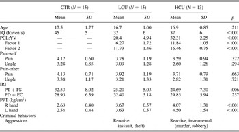
All participants were Chinese. Twenty-eight male adolescent offenders (aged 15–18 years), who had engaged in various crimes (e.g., murder, assault, robbery, or theft), from a high-security juvenile detention center, underwent screening for participation in the study (Table 1). All met the DSM-IV criteria for conduct disorder. The offenders were categorized into two groups from their total scores on the Hare Psychopathy Checklist Youth Version (PCL:YV; Forth, Kosson & Hare, Reference Forth, Kosson and Hare2003). Fifteen individuals were characterized as low CU (LCU; total score of PCL:YV ≤25), and 13 individuals as high CU (HCU; ≥30). The cut score was determined by the PCL:YV technical manual and previous empirical reports (e.g., Kiehl et al., Reference Kiehl, Smith, Hare, Mendrek, Forster and Brink2001; Richell et al., Reference Richell, Mitchell, Newman, Leonard, Baron-Cohen and Blair2003). The PCL:YV evaluation included interview, institutional data, psychiatric profiles, and behavior reports. Individuals with mental deficiencies, alcohol or other drug abuse, and sexual offenders were not included in the study. In addition, 17 noncriminal typically developing male adolescents matched on age and sex, who had no versatile violent history, official criminal record, and no neurological or psychiatric illnesses, were recruited as a control group (CTR) through bulletin board announcements in the community. Two of the controls were excluded from data analysis because of excessive artifacts during EEG recording. All subjects were right-handed without color blindness. The study was approved by the local Ethics Committee of the National Yang-Ming University Hospital (Taipei, Taiwan), and all participants provided informed consent according to the Declaration of Helsinki.
Demographic characteristics, dispositional measures, subjective rating, and pressure pain threshold of the study participants

Note: CTR, control group; LCU, group with low callous–unemotional traits; HCU, group with high callous–unemotional traits; PCL:YV, Factor 1, interpersonal/affective, Factor 2, behavioral/antisocial; IRI, interpersonal reactivity index; PT, perspective taking; FS, fantasy; EC, empathic concern; PD, personal distress; Pain self, watching someone accidentally in pain; Pain other, watching someone being intentionally hurt by another; PPT, pressure pain threshold; L, left; R, right.
PCL:YV
The PCL:YV assesses interpersonal, affective, and behavioral features related to psychopathy in male and female offenders aged 12 to 18. The PCL:YV consists of 20 items, each one rated on a 3-point ordinal scale (2, 1, or 0) based on the degree to which the personality and behavior of the adolescent matches the item description marked in the manual. Within the research context, the PCL:YV provides a standardized method of assessing the presence of CU traits, which is central to the construct of psychopathy. Reliability information on the PCL:YV administration was achieved by the following:
1. We reviewed the collateral information before conducting the interview. This step provides a basis for evaluating the responses and statements during the interview (e.g., denial, lying, distortions).
2. In resolving the discrepancies between the information provided in the interview and that obtained through collateral sources, a greater emphasis was placed on the most credible source of information.
3. Once all collateral and PCL:YV interview information was collected and reviewed, each item was carefully scored on the PCL:YV QuickScore form.
Visual stimuli
The visual stimuli consisted of 124 digital static color pictures depicting individuals in painful or nonpainful situations. The stimuli had been validated for pain intensity and perceived agency (intentional vs. nonintentional) and used in four fMRI studies with adult, adolescents (Akitsuki & Decety, Reference Akitsuki and Decety2009) and developing children (Decety et al., Reference Decety, Michalska and Akitsuki2008; Decety & Michalska, Reference Decety and Michalska2010); and adolescents (Decety et al., Reference Decety, Michalska, Akitsuki and Lahey2009; see supplementary materials online at http://journals.cambridge.org/dpp). The pictures were shot from the third-person perspective and edited to the same size (205 × 154 pixels). The stimuli were presented in the center of a gray background of a 17-in. color monitor with a visual angle of 5.05 degrees × 3.8 degrees at a viewing distance of 100 cm. Four categories of stimuli were included:
1. One person is in a painful situation accidentally caused, for example, an individual dropping a heavy bowl on her hand (pain-self).
2. One person is involved in a nonpainful situation, for example, opening a door (no pain-self).
3. One person is in a painful situation, intentionally caused by another individual, for example, stepping purposely on someone's toe (pain-other).
4. Two individuals are present and interacting (e.g., giving a flower), but there is no pain in the interaction (no pain-other).
General procedures
Before the EEG recordings, participants were required to fill in the Interpersonal Reactivity Index (Davis, Reference Davis1996), a self-report dispositional measure of empathy. Then, they underwent the assessment of pressure pain threshold (PPT). This is a objective test in which gradual pressure is applied to an area of the skin (left and right index proximal interphalangeal muscle) with an algometer, causing a gradual displacement of the skin surface, until the subject reports feeling pain (see supplementary materials online at http://journals.cambridge.org/dpp).
During the EEG recordings, the subjects were presented with static pictures depicting body parts (hands and feet) in painful and nonpainful situations, which were accidentally or intentionally caused. Each subject participated in six runs, where every run consisted of 160 trials (40 trials for each condition) starting with instructions (5 s) and lasting about 7 min. Between runs, subjects could freely relax until they wanted to move on. Instructions reminded the participants to look attentively at the visual stimuli.
After the EEG recordings, the same stimuli that had been shown during EEG sessions were presented to the participants on a computer screen. They were asked to rate how painful and how unpleasant each situation was by using the Face Pain Scale—Revised (Bieri, Reeve, Champion, Addicoat, & Ziegler, Reference Bieri, Reeve, Champion, Addicoat and Ziegler1990), which contains six faces expressing neutral to extremely painful expressions.
PPT
The PPT was assessed by a hand-held pressure algometer (UTECH Pain Track, Zevex International, Salt Lake City, UT), which consists of an ergonomic dynamometer with a flat stainless steel circular probe (1.52 cm2) connected to a commander console. PPT was determined at the dorsal side of the proximal phalanx of the index finger on both of the left and right hands. The pressure algometer was placed perpendicularly against the skin. Participants were instructed to say “yes” at the moment they started feeling pain or discomfort. When the participant said “yes,” the pressure was stopped and the threshold value was recorded. This procedure was repeated three times, alternating the left and right sides, to improve reliability, so that three PPT evaluations were obtained for each location. The mean of the three evaluations was calculated for each side.
EEG apparatus and recordings
For the CTR group, the EEG was continuously recorded with a 64-channel system, using scalp electrodes mounted on an elastic cap in accordance with the extended 10–20 system, with the addition of two mastoid electrodes. Because of the constrained experimental time in the juvenile detention center, the groups of LCU and HCU had EEG recorded from 32 scalp electrodes.
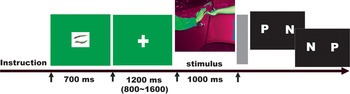
The paradigm of EEG recordings was presented with E-Prime software (Psychology Software Tools, Inc., Pittsburgh, PA; Figure 1; see supplementary materials online at http://journals.cambridge.org/dpp). Each trial consisted of the stimulus presentation (1000 ms), followed by an eye-closed sign and a fixation mark. The eye-closed sign was displayed on a gray background (=1.73 degree horizontal × 1.26 degree vertical visual angles) at the center of the screen and lasted 700 ms in order to minimize eye blinking during baseline and stimulus presentation (van Heijnsbergen, Meeren, Grèzes, & de Gleder, Reference Van Heijnsbergen, Meeren, Grèzes and de Gelder2007). Participants were instructed to blink only when the eye-closed sign was presented. The fixation mark was a white cross sign (=1.0 degree horizontal and 1.0 degree vertical visual angles) on a gray background with a jittered duration between 800 and 1600 ms (1200 ms on average). Then, a gray screen (300 ms) followed the picture presentation in order to prune away the backlash of the error-related negativity and late-response potential. Finally, there was a response window for the pain judgment task during which the participant chose one of two buttons according to the scenario of the stimuli (pain or no pain). The button associated with each scenario was randomized across trials, and the subjects were told which button corresponded with which scenario during the response window; that is, “P N” indicated that the left button was the response for pain, whereas “N P” indicated that the right button was the response for pain. This was done to reduce the subject's expectancy and preparatory responses. Subjects responded to each trial by a button pressing using left or right index finger. The next trial beginning with an eye-closed sign followed immediately thereafter.
The paradigm structure of the event-related potential recordings. [A color version of this figure can be viewed online at http://journals.cambridge.org/dpp]

The electrode at the right mastoid was used as reference. Eye blinks and vertical eye movements were monitored with electrodes located above and below the left eye. The horizontal electrooculogram was recorded from electrodes placed 1.5 cm lateral to the left and right external canthi. A ground electrode was placed on the forehead. Electrode/skin impedance was kept at <5 kΩ. Signals were sampled at 500 Hz (0.1–100 Hz bandpass) and were epoched over an analysis time of 1000 ms, including 200 ms of prestimulus delay used for baseline correction. An automatic artifact rejection system excluded from the average all trials containing transients exceeding ±90 µV at any recording channel, including the electrooculogram. Further, the quality of ERP traces was ensured by careful visual inspection in every subject and trial, excluding all epochs containing blink artifacts and applying appropriate digital, nonphase shift low-pass filters at 30 Hz. Each event in the paradigm (visual) was associated with a digital code (trigger) that was sent to the continuous EEG, allowing off-line segmentation and average of selected EEG periods for analysis. ERPs were processed and analyzed by Neuroscane (Compumedics, Hamburg, Germany).
Statistical analysis
The LPP, a well-studied component as a neurophysiological marker for emotion regulation in children and adolescents, was selected within a time window of 400 to 800 ms. Mean ERP voltages were obtained from each grand average peak (±20 ms), starting from the onset of each visual stimulus and continuing 800 ms poststimulus. Statistical analysis, using two-way mixed analyses of variance (ANOVAs) with one within-subject factor and one between-subject factor: (a) stimulus (pain-self vs. no pain-self; pain-other vs. no pain-other) and (b) group (CTR vs. LCU vs. HCU), followed by post hoc Bonferroni analysis, were computed at electrodes selected from the frontal, central, and parietal regions. The dependent variables were the mean ERP amplitudes of the electrodes from each brain region during each ERP component (N120, P3, N360, LPP). Degrees of freedom were corrected using the Greenhouse–Geisser method for correlated measures.
To assess the sensorimotor resonance induced by the perception of the stimuli, the ~8- to 12-Hz range (mu) power spectrum densities were analyzed from the averaged epochs by Matlab 7.0 procedures. Reflection (i.e., symmetrical extension) extended the data in the prestimulus baseline (100 ms) to match the poststimulus data set. This approach eliminates the discontinuity by tacking on the end points in reverse order (Semmlow, Reference Semmlow2004). Segments with equal data length were calculated for comparisons between the stimuli (pain-self vs. no pain-self; pain-other vs. no pain-other) between groups (CTR vs. LCU vs. HCU). Hamming window was applied to smooth the truncation for reducing the ripple effect. The ~10-Hz power was computed from the adjacent frequencies (±4 Hz) of the individualized largest power around 8–12 Hz and from the mean over electrode C3, central region, and C4. The ratio of the power during each stimulus relative to the power during the prestimulus baseline was to control variability in absolute mu power as result of individual differences, for example, scalp thickness and electrode impedance, as opposed to pain empathy-related activity. The log transformation (logarithm to the base 2) was further calculated for each ratio to correct for the inherent nonnormality of ratio data as a result for lower bounding (Fan, Decety, Yang, Liu, & Cheng, Reference Fan, Decety, Yang, Liu and Cheng2010). The negative value means mu rhythm suppressions. Statistical analysis were conducted with a two-way mixed ANOVA with the factors (a) stimulus (pain-self vs. no pain-self; pain-other vs. no pain-other) and (b) group (CTR vs. LCU vs. HCU), followed by post hoc Bonferroni analysis, computed with the dependent variable as the log transformed ~10-Hz power ratio during each stimulus.
Results
Demographics, dispositional measures, and pain threshold assessments
Youths with LCU and HCU were group matched to the CTR on age, gender, and handedness. Scores on the Interpersonal Reactivity Index revealed a significant group difference in the cognitive dimension, representing antecedents of experiencing emotions in response to emotions felt by others (p = .006). This difference mainly comes from higher rating of the CTR relative to the LCU and HCU (p = .017 and .014).
Significant differences in pain thresholds were found across groups: left hand, F (2, 40) = F (2, 40) = 14.17, p < .001; right, F (2, 40) = 11.25, p < .001. Post hoc analysis on the left hand showed higher PPT in participants with HCU relative to the CTR (p < .001) and the participants with LCU (p = .036). On the right hand, the PPT was higher in participants with HCU and participants with LCU when compared to the CTR participants (p < .001, p = .009). This finding suggests that young offenders are less sensitive to physical pain, especially those with higher CU traits.
During EEG recordings, all participants were able to correctly recognize the painful situations in the pictures (CTR: 87%; LCU: 85%; HCU: 90%). This ensures that the participants watched the stimuli attentively, and that group differences in ERPs, if any, did not result from differences in attention and cognitive understanding. For the subjective evaluations of the stimuli, no significant difference was found between the three groups for both pain intensity and unpleasantness on the pain-self stimuli (p = .325 and .310) and the pain-other stimuli (p = .678 and .766).
Electrophysiological data
Perceiving one person accidentally in pain versus not in pain (pain-self vs. no pain-self)
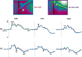
Grand-averaged ERPs of the stimuli depicting individuals being accidentally hurt compared with individuals in nonpainful situations are shown in Figure 2. In the CTR group, these stimuli evoked a negative component between 100 and 140 ms (N120), followed by a positive deflection between 160 and 200 ms (P180), then a negative wave with the maximum amplitude peaking between 210 and 250 ms (N230), then another positive deflection peaking at 300 ms (P3), followed by a negative deflection peaking at 360 ms (N360), and a long-latency positivity between 400 and 800 ms (LPP, peaking at 600 ms) over the frontal and central areas. These ERP components in the CTR are consistent with previous studies of pain empathy (Decety et al., Reference Decety, Yang and Cheng2010; Fan & Han, Reference Fan and Han2008; Han et al., Reference Han, Fan and Mao2008). In addition, a positive wave peaking at 120 ms and a long-latency positivity between 400 and 800 ms (LPP) occurred over the parietal and the occipital areas.
Cortical-evoked responses to perceiving individuals in pain versus no pain accidentally caused (pain-self, red, vs. no pain-self, blue, online only) in the controls (CTR) versus the group with low callous–unemotional traits (LCU) versus the group with high callous–unemotional traits (HCU) traits. The frontal N120 and the central P3 were differentiated in the CTR (marginal p = .053, gray arrow) and the LCU (p < .05, black arrow), not in the HCU (p > .05, white arrow). The central late-positive potential (LPP) was differentiated in the CTR, not in the LCU and the HCU. [A color version of this figure can be viewed online at http://journals.cambridge.org/dpp]

Frontal N120/P3
ANOVAs on the mean ERP amplitudes show a group effect at N120, F (2, 40) = 19.66, p < 0.001, and an effect of pain at N120, F (1, 40) = 7.69, p = .008, and P3, F (1, 40) = 6.372, p = .016. Post hoc analysis showed that the frontal N120 in the CTR and LCU had more negative deflection in response to painful relative to nonpainful stimuli (p = .053 and 0.043) despite of a marginal effect in the CTR, but no such response was detected in the HCU (p = .454).
Central N120/P3
There was a group effect at N120, F (2, 40) = 12.985, p < .001, and an effect of pain at N120, F (1, 40) = 4.164, p = .048, and P3, F (1, 40) = 8.458, p = .006. The painful stimuli elicited larger central P3 amplitudes than the nonpainful stimuli in the CTR (p = .016) and in the LCU (p = .046), but not in the HCU (p = .561). These findings suggest that individuals with HCU lack frontal N120 and central P3 to pain empathy, whereas the LCU preserve such a response.
LPP
The central LPP showed a group effect, F (2, 40) = 6.927, p = .003, and the parietal LPP an effect of pain, F (1, 40) = 4.527, p = .040. The painful stimuli elicited larger central LPP amplitudes than nonpainful stimuli in the CTR (p = .034), but not in the LCU (p = .424) or in the HCU (p = .776). The presence of central LPP differentiates the controls participants from the offenders.
Perceiving people intentionally hurt versus interacting without pain (pain-other vs. no pain-other)
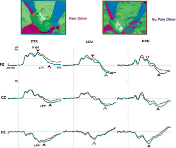
Grand-averaged ERPs to the stimuli depicting situations in which an individual intentionally hurt another are shown in Figure 2. In the CTR group, these stimuli evoked a negative component between 100 and 140 ms (N120), followed by a positive deflection between 160 and 200 ms (P180), then a negative wave with the maximum amplitude peaking between 230 and 270 ms (N250), then another positive deflection peaking at 300 ms (P3), followed by a negative deflection peaking at 360 ms (N360), and a long-latency positivity between 400 and 800 ms (LPP, peaking at 620 ms) over the frontal and central areas. In addition, a positive wave peaking at 140 ms, followed by a negative deflection at 220 ms, and a long-latency positivity between 300 and 800 ms (LPP, peaking at 400 ms) occurred over the parietal electrode.
Frontal N120/N360
ANOVAs showed a group effect at N120, F (2, 40) =16.415, p < .001, and N360, F (2, 40) = 10.288, p < .001, and an effect of pain at N360, F (1, 40) = 13.265, p = .001 (Figure 3). Post hoc analysis indicated that the frontal N360 amplitudes were stronger for painful relative to nonpainful stimuli in the CTR (p = .026) and LCU (p = .026), but not in the HCU (p = .191).
Cortical-evoked responses to perceiving individuals intentionally hurt by another versus no pain in the interaction (pain-other, red, vs. no pain-other, blue, online only) in the controls (CTR) versus the group with low callous–unemotional traits (LCU) versus the group with high callous–unemotional traits (HCU). The frontal N360 was differentiated in the CTR and LCU (p < .05, black arrow), not the HCU (p > .05, white arrow). It is important that the central and parietal late-positive potential (LPP) was differentiated in the CTR and the HCU, not the LCU. [A color version of this figure can be viewed online at http://journals.cambridge.org/dpp]

LPP
There was a group effect over the parietal region, F (2, 40) = 6.449, p = .004, and an effect of pain over the frontal, F (1, 40) = 26.871, p < .001, central, F (1, 40) = 24.506, p < .001, and parietal, F (1, 40) = 10.682, p = .002, areas. Notably, the frontal, central and parietal LPP reached a significant difference in the CTR (p = .002, .011, and .022) and HCU (p = .002, p < .001, p = .014), but not in the LCU (p = .081, .094, and .595). Individuals with HCU seemed to preserve a LPP response to situation that involved understanding social context.
Correlation between central LPP and psychopathic traits
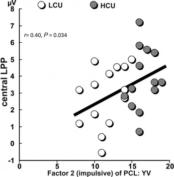
To explore the extent to which the electrophysiological responses elicited by the empathy-eliciting stimuli were associated with the severity of CU traits, multiple regression analysis was calculated for the ERPs and PCL:YV scores in the LCU and HCU groups collapsed together (Figure 4). For pain-self, neither the Factor 1 (emotional; r = .09, p = .619) nor the Factor 2 (impulsive; r = .13, p = .508) were correlated with the central LPP. It is interesting that for pain-other, the Factor 2 (r = 0.40, p = .034) was significantly positively correlated with the central LPP.
Correlations between the event-related potential response to the perception of stimuli depicting harm intentionally inflicted (social context) and psychopathic traits in the low callous–unemotional (LCU) and high callous–unemotional (HCU) groups collapsed together. The amplitude of the central late-positive potential (LPP) was positively correlated with the Factor 2 (impulsive) score of Hare Psychopathy Checklist Youth Version (PCL:YV).

Correlation between frontal N120/LPP and pain thresholds
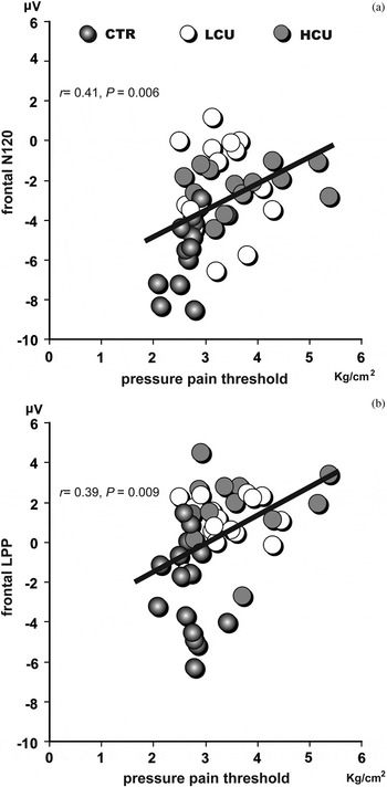
To determine whether the neural response to empathy stimuli is associated with the sensitivity to actual pain, a correlation analysis between the ERPs elicited by the empathy-eliciting stimuli and the PPT was conducted in all groups collapsed together (Figure 5). During pain-self and pain-other, less negative-going deflection of frontal N120 was associated with higher pain thresholds (left hand: r = .49, p = .001; r = .42, p = .005; right hand: r = .42, p = .005; r = .41, p = .006). Similarly, stronger frontal LPPs for pain-self and pain-other were coupled with higher pain thresholds (left: r = .35, p = .021; r = .38, p = .011; right: r = .37, p = .014; r = 0.39, p = .009). In addition, the mediation analysis confirmed no mediated effect by which the level of CU traits affects the deflection of frontal N120 (p = .833) and LPP (p = .640) through PPTs.
Correlations between the event-related potential response to the perception of stimuli depicting harm intentionally inflicted (social context) and the pressure pain threshold (PPT) in all three groups collapsed together. (a) Less negative-going deflections of frontal N120 were associated with higher pain thresholds. (b) Stronger frontal late-positive potentials (LPPs) were coupled with higher pain thresholds.

Correlation between frontal N120/LPP and subjective pain ratings
The correlation computed between the ERP amplitudes elicited by the painful stimuli and the Face Pain Scale—Revised scores showed that the frontal N120 for pain-self was not correlated with the ratings on pain intensity in the LCU (r = .17, p = .549) and HCU (r = −.29, p = .329; see Figure S.1 online at http://journals.cambridge.org/dpp). However, the frontal LPP for pain-other was significantly correlated with ratings of pain intensity (r = .77, p = .001) in the LCU, not in the HCU (r = −.14, p = .644).
Sensorimotor resonance
A main effect of the mu rhythm wave suppression was found for pain-self versus no pain-self, F (1, 38) = 9.983, p = .003, and for the group, F (2, 38) = 3.309, p = .047, but no interaction reached significance, F (2, 38) = 1.007, p = .375. Post hoc analysis indicated that the effect of pain was mainly driven from the CTR (p = .018). The HCU relative to the LCU showed stronger mu suppression (p = .043) even though both of them did not differ from the CTR (p = .744 and .553). For pain-other versus no pain-other, only the group factor reached significance, F (2, 38) = 4.370, p = .020. Neither the stimulus, nor the interaction had any effect, F (1, 38) = 0.385, p = .539; F (2, 38) = 1.308, p = .364. The effect came from the fact that the HCU than the LCU displayed stronger mu suppression (p = .016), not from their comparisons with the CTR (p = .359 and .597).
Discussion
Impaired social–emotional processing in children and adolescents is known to increase the risk of offending behaviors. Despite the indisputable importance of social factors in antisocial behavior, the understanding of antisocial behavior in youth should benefit from understanding its neurobiological bases. The goal of this study was to better understand the contribution of neural processing associated with the perception of others' distress in children and adolescents with disruptive behavior disorders by using a well-validated experimental procedure that assesses the component processes (affective arousal and emotion understanding) of empathy.
The results of our study demonstrate atypical neural processing in response to empathy-eliciting stimuli in young offenders compared with control adolescents. It is interesting that young offenders, compared to controls, showed higher pain thresholds, which indicates that the perception of noxious stimuli is perceived as less intense for these individuals. The evoked cortical response to stimuli depicting people in pain was different between the two groups of offenders. Although participants with LCU showed a typical frontal N120 and central P3 response, no such responses were detected in participants with HCU, and no central LPP was detected in either groups. When perceiving situations that involved a social interaction leading to harmful consequences (pain-other vs. no pain-other), the LPP was found to be reduced in participants with LCU but retained in participants with HCU. These results suggest a disruption in the late processing of empathy in participants with LCU as indicated by a lack of central LPP. Participants with HCU were impaired both in the early and late processing of empathy, as reflected by the decreased frontal N120, central P3, and LPP in response to stimuli depicting pain. It is important that in the presence of a social context (pain caused intentionally), which requires a cognitive understanding of the situation, participants with HCU retained the LPP response. Stronger LPP amplitudes were associated with higher psychopathic traits, and higher pain thresholds. It is important that the affective arousal deficit in participants with HCU cannot be explained by a lack of sensorimotor resonance, as measured by mu wave suppression, which was present in all participants. This finding indicates that affective arousal is not mediated by the mirror neuron system.
While watching someone accidentally in pain, participants with LCU showed a frontal N120 response to similar the CTR, whereas the individuals from HCU group did not display such an evoked response. Consistent with previous electrophysiological studies (Decety et al., Reference Decety, Yang and Cheng2010; Fan & Han, Reference Fan and Han2008; Han et al., Reference Han, Fan and Mao2008), the neural dynamics underpinning pain empathy processing occurs as early as 120 ms after stimulus onset over the frontal area. This response has been interpreted as reflecting an automatic arousal reaction. In addition, a number of studies demonstrated that this early frontal negativity can be attributed to negativity bias to unpleasant stimuli (Carretie et al., Reference Carretie, Hinojosa, Martin-Loeches, Mercado and Tapia2004; Doallo, Cadaveira, & Rodriguez Holguin, Reference Doallo, Cadaveira and Rodriguez Holguin2007; Olofsson et al., Reference Olofsson, Nordin, Sequeira and Polich2008; Palomba et al., Reference Palomba, Angrilli and Mini1997). Perceiving the difference between negative and neutral stimuli occurs very rapidly because it plays a crucial role in survival (Cacioppo & Gardner, Reference Cacioppo and Gardner1999; Vuilleumier & Pourtois, Reference Vuilleumier and Pourtois2007). Given that the perception of others' pain elicited strong affective arousal response in the control participants as well as in participants with LCU, but not in youth with HCU, it may be suggested that participants from this latter group have a reduced negativity bias to perceiving painful and distressful situations in others. This is important, because affective arousal is considered to be the first component of empathy in place during development (Decety, Reference Decety2010b). It refers to the automatic discrimination of a stimulus, or features of a stimulus, as appetitive or aversive, hostile or hospitable, pleasant or unpleasant, and threatening or nurturing, which has evolved to differentiate hostile from hospitable stimuli and to organize adaptive responses to these stimuli. Strong bidirectional connections between the amygdala and sensory cortices associated with complex processing of stimuli with emotional significance, as well as connections with the hypothalamus and hippocampus (Ghashghaei & Barbas, Reference Ghashghaei and Barbas2002) are the fundamental neural underpinnings of affective arousal.
In the young offenders, the impaired early affective response to others' pain seems to parallel their relative insensitivity to physical pain, as assessed by their pain threshold. The offenders exhibited less sensitivity to pain than the CTR as indicated by higher thresholds of pressure pain. It is important that more positive-going deflections of the frontal N120, which infers less degree of early emotional arousal of pain empathy, were closely coupled with higher pain thresholds, reflecting more insensitivity to actual pain.
Similarly, the central P3 to pain empathy stimuli was detected in participants with LCU but not in individuals with HCU. The P3 is considered as an index of the allocation of neural resources for attention capture and stimulus encoding (Polich & Comerchero, Reference Polich and Comerchero2003; Reese & Polich, Reference Reese and Polich2003). Here, the overlapping P3 responses to painful versus nonpainful stimuli account for the fact that participants with HCU did not require enhanced attention allocation for processing aversive stimuli. One recent meta-analysis found that antisocial behavior was associated with P3 amplitude decrements during the continuous performance task, in which participants are asked to respond to target stimuli and ignore nontargets. No such decrements, however, occurred in individuals with psychopathic tendency (Gao & Raine, Reference Gao and Raine2009). Further, P3 reduction could be an endophenotypic maker of the neurobiological vulnerability to externalizing, which might be associated with antisocial features of psychopathy (Hicks et al., Reference Hicks, Bernat, Malone, Iacono, Patrick and Krueger2007; Patrick et al., Reference Patrick, Bernat, Malone, Iacono, Krueger and McGue2006). Our results suggest that higher psychopathic traits in participants with HCU influence the P3 amplitudes induced by pain empathy.
The central LPP elicited by the perception of stimuli depicting people accidentally in pain was impaired in young offenders. The positive-going ERP following the presentation of emotional compared to neutral pictures is defined as LPP, beginning as early as 400 ms, maximal at posterior–superior sites, and reflecting facilitated attention to emotional stimuli (Cuthbert, Schupp, Bradley, Birbaumer, & Lang, Reference Cuthbert, Schupp, Bradley, Birbaumer and Lang2000; Schupp et al., Reference Schupp, Cuthbert, Bradley, Cacioppo, Ito and Lang2000). One ERP study reported that the LPP indicates late cognitive processing of empathy, which was enhanced by a pain judgment task, but diminished by a limb counting task (Fan & Han, Reference Fan and Han2008). Here, our results demonstrate that the central LPP was unresponsive to pain empathy in adolescents with CU traits.
It is important that adolescents with HCU did not seem to be impaired in perceiving and understanding social interactions, as reflected by an intact LPP response to situations in which an individual intentionally harmed another. First, this result can be ascribed to differential emotion regulation related to various degrees of CU traits. A stronger LPP for painful stimuli may reflect increased attentional processes engaged by motivationally relevant stimuli (Dennis & Hajcak, Reference Dennis and Hajcak2009; Hajcak, Moser & Simons, Reference Hajcak, Moser and Simons2006; Hajcak & Nieuwenhuis, Reference Hajcak and Nieuwenhuis2006). The reappraisal of unpleasant stimuli would decrease the LPP magnitudes. Second, retained LPP may indicate an enhanced response to environmental cues in youth with higher CU traits. Increased, rather than decreased amygdala activation in response to negative stimuli, has been detected in adolescents with early-onset conduct disorder (Herpertz et al., Reference Herpertz, Huebner, Marx, Vloet, Fink and Stoecker2008) as well as adolescents with aggressive conduct disorder and psychopathic tendencies (Decety et al., Reference Decety, Michalska, Akitsuki and Lahey2009). Here, our results show that the LPP response to stimuli depicting pain-self was similar in participants with LCU and participants with HCU, but different for stimuli depicting pain-other. This suggests that young offenders with higher CU traits do not have a deficit in understanding others' intentions in a social context.
Furthermore, the central LPP response in the context of the perception of pain in a social interaction was found to be correlated with our participants' psychopathic traits. A previous ERP study reported that offenders with psychopathic traits showed P3 impairments in the standard oddball task, but not in other tasks (Gao & Raine, Reference Gao and Raine2009). The task type was a moderating factor for the association of P3 amplitude decrements and prolonged latencies with antisocial behavior. Here, the central LPP selectively elicited by the perception of pain-other (but not by pain-self), was positively correlated with the Factor 2 (impulsive) score of PCL:YV.
Our study has a number of limitations that need to be acknowledged. One limitation is that delinquent participants are differentiated by more than their CU traits. Both LCU and HCU groups also substantially differ on the antisocial and behavioral dimensions of Factor 2 (see Table 1), which may hamper the interpretations regarding the relationship between CU traits and empathic processing. The CU traits seem to be related to a particularly severe, aggressive, and stable pattern of antisocial behavior (Frick & White, Reference Frick and White2008). In addition, the small sample size and the homogeneity of the ethnicity (non-Caucasian) can also be potential limitations including the absence of interaction effect between group and pain. The adolescent offenders with a broad age range presumably have already experienced some alternations in brain development as a result of their antisocial behaviors. Given the cross-sectional design, it is unclear whether or which neurobiological deficits predated antisocial behavior. Even though participants with possible confounding risk behaviors (e.g., substance use) and head injuries often found in juvenile offender populations were excluded, this may not have exhausted other neurobiological antecedents of antisocial behavior. Further studies that more tightly control CU traits and the age range in larger sample sizes are thus warranted.
Overall, the current study demonstrates selective impairment in components of empathy associated with specific abnormal neural processing dynamics in young offenders with disruptive behavior disorders. Although participants with LCU were impaired only in the late processing of empathy-eliciting stimuli, participants with HCU were impaired in both early affective arousal and late cognitive evaluation. It is important that adolescents with HCU showed no deficits in the perception of intentionality, as indicated by their normal LPP responses to social situations in which an individual was intentionally harmed. This finding supports the idea that neurobiological impairments in individuals with CU traits who display little empathy are not due to a reduced ability to understand the emotions of others (Shirtcliff et al., Reference Shirtcliff, Vitacco, Graf, Gostisha, Merz and Zahn-Waxler2009). Rather, the current evidence suggests that they show little arousal to others' distress. Juveniles with psychopathic tendencies and high callous unemotional traits may very well understand the emotional state of others without “sharing” their feelings. The sharing of vicarious negative arousal provides a strong signal that can promote empathic concern (Decety & Michalska, Reference Decety and Michalska2010). To be motivated to help another, one needs to be affectively aroused, and to be able to anticipate the cessation of mutually experienced personal distress or discomfort. This signal may be lacking in these individuals, as it is already established with adult psychopaths who exhibit weaker psychophysiological reactions to emotional stimuli and poor passive avoidance learning (Kosson, Lorenz, & Newman, Reference Kosson, Lorenz and Newman2006), and this may contribute to vulnerability to engage in antisocial behavior.
Conclusion
Research in affective neuroscience with children and adolescent is important, not only because it advances the understanding of the neurophysiological mechanisms underlying individual differences in the etiology of aggressive and antisocial behavior, but it also benefits treatment programs that are often driven more by theory than empirical evidence. One of the major problems in trying to establish whether empathy can be increased with interventions is related to conceptual difficulties in defining the empathy construct and how measurement difficulties might be a factor in explaining the disparities noted in the empirical research (Day, Casey, & Gerace, Reference Day, Casey and Gerace2010). More recent models of empathy that articulate theory and empirical data from affective neuroscience, developmental psychology, and neuropsychiatric disorders view this ability as a complex construct that involves the interaction of multiple components including affective arousal, emotion understanding and self-regulation (Decety, Reference Decety2010a; Decety & Jackson, Reference Decety and Jackson2004; Decety & Meyer, Reference Decety and Meyer2008; Singer & Lamm, Reference Singer and Lamm2009). Neurodevelopmental research indicates that affective arousal is a necessary (although not sufficient) precursor to mature empathic understanding and morality (Blair, Reference Blair2005; Decety & Michalska, Reference Decety and Michalska2010; Decety, Michalska, & Kinzler, Reference Decety, Michalska and Kinzler2011). The rationale behind empathy interventions is that increased empathy will have an inhibitory effect upon the individual's motivation to offend as a response to the cognitive or affective dissonance experienced by the offender, which in turn, serves to reduce the risk of aggression (Hildebrain & Pithers, Reference Hildebrain, Pithers and Laws1989). Findings from the current study showing that young offenders with HCU understand the harm done to victims, but do not seem to process cues of distress as a negative signal, as well as evidence from one fMRI study that reported activation of reward areas in children with aggressive conduct disorder and psychopathic tendencies when watching harm done to others (Decety et al., Reference Decety, Michalska, Akitsuki and Lahey2009) seem to document a dissociation between affective sharing and emotion understanding. A more detailed distinction between the component processes of empathy, their neural underpinnings, and their atypical expression in juvenile offenders, is thus of critical interest for intervention, in assisting to differentially target affective, emotion regulation and theory of mind. Finally, these data supports the view that treatment provision should be designed specifically for offenders with marked psychopathic traits rather than fitting them into treatment planned primarily for ordinary offenders.
Supplementary Materials and Methods
The supplementary material referred to in this article can be found online at http://journals.cambridge.org/dpp