The coexistence of structural and idiopathic epilepsies is an uncommon and rarely described phenomenon, Reference Diehl, Wyllie, Rothner and Bingaman1–Reference Asadi-Pooya, Malekpour, Taherifard, Mallahzadeh and Farjoud Kouhanjani4 which may pose significant diagnostic challenges and complicate therapeutic strategies. Dysembryoplastic neuroepithelial tumors (DNET) are a well-recognized cause of drug-resistant focal epilepsy. Surgical resection of DNET generally results in a high rate of seizure freedom, with favorable outcomes reported in 70%–90% of cases. Reference Raymond, Halpin and Alsanjari5 Epilepsy with eyelid myoclonia (EEM), previously referred to as Jeavons syndrome, is a rare form of genetic generalized epilepsy (GGE) characterized by the classic triad of (1) eyelid myoclonia, with and without absences, (2) seizures triggered by eye closure and (3) photosensitivity. Reference Striano, Capovilla and Sofia6,Reference Specchio, Wirrell and Scheffer7 We herein report a patient presenting with the rare coexistence of drug-resistant focal epilepsy attributable to DNET alongside EEM, undergoing lesionectomy.
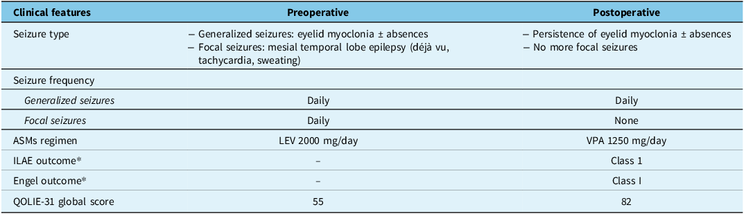
A previously healthy 17-year-old male initially presented with a history of sporadic, seemingly generalized tonic-clonic seizures (GTCS), occurring primarily while watching television, with onset at age 13. At 15 years of age, treatment with valproic acid (VPA) up to 1250 mg/day was initiated, resulting in complete control of GTCS. Two years later, at the age of 17 years, he presented to our Epilepsy Center reporting monthly seizures characterized by déjà vu phenomena, sudden unprovoked fear, palpitations, followed by confusion and drowsiness. Neurological and neuropsychological evaluations were unremarkable. Video-electroencephalogram (EEG) recording showed eyelid myoclonia, sometimes associated with absences, consistently triggered by eye closure and linked to diffuse polyspike-wave discharges, predominantly over posterior cortical areas (Figure 1A). Intermittent photic stimulation elicited generalized photoparoxysmal responses across a frequency range of 9–35 Hz (Figure 1B). These responses were abolished by the use of optical filter lenses. Interictal EEG also revealed the presence of focal epileptic abnormalities localized to the right temporal lobe. Brain MRI showed a multicystic lesion in the left mesial temporal region, characterized by hypointensity on T1-weighted images and hyperintensity on T2-FLAIR sequences, without contrast enhancement, suggestive of a DNET (Figure 2A–D). Treatment with multiple anti-seizure medications (ASMs), including carbamazepine, lamotrigine (LTG), levetiracetam (LEV), lacosamide and VPA, failed to control seizures. The patient was subsequently admitted for prolonged video-EEG monitoring, during which three stereotyped seizures, marked by déjà vu, tachycardia and intense sweating, were captured, each associated with corresponding ictal discharges from the left temporal region (Figure 1C). Eye closure-induced epileptic discharges and eyelid myoclonia, sometimes with associated absences, were also documented. Quality of life (QoL) was assessed through the QoL in Epilepsy Inventory (QOLIE)-31, yielding a global score of 57. Due to his refractory temporal lobe seizures, the patient underwent surgical resection of DNET. Complete excision of the DNET was achieved (Figure 2E, F). Histopathological analysis confirmed a grade 1 DNET. Following surgery, the patient became free from temporal seizures in the following 9 years of follow-up (postsurgical outcome for focal seizures: ILAE Class 1; Engel Class I). However, eyelid myoclonia, isolated or concomitant to absences, persisted despite treatment with VPA 1250 mg/day. A routine ambulatory EEG recording performed 30 days after surgery demonstrated persistence of eyelid myoclonia and photoparoxysmal response, with no evidence of residual temporal focal abnormalities. All subsequent ambulatory EEGs obtained during the 9-year follow-up period consistently demonstrated the same findings. Nevertheless, the patient reported a significant improvement in his QoL, with the QOLIE-31 global score increasing to 82 (Table 1).
Presurgical EEG recording. The figure illustrates key electroencephalographic findings recorded prior to surgery. Panel A shows diffuse polyspike-and-wave (PSW) discharges (✻) occurring immediately after eye closure, associated with eyelid myoclonia (EM). In panel B, a generalized photoparoxysmal response (PPR) (red ★) is elicited by intermittent photic stimulation at 21 Hz. Panel C displays an ictal discharge arising from the left temporal region (blue box), recorded using additional zygomatic electrodes (T1: left zygomatic; T2: right zygomatic).

Brain MRI. The figure shows a multicystic lesion located in the left mesial temporal region (red arrows), visible on coronal inversion recovery (A), coronal T2-weighted (B) and axial FLAIR (C) brain MRI sequences, consistent with a dysembryoplastic neuroepithelial tumor. No contrast enhancement was observed following gadolinium administration on axial T1-weighted images (D). Postoperative MRI (E, F) shows the complete resection of the lesion (white arrows).

Comparison of pre- and postoperative clinical findings

Note: ASMs = anti-seizure medications; LEV = levetiracetam; QOLIE-31 = Quality of Life in Epilepsy Inventory-31; VPA = valproic acid.
* ILAE and Engel classifications refer only to temporal lobe seizures; eyelid myoclonia and absences were not considered in the surgical outcome.
To our knowledge, this is the first report of a patient with surgically confirmed coexistence of symptomatic focal epilepsy caused by a DNET and EEM. The patient presented with two distinct epileptic conditions: a structural focal epilepsy and a recognized GGE. This case highlights that the occurrence of a concomitant generalized epilepsy syndrome does not represent an absolute contraindication for surgery in a patient with drug-resistant structural epilepsy. In fact, our patient experienced a striking improvement in his QoL following resection of the DNET, despite the persistence of eyelid myoclonia with or without absences. The substantial increase in the QOLIE-31 score emphasizes that meaningful gains in daily functioning and psychosocial well-being can occur even without complete seizure freedom, underscoring the importance of evaluating surgical outcomes not only in terms of seizure cessation but also in terms of holistic patient-centered benefits.
The patient fulfilled the proposed criteria for EEM, presenting eyelid myoclonia (brief, typically <1 to 3 s, repetitive eyelid jerks with upward eye deviation and head extension), both isolated and associated to absences, in addition to epileptic abnormalities (brief bursts of fast – 3–6 Hz – irregular, generalized polyspike-and-wave complexes) occurring after eye closure and photosensitivity. Reference Striano, Capovilla and Sofia6,Reference Specchio, Wirrell and Scheffer7 EEM is classified among GGE, with a positive family history reported in approximately 25%–80% of cases, predominantly involving generalized seizures in affected relatives. Reference Specchio, Wirrell and Scheffer7 While most patients lack identification of a single pathogenic gene variant, monogenic gene mutations, including SYNGAP1, CHD2, KIAA2022/NEXMIF and RORB, have been reported, particularly in cases where this syndrome occurs in the setting of a developmental and epileptic encephalopathy. Reference Specchio, Wirrell and Scheffer7 Despite EEM being classified among GGE, its underlying mechanisms remain debated. Recent evidence demonstrated occipital hyperexcitability with rapid bilateral cortical recruitment, supporting the hypothesis that generalized discharges and photosensitivity may arise from an occipital-driven network. Reference Gélisse, Gallegos, Nilo, Macorig, Genton and Crespel8
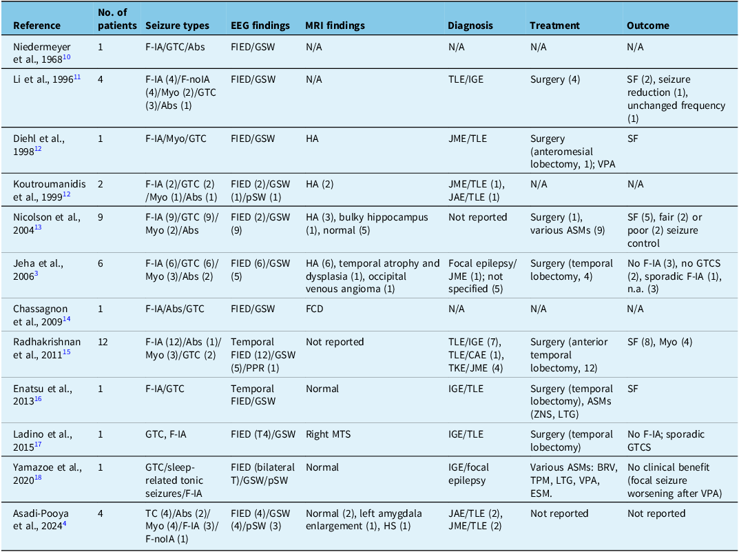
In parallel, the patient also suffered from drug-resistant focal seizures arising from the temporal lobe due to a DNET. Surgical removal of the DNET resulted in complete control of these seizures. Although additional invasive monitoring was not performed, the long-term postoperative clinical and EEG findings confirm complete resolution of focal seizures. While involvement of occipitotemporal or insular network involvement has been suggested in the pathophysiology of EEM, Reference Zawar, Franic and Pestana Knight9 the distinct electroclinical features and divergent postoperative outcomes in our case (full remission of focal seizures with persistence of generalized eyelid myoclonia) support the coexistence of two independent epileptic mechanisms. Recognizing and correctly distinguishing between coexisting generalized and focal structural epilepsies is of paramount importance, since it introduces unique diagnostic and therapeutic challenges. Reference Raymond, Halpin and Alsanjari5 Indeed, in patients with evident structural lesions, the diagnosis of a concurrent generalized epilepsy may sound improbable and be overlooked. The present case highlights that effective seizure control can be achieved through surgical intervention in patients with medically intractable focal seizures, even when a concurrent generalized epilepsy coexists. Reference Jeha, Morris and Burgess3,Reference Asadi-Pooya, Malekpour, Taherifard, Mallahzadeh and Farjoud Kouhanjani4 The coexistence of focal structural epilepsy and IGE in the same patient is a rare occurrence, occurring in approximately 0.2% of all epilepsy patients. Reference Jeha, Morris and Burgess3 Table 2 summarizes the few published studies describing such cases. Reference Jeha, Morris and Burgess3,Reference Asadi-Pooya, Malekpour, Taherifard, Mallahzadeh and Farjoud Kouhanjani4,Reference Niedermeyer10–Reference Yamazoe, Dubeau, von Ellenrieder, Zazubovits and Gotman18 Most involve pediatric patients, predominantly with mesial temporal lobe epilepsy, as in our case, coexisting with generalized syndromes, including juvenile myoclonic or absence epilepsy. In line with our findings, several reports have documented favorable surgical outcomes. Reference Jeha, Morris and Burgess3,Reference Enatsu, Wyllie, Kotagal and Bingaman16 Effective seizure control can be attained with tailored treatment strategies and the appropriate selection of ASMs. Some ASMs typically prescribed for focal seizures (e.g., carbamazepine, phenytoin, vigabatrin, lacosamide) may exacerbate generalized epilepsy, including EEM, due to their mechanism of action on voltage-gated sodium channels. Reference Thomas, Valton and Genton19 This potential for exacerbation highlights the importance of careful drug selection in patients presenting with both focal and generalized epilepsies. Broad-spectrum ASMs, such as VPA, LTG, LEV, topiramate, phenobarbital and perampanel, are therefore preferred in these patients, as they are effective against multiple seizure types without increasing the risk of worsening generalized epileptic activity. Choosing appropriate ASMs in this context is essential to achieve optimal seizure control and to minimize iatrogenic exacerbation of generalized epilepsy. Reference Thomas, Valton and Genton19
Review of previous studies describing patients with coexisting focal epilepsy and idiopathic generalized epilepsy

Note: References are ordered by publication date. () = number of patients; Abs = absence seizures; ASMs = anti-seizure medication; CAE = childhood absence epilepsy; ESM = ethosuximide; F-IA = focal seizures with impaired awareness; F-noIA = focal seizures without impaired awareness; FCD = focal cortical dysplasia; FIED = focal interictal epileptiform discharges; GSW = generalized spike-waves; GTCS = generalized tonic-clonic seizures; HA = hippocampal atrophy; HS = hippocampal sclerosis; IGE = idiopathic generalized epilepsy; JAE = juvenile absence epilepsy; JME = juvenile myoclonic epilepsy; LTG = lamotrigine; MTS = mesial temporal sclerosis; Myo = myoclonic seizures; PPR = photoparoxysmal response; pS = polyspikes; TC = tonic-clonic seizures; TLE = temporal lobe epilepsy; TPM = topiramate; SF = seizure free; VPA = valproic acid; ZNS = zonisamide.
In conclusion, this case report highlights that epilepsy surgery should be considered in patients with coexisting focal structural and GGE when focal seizures are drug-resistant and the epileptogenic zone is well localized. The presence of a generalized epilepsy syndrome should not automatically preclude surgical candidacy in patients with disabling drug-resistant focal epilepsy.
Author contributions
RC: writing – original draft, writing – review & editing, data curation, investigation, visualization; AP: writing – original draft, writing – review & editing, data curation, investigation, visualization; SG: data curation, investigation, writing – review & editing; VC: data curation, investigation; GGT: data curation, investigation; CCP: data curation, investigation; AM: data curation, investigation; AA: data curation, investigation; AP: data curation, investigation; MC: data curation, investigation; UA: data curation, investigation, visualization, supervision; EF: writing – original draft, writing – review & editing, conceptualization, data curation, investigation, visualization, supervision.
Funding statement
This work was supported by #NEXTGENERATIONEU (NGEU) and funded by the Ministry of University and Research (MUR), National Recovery and Resilience Plan (NRRP), project MNESYS (PE0000006) – a multiscale integrated approach to the study of the nervous system in health and disease (DN. 1553 11.10.2022).
Competing interests
The authors have no competing interests to declare that are relevant to the content of this article.
Ethical standard
The study was performed in accordance with the ethical standards laid down in the 1964 Declaration of Helsinki and its later amendments.
Written informed consent has been obtained from the patient.