Background
Mild cognitive impairment (MCI) is defined as subjective and objective declines in cognition and function greater than expected for an individual's age and education level and that does not meet criteria for a diagnosis of dementia (Petersen, Reference Petersen2004). Recently, there has been increasing interest in the characteristics of the early cognitive changes before the development of dementia. MCI is recognized as a pathological condition interposed between the cognitive changes of normal aging and what might constitute very early dementia (Petersen, Reference Petersen2004). It has been reported that a substantial portion of MCI patients have some neuropsychiatric symptoms such as mood or behavioral symptoms. Among these symptoms, the most frequent neuropsychiatric symptom is related to depression (Ellison et al., Reference Ellison, Harper, Berlow and Zeranski2008).
Even though a large proportion of elderly people with MCI experience depressive symptoms, little is known about the neurocognitive characteristics of this subgroup and, in particular, whether their pattern of cognitive decline differs from that of MCI without depressive symptoms (Hudon et al., Reference Hudon, Belleville and Gauthier2008). An understanding of the differences in neurocognitive characteristics between these two groups is critical because depression could contribute to the cognitive heterogeneity of MCI, which is a crucial issue in understanding MCI (Hudon et al., Reference Hudon, Belleville and Gauthier2008). In addition, differences in the cognitive deficits in MCI patients with depressive symptoms from those of MCI without a depressive component suggest that the pathophysiology of the two subgroups is different and might compel distinct approaches in treatment or prevention (Steffens et al., Reference Steffens2006).
However, there is little research investigating the relationships between depression and its neurocognitive characteristics in MCI patients. One study reported greater deficits in immediate and delayed memory in depressed MCI patients compared with non-depressed MCI patients (Johnson et al., Reference Johnson2013). Another study showed that poorer immediate and delayed memory was associated with depression in amnestic MCI (Brunet et al., Reference Brunet2011). Yet, another study showed that depressed MCI patients exhibited greater deficits in executive function, but not in memory, compared to non-depressed MCI patients (Hudon et al., Reference Hudon, Belleville and Gauthier2008).
In the three previous studies, these partially inconsistent findings might be due to difference in assessment methods. And previous studies were limited by a small sample size or a limited number of neuropsychological tests. In the present study, the diagnosis of depression was made according to the DSM-IV-TR criteria (APA, 2000) and confirmed by board-certified psychiatrists. And the authors of the present study recruited a larger sample size of MCI participants, and the neurocognitive functions of the participants were assessed using comprehensive neuropsychological battery with numerous cognitive subsets. This study was conducted to examine the cross-sectional associations of neurocognitive functions with depression by comparing the results of neuropsychological tests between depressed MCI patients (MCI/D+) and non-depressed MCI patients (MCI/D−). The authors hypothesized that MCI/D+ patients would demonstrate poorer performance than MCI/D− patients on several neurocognitive tests.
Methods
Participants and assessments
The sample consisted of 153 participants diagnosed with MCI. All MCI participants were recruited through advertisement and screened as outpatients at one university hospital-memory clinic. MCI was determined through consensus diagnosis on the basis of Mayo Clinic criteria: memory complaints, objective memory impairment for age, intact activities of daily living, preserved general cognitive function, and without dementia (Petersen, Reference Petersen2004). Depressed participants met criteria for major depressive disorder according to the Diagnostic and Statistical Manual of Mental Disorders, Fourth Edition, (APA, 2000). The diagnosis of MCI and major depressive disorder was made after interview using the Mini International Neuropsychiatric Interview (Lecrubier et al., Reference Lecrubier1997) and was confirmed by two board-certified psychiatrists. The MCI participants who did not meet criteria for major depressive disorder were classified as MCI/D− group.
Exclusion criteria were as follows: (1) any possible brain MRI lesion that could impact cognitive function; (2) history of cerebrovascular disorder; (3) previous intracranial surgery; (4) alcohol or drug use disorder; (5) underlying medical disease that could affect the study evaluation; and (6) current psychotic symptom.
This research was conducted using an Institutional Review Board (Korea University Ansan Hospital) – approved protocol and all participants provided written informed consent.
Measures
All participants were administered a number of measurements to provide quantification of clinical characteristics and general cognitive and functional status. Depressive symptoms were evaluated using the Patient Health Questionnaire-9 (PHQ-9) (Spitzer et al., Reference Spitzer, Kroenke and Williams1999). Global cognitive function of participants was evaluated using the Mini-Mental State Examination (MMSE) (Folstein et al., Reference Folstein, Folstein and Mchugh1975) and the Montreal Cognitive Assessment (MoCA) (Nasreddine et al., Reference Nasreddine2005). General functional status and severity of functional disability were clinically determined using the Global Deterioration Scale (GDS) (Reisberg et al., Reference Reisberg, Ferris, De Leon and Crook1982). The Barthel Index (Mahoney and Barthel, Reference Mahoney and Barthel1965) and the Korean Instrumental Activities of Daily Living (K-IADL) (Kang et al., Reference Kang, Choi, Lee, Kwon, Na and Han2002) were used to evaluate individual status regarding basic activities of daily living and instrumental activities of daily living, respectively.
Based on a previous meta-analysis investigating the impact of depression on neurocognitive functions (Lim et al., Reference Lim2013), the authors selected a subset of neurocognitive tests:
-
1. Verbal memory: memory registration and memory recall (Kang and Na, Reference Kang and Na2003).
-
2. Executive function: Contrasting Program, Go-no-Go test, Fist-edge-palm test, Controlled Oral Word Association Test (COWAT), Stroop Test (Lee et al., Reference Lee2002), Trail-Making Test B (TMT-B) (Reitan, Reference Reitan1955), and Wisconsin Card Sorting Test (WCST) (Heaton, Reference Heaton1993).
-
3. Attention/Processing speed: Trail-Making Test A(TMT-A) (Reitan, Reference Reitan1955), Digit Symbol Test (Wechsler, Reference Wechsler1958), Continuous Performance Test (CPT), Digit Span Forward and Digit Span Backward (Kang and Na, Reference Kang and Na2003).
-
4. Visual Memory: Contructional Praxis and Constructional Praxis Recall (Lee et al., Reference Lee2002).
-
5. Language and other functions: Boston Naming Test (BNT), ideomotor praxis, buccofacial praxis (Kang and Na, Reference Kang and Na2003).
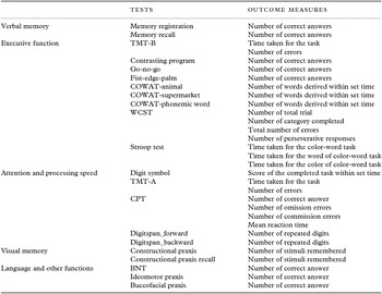
Table 1 presents the chosen individual neuropsychological tests and their outcome variables.
Neuropsychological tests and outcome measures

Note. MMSE = Mini-Mental Status Examination; MoCA = Montreal Cognitive Assessment; TMT-B = Trail-Making Test-B; COWAT = Controlled Oral Word Association Test; WCST = Wisconsin Card Sorting Test; TMT-A = Trail-Making Test-A; CPT = Continuous Performance Test; BNT = Boston Naming Test.
MCI patients were classified as: (1) amnestic MCI if they had a prominent memory impairment (1SD less than the mean when adjusted for age and education) in any one of memory function tests either alone or with other cognitive impairments or as (2) non-amnestic MCI if a single non-memory domain was impaired alone or in combination with other non-memory deficits when adjusted for age and education.
Statistical analysis
Sociodemographic variables and clinical characteristics
Continuous variables (age, education, PHQ-9 score) were compared between MCI/D+ and MCI/D− patients using Student's t-test. The distributions of gender and MCI subtype between the two groups were compared using χ 2 test.
General cognitive and functional statuses
MMSE score, MoCA score, GDS score, Barthel index score, and K-IADL score were compared between MCI/D+ and MCI/D− patients using Student's t-test.
Neuropsychological subtests
The results of individual neuropsychological subtests were compared using Student's t-tests. Effect sizes were calculated using Cohen's d (Cohen, Reference Cohen1988). Subgroup analyses according to MCI subtype (amnestic MCI vs. non-amnestic MCI) were conducted using Student's t-test to compare the results of neuropsychological subtests based on depression.
The statistical analyses were executed using SPSS 20.0 (SPSS Inc., Chicago, IL, USA).
Results
Sociodemographic variables and clinical characteristics
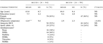
The MCI sample consisted of 153 participants with a mean age of 64.6 (SD = 8.6) years, and a mean length of education of 8.0 (SD = 4.5) years. The sample consisted of 28 men and 125 women. Descriptive statistics for the MCI/D+ and MCI/D− groups are shown in Table 2. The analyses used Student's t-test to confirm neither age nor years of education was not significantly different between the MCI/D+ and MCI/D− groups (t = 1.5, p = 0.13; t = 1.0, p = 0.33, respectively). The analyses used χ 2 test confirm that there was no significant difference in sex between the MCI/D+ and MCI/D− groups (p = 0.61). The MCI/D+ group scored significantly higher on the PHQ-9 (p < 0.001) compared with the MCI/D− group. There were no significant differences among the groups based on MCI subtypes (amnestic MCI and non-amnestic MCI).
Sociodemographic variables and clinical characteristics of MCI/D+ and MCI/D− participants

Note. MCI/D+ = depressed mild cognitive impairment patients; MIC/D− = non-depressed mild cognitive impairment patients; M = male; F = female; PHQ-9 = Patient Health Questionnaire-9; GDS = Geriatric Depression Rating Scale; CDR = Clinical Dementia Rating; GDS = Global Deterioration Scale; NS = not significant.
*p < 0.05.
**p < 0.01.
***p < 0.001, compared to the MCI/D−.
a Percentages do not add up to 100 because some patients were taking two or more antidepressant medications.
General cognitive and functional status
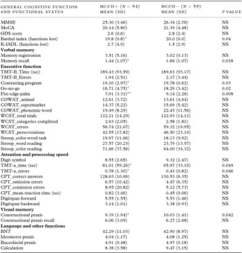
There were no significant difference between the MCI/D+ and MCI/D− groups for MMSE total score or MoCA total score. The MCI/D+ group scored significantly lower on the Barthel index (p = 0.04) compared with the MCI/D− group. There were no significant differences between the two groups for GDS score or IADL scale (Table 3).
Results of neuropsychological tests

Note. MCI/D+ = depressed mild cognitive impairment patients; MIC/D− = non-depressed mild cognitive impairment patients; MMSE = Mini-Mental Status Examination; MoCA = Montreal Cognitive Assessment; TMT-B = Trail-Making Test-B; COWAT = Controlled Oral Word Association Test; WCST = Wisconsin Card Sorting Test; TMT-A = Trail-Making Test-A; CPT = Continuous Performance Test; BNT = Boston Naming Test; NS= not significant.
*p < 0.05.
**p < 0.01.
***p < 0.001, compared to the MCI/D−.
Neuropsychological tests
Verbal memory
Performance on the memory recall test (p = 0.018, cohen's d = 0.39) was significantly impaired in MCI/D+ patients, but the result of the memory registration test was not significantly different between MCI/D+ and MCI/D− patients (Table 3).
Executive function
MCI/D+ patients showed significantly reduced performance in the Contrasting Program (p = 0.03, cohen's d = 0.32), Go-no-go test (p = 0.02, cohen's d = 0.38), and Fist-edge-palm test (p = 0.008, cohen's d = 0.43) compared with MCI/D− patients. Performances on the TMT-B, COWAT (animal, supermarket, phonemic word), and Stroop Test were not different between MCI/D+ patients and MCI/D− patients (Table 3).
Attention/processing speed
MCI/D+ patients took significantly more time and made more errors in the TMT-A test (p = 0.049, cohen's d = 0.31; p = 0.046, cohen's d = 0.32, respectively). On the other hand, performances on the Digit Symbol test, CPT, Digit Span Forward test, and Digit Span Backward test were not significantly different between the two groups (Table 3).
Visual memory
Performance on the Constructional Praxis test (p = 0.042, cohen's d = 0.38) was significantly impaired in MCI/D+ patients, but the result of Constructional Praxis Recall was not significantly different between MCI/D+ and MCI/D− patients (Table 3).
Language and other functions
Performances in the BNT, ideomotor praxis test, and buccufacial praxis and calculation test were not significantly different between the two groups (Table 3).
Amnestic mci and non-amnestic mci
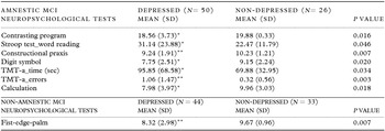
Subgroup analyses according to MCI subtype (amnestic MCI and non-amnestic MCI) were conducted to examine if the results of neuropsychological tests were different based on depression in amnestic MCI and non-amnestic MCI participants. The results, including the p-values of the tests for which the results were significantly different are shown in Table 4. The results show that amnestic MCI patients who were depressed performed worse on Contrasting Program (p = 0.016), Stroop Test-Word Reading Task (p = 0.046), Constructional Praxis (p = 0.007), Digit Symbol Test (p = 0.020), reaction time in TMT-A (p = 0.034), number of errors in TMT-A (p = 0.003), and calculation (p = 0.018) than those who were not depressed. Non-amnestic MCI patients who were depressed performed worse on measures of the Fist-edge-palm test (p = 0.007) than their non-depressed counterparts.
Results of neuropsychological tests in the amnestic MCI and non-amnestic MCI subgroups

Note. TMT-A = Trail-Making Test-A; NS = not significant.
*p < 0.05.
**p < 0.01.
***p < 0.001, compared to non-depressed counterparts.
Discussion
The objective of this study was to compare neurocognitive functions and performances of neuropsychological tests between depressed and non-depressed participants with MCI. Two subgroups of participants with MCI were distinguished according to the presence (MCI/D+) or absence (MCI/D−) of concomitant depression. All participants were evaluated for general cognitive and functional status, and then performed comprehensive neuropsychological tests that measured all cognitive domains. The results of the current study indicated that depression is significantly related to greater deficits in neurocognitive functions in MCI patients compared with non-depressed MCI patients. In the analyses of neuropsychological test results between MCI/D+ and MCI/D− groups, significant impairments were shown in the Contrasting Program, Fist-edge-palm test, Go-no-go test, TMT-A, memory recall test, and Constructional Praxis. And overall, the effects of depression on the performance of these six tests were small to medium. The analysis shows that the largest effect of depression occurs when performing the Fist-edge-palm test.
The Contrasting Program and Fist-edge-palm test are motor sequencing tests that evaluate prefrontal integrity (Sander, Reference Sander2010). The Go-no-go test is a response suppression task, and poor test results reflect failures in response control (Sander, Reference Sander2010). Poor performance on the Contrasting Program, Go-no-go test, and Fist-edge-palm test suggest frontal executive dysfunction. TMT-A is a timed test evaluating processing speed (Sanchez-Cubillo et al., Reference Sanchez-Cubillo2009). Poor performance on memory recall test suggests impairment in delayed recall of verbal memory (Ashford et al., Reference Ashford, Kolm, Colliver, Bekian and Hsu1989). Lastly, the Constructional Praxis is a measure of free visual recall (Spangenberg et al., Reference Spangenberg, Henderson and Wagner1997).
MCI/D+ patients had significantly greater deficits in neurocognitive functions including executive function (Contrasting program, Go-no-go test, and Fist-edge-palm test), verbal memory (memory recall test), visual memory (Constructional Praxis), and attention/processing speed (reaction time and the number of errors in TMT-A). Similar but more extensive findings were obtained from the present study, compared with previous studies. Some differences between the present study and some previous studies may arise from the different definition of depression (MDD vs. self-reporting depressive symptoms).
Several studies have shown that depression is related to deficits in executive functions (Butters et al., Reference Butters2004), memory (Nebes et al., Reference Nebes2000), and processing speed (Nebes et al., Reference Nebes2000) in cognitively normal participants. A meta-analysis investigating the cognitive domains and neuropsychological subtests affected by depression showed that depression is related to impairment in attention (Digit Span Test, CPT), processing speed (Trail Making Test-A, Digit Symbol Test), executive function (Stroop Test, WCST, Verbal Fluency), and memory (immediate verbal memory) (Lim et al., Reference Lim2013).
The reason for differences in results among tests within the same cognitive domain could be due to differences in the sensitivity of each neuropsychological subtests. In this regard, the above tests showing significant differences between the two groups (Contrasting Program, Go-no-go, Fist-edge-palm, memory recall test, Constructional Praxis test, and TMT-A) could be sensitive tests for identifying the effect of depression on neurocognitive functions in MCI patients.
The biological implication of significant impairment of specific cognitive domains in MCI/D+ patients compared with MCI/D− patients is important for understanding the pathophysiology of depression and its relation with cognitive decline. Impairment in executive function, memory, and attention/processing speed in depressed MCI patients might be due in part to frontostriatal disconnection. “Vascular Depression” hypothesis proposed that cerebrovascular disease disrupting fiber tracts within frontostriatal circuits may predispose or precipitate some geriatric depressive syndromes and the hallmark of MRI-defined vascular depression is the presence of white matter lesions identified as white matter hyperintensities(WMH) in the frontal and temporal lobe (Taylor et al., Reference Taylor, Aizenstein and Alexopoulos2013). A large body of literature describes increased numbers of WMH in late-life depression, (Schermuly et al., Reference Schermuly2010) and studies examining the relationship between WMH and cognition in late-life depression have found associations with executive dysfunction, memory impairment, and slow processing speed (Hickie et al., Reference Hickie, Scott, Mitchell, Wilhelm, Austin and Bennett1995; Kramer-Ginsberg et al., Reference Kramer-Ginsberg1999; Murata et al., Reference Murata2001; Sheline et al., Reference Sheline2008). The present study showed that the pattern of cognitive deficits in depressed MCI patients was similar to that of vascular depression. In MCI patients, depression may contribute to cognitive decline and progression to dementia through vascular disease.
Subgroup analyses were conducted to investigate whether MCI subtype influenced the results of the current study. Our results suggest that depressed amnestic MCI participants showed more neurocognitive deficits in executive function (Contrasting Program, Stroop Test), attention/processing speed (Digit Symbol, TMT-A), visual memory (Constructional Praxis), and language-related function (calculation) than their non-depressed counterparts. Depressed non-amnestic MCI participants did not show significant differences in any of the tests except the Fist-edge-palm test compared with their non-depressed counterparts. This discrepancy in the number of neurocognitive tests significantly affected by depression between the two subtypes of MCI is clinically interesting. The amnestic MCI subtype likely represents a prodromal form of Alzheimer's disease (Petersen, Reference Petersen2004), and the non-amnestic MCI subtype has a higher likelihood of progressing to non-Alzheimer's disease dementia such as dementia with Lewy bodies (Boeve et al., Reference Boeve2004). Our findings suggest that depression more strongly affects neurocognitive deficits in amnestic MCI than in non-amnestic MCI and might have a stronger association with Alzheimer's disease than other types of dementia. However, the results from each MCI subtype should be interpreted with caution due to the relatively small sample size.
Whether or not and the degree to which neurocognitive impairment in depressed MCI patients is reversible with successful treatment of depression is still uncertain. A few previous studies have found neurocognitive functions to improve and even return to normal levels when depression is successfully treated (Abas et al., Reference Abas, Sahakian and Levy1990). If so, neurocognitive impairment might merely represent the consequence or epiphenomenon of depression and so can be recovered by simply focusing on depression treatment.
In contrast, other authors have suggested that depression could be a causal risk factor of progression to dementia mediated by several mechanisms (Richard et al., Reference Richard2013). Alternatively, depression could affect the cognitive threshold for dementia, resulting in an earlier onset (Butters et al., Reference Butters2008). If this causal relationship does exist, it is important to treat depression in MCI patients, and it may be possible to return neurocognitive impairment to a normal level or to stop or delay the progression of MCI to dementia.
However, previous studies have demonstrated persistent neurocognitive impairment in fully remitted patients (Nebes et al., Reference Nebes2003). One possible reason for persistent neurocognitive impairment is a co-morbid dementing illness; depression in MCI patients might be a predictor or an early manifestation of dementia (van Reekum et al., Reference Van Reekum, Simard, Clarke, Binns and Conn1999). If so, even if depression is fully remitted, neurocognitive impairment might be persistent or progress to dementia. In this case, clinicians should consider early intervention for cognitive decline while also treating depression.
Our study design is cross-sectional, so the authors could not examine the exact mechanisms linking depression and progression to dementia. Longitudinal follow-up is ongoing, and future studies may determine the results of depression and cognitive impairment after treatment of depression in MCI patients and the influence of the relationship of depression and MCI on disease progression.
The results of the present study showed that MCI/D+ and MCI/D− differed at the cognitive profile. This may suggest that MCI/D+ belongs to the independent nosological entity at high risk of evolving into dementia. However, since many factors can influence the impact of depression on cognitive deficits, it will be necessary to conduct longitudinal studies and biomarkers studies to verify this hypothesis. Such works would provide important insights to understand the associations between depression and MCI as well as the heterogeneity of MCI.
The present study has several limitations. First, Table 3 shows the main results and several p-values that are nominally significant (p < 0.05). However, since the authors have tested multiple hypotheses, the risk of a false positive finding could be inflated. Therefore, these significant associations should be regarded as interesting leads for further study rather than firm evidence of strong association. Second, the present study is also limited by the heterogeneity of whole participants. The authors recruited both amnestic and non-amnestic MCI and this factor could have obscured differences between groups. And it is difficult to draw conclusions from the subgroup analyses due to limited sample size of subgroup (amnestic MCI and non-amnestic MCI). The data of subgroup analyses need to be confirmed with a much larger sample size. The third limitation was the absence of information about age of depression onset, illness duration of depression and the number of depressive episodes. The current results could be influenced by these factors. Fourth, MCI/D+ patients were not free of psychotropic drug intake, as more than half of the patients were treated with at least one antidepressants. Consequently, these psychopharmacological agents also might have influence the current results. Fifth, the authors divided the whole participants into two groups based on the diagnosis of major depressive disorder, so the negative group included the participant with minor depression or single depressive symptom. This may have influenced current results. Finally, all patients were recruited from one university hospital memory clinic. Therefore, our findings are likely not generalizable to all MCI patients. It will be interesting in future studies to assess whether our results are confirmed using a more diverse sample.
In conclusion, the present study suggests that depression negatively affects the neurocognitive functions of MCI patients. This study showed several subsets of neuropsychological tests (executive function, verbal and visual memory, and attention/processing speed) were significantly impacted in depressed MCI patients compared with non-depressed MCI patients. The present study also adds to a growing body of evidence showing that MCI is not a homogeneous clinical entity, and that subgroups could possibly be identified. Further, once our prospective data are analyzed, the authors might be able to examine clinical outcome and prognosis of MCI/D+ patients following antidepressant therapy compared with MCI/D− patients and the relationship between depression, MCI, and its progression. Further neurobiological studies are needed to help clarify the mechanisms of depression-related changes in cognitive deficits in MCI patients.
Conflict of interest
This study was supported by a grant of the Korean Health Technology R&D Project, Ministry of Health & Welfare, Republic of Korea (A070001).
Description of authors’ roles
Changsu Han contributed to the study design, project management of the study, data collection, and data interpretation; Hyunseok Dong carried out the statistical analysis, data interpretation, and writing the paper; Sang Won Jeon, Seoyoung Yoon, Hyun-Ghang Jeong, Young-Hoon Ko, Chi-Un Pae, Seung Hyun Kim, Ashwin A. Patkar, and David C. Steffens contributed to the drafting of the paper and provided a review of it prior to submission; Yu Jeong Huh carried out the neuropsychological testing and data collection.
Acknowledgments
We gratefully acknowledge the assistance of Ms Taehee Kim in coordinating the study.