Introduction
The quantitative morphology of the face is of growing importance. In rhinoplasty surgery, standard, uniform colour photographs prior to and following surgery are the accepted means of storing an image of the nose and facial features. They allow pre-operative planning and provide a definitive record, enabling evaluation of the nose by both the surgeon and the patient. In addition, they form a valuable teaching tool and are a vital part of the medical and legal record.
However, one of the most significant limitations of such photographs in the planning of facial plastic procedures is the limitation of working with a two-dimensional likeness, while attempting to create a mental image of what is, in reality, a three-dimensional object. Such a limitation may significantly contribute to the dissatisfaction with the aesthetic outcome of surgery which is sometimes felt by both the patient and the surgeon.
The reliability of optical surface scanners in three-dimensional documentation of the face has already been reported.Reference Kusnoto and Evans1–Reference Aung, Ngim and Lee4 Recent software development has extended the use of such systems; they can now be used to accurately analyse surgery outcomes, as well as to enable surgical simulation of the probable changes following operative intervention.Reference Moss, Ismail and Hennessy5
We have been considering the application of this technique in the assessment of rhinoplasty patients, and we plan to use the technique to document cosmetic change following simple septoplasty surgery. In order to demonstrate this, we here present the pre- and post-operative optical scanning results from a patient undergoing corrective septorhinoplasty.
Technique
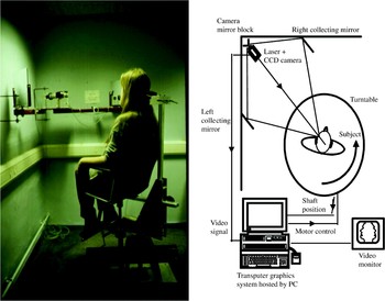
The technique records on-line the shape of a line of laser light projected onto the face (Figure 1). This line is then viewed obliquely by a closed circuit video camera via mirrors either side of the face. This viewing system minimises the loss of data due to shadowing of parts of the face by prominent features, principally the nose. Simple geometric analysis using the principles of triangulation allows this shape to be transformed into a set of three-dimensional co-ordinate points along the line illuminating the face. By rotating the subject under computer control, the whole of the face is scanned. Such a scan involves only a short exposure to a low intensity beam of laser light and is not hazardous. The reproducibility of this form of laser scanning has been previously demonstrated by Benn et al. Reference Benn, Ruff, Cartledge, Sauret, Copas and Linney7
Optical scanner. CCD = closed circuit digital; PC = personal computer. Reprinted with permission.Reference Linney, Campos and Richards6

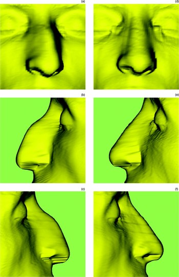
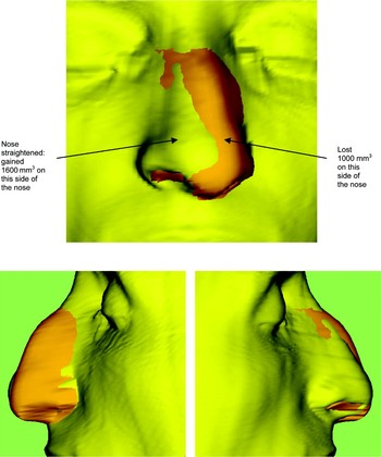
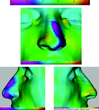
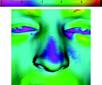
Our patient was scanned pre-operatively before his corrective septorhinoplasty, and then one week post-operatively after splint removal (Figure 2). As it was unlikely that the patient would be scanned in exactly the same position on consecutive occasions, the scans were realigned (i.e. registered) prior to any comparisons being made. This was done by interactively ‘painting’ areas that were known to be unchanged between scans and by using physical markers positioned on the skin. An iterative closest point algorithm was then used to register the unchanged surfaces or markers; after registration, the difference between the pre- and post-operative scans (i.e. a three-dimensional ‘difference map’) was visualised and colour-coded. The volume differences over various regions of interest could then be computed (Figure 3). In addition, differing areas of interest could also be individually visualised. This can be seen in Figure 4, in which coding on a continuous colour scale was utilised; warm colours (green to red) were used to represent positive surface displacement and cold colours (blue to pink) to represent negative displacement. Turquoise areas represented surfaces that had not changed.
Laser optical scans: pre-operative ((a) to (c)) and one week after corrective septorhinoplasty ((d) to (f)).

The two initial scans superimposed: orange = pre-operative; yellow = post-operative.

Post-operative scan analysis: warm colours (green to red) represent positive surface displacement (in millimetres); cold colours (blue to pink) represent negative displacement. Turquoise areas represent surfaces that have not changed.

Finally, the process was repeated at one year; the results of this analysis are shown in Figure 5.
Repeat scan analysis, one year post-operative.

Discussion
The scans clearly show that the patient's dorsal hump has been well reduced and the nose straightened, with a resulting 1600 mm3 gain on the right side and a 1000 mm3 loss on the left side of the nose. Tip projection has also been achieved. Figure 4 shows the exact areas on the nose and face that have moved, and in which direction. Post-operative swelling of the cheeks is seen initially but, after one year, this swelling has settled especially over the dorsum of the nose (blue area); the nose itself remains straight (Figure 5).
In this report, we wish simply to present these images in order to demonstrate the measurement of the changes achieved by surgery. To date, we have not used the technique to predict outcome from different interventions prior to surgery. However, such manipulation is now possible, and this form of editing of the anatomical surface would allow both the patient and the surgeon to predict the probable results of different surgical manoeuvres. This, we think, would probably result in more realistic expectations of outcome for all involved. The technique also has other possible uses. We are presently considering its application in the assessment of the degree of cosmetic change that follows septal surgery in adults. It is known that such changes may occur; however, good, prospective studies with subjective and objective data are rare,Reference Schwab and Pirsig8–Reference Jones10 and different rates of change have been reported ranging from 0 to 21 per cent.Reference Vuyk and Langenhuijsen11–Reference Low and Willatt13 To date, such studies have all relied on standardised photographs and have been subjective, relying as they have on the impressions of differing numbers of observers. As we have shown in this report, the use of an optical surface scanner would allow wholly objective quantification of any such changes, and might well allow such changes to be predicted in certain situations; this would also allow better data to be available for informed consent.
Conclusion
In conclusion, we present the results of using an optical surface scanner in the evaluation of nasal architecture before and after rhinoplasty surgery. This technique allows objective quantification of facial features, and this may well prove useful in the future in predicting change following surgical intervention.