Introduction
Infectious bronchitis is a highly contagious upper-respiratory tract disease of chickens and causes a severe economic burden on the poultry industry worldwide (Fraga et al., Reference FRAGA, BALESTRIN, IKUTA, FONSECA, SPILKI, CANAL and LUNGE2013; Umar et al., Reference UMAR, ULLAH, YAQOOB, SHAH and DUCATEZ2014). The causative agent, infectious bronchitis virus (IBV), has been found in many types of birds. Clinical signs of the disease in chickens include watery eyes, mucus in the nares and trachea, gasping, coughing and tracheal rales. The disease leads to a decrease in egg production and egg quality, and some strains of the virus can cause an interstitial nephritis. The virus replicates in epithelial cells causing lesions in the nasal turbinates, trachea, kidney, gonads, oviduct, lungs and air sacs. Lesions can also be found in the gastrointestinal tract, bursa of Fabricius and caecal tonsils (Jackwood, Reference JACKWOOD2012; Cook et al., Reference COOK, JACKWOOD and JONES2012).
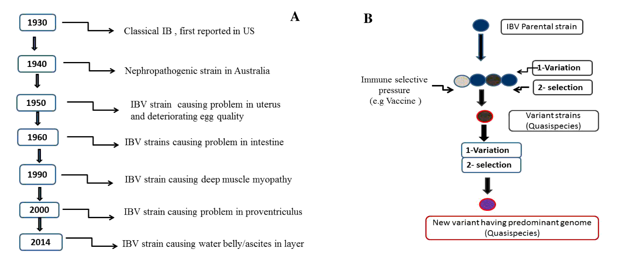
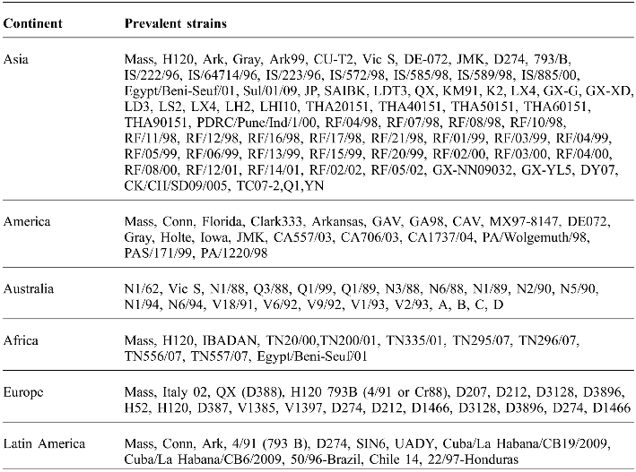
The viral distribution is virtually global. It was first described in the 1930s in the USA and more than 50 variants or serotypes have been documented globally. The time line discovery of IBV strains prevalent in different continents of the world are shown in Figure 1A and Table 1 respectively (Dhama et al., Reference DHAMA, SINGH, BARATHIDASAN, DESINGU, CHAKRABORTY, TIWARI and KUMAR2014).
Rapid evolution in IBV is facilitated by strong selection, large population sizes and high genetic diversity within hosts and transmission bottlenecks between hosts. Genetic diversity within the host arises primarily by mutation, which includes substitutions, insertions and deletions. Mutations are caused both by the high error rate and limited proof reading capability of the viral RNA-dependent RNA-polymerase, and by recombination. Recombination generates new haplotype diversity by recombining existing variants. These genetic variations occur continuously in nature and lead to the emergence of multiple phenotypes in terms of pathotypes and immuno-types (Cavanagh, Reference CAVANAGH2007). Specifically, virus neutralisation tests revealed the existence of a number of serotypes, which are poorly cross-protective (Cavanagh, Reference CAVANAGH2007; Marandino et al., Reference MARANDINO, PEREDA, TOMÁS, HERNÁNDEZ, IRAOLA, CRAIG, HERNÁNDEZ, BANDA, VILLEGAS, PANZERA and PÉREZ2015). Population genetics emphasises the comprehension of main evolutionary forces e.g. natural selection, migration, genetic drift, mutation and genetic structure of subject (Abro et al., Reference ABRO, RENSTRÖM, ULLMAN, BELÁK and BAULE2012a). Likewise, genomic transcription of IBV shows high error rate (Jackwood et al., Reference JACKWOOD, HALL and HANDEL2012), generating a phenomenon of quasispecies, which is significant for the evolution and persistence of virus as shown in Figure 1B (Montassier, Reference MONTASSIER2010; Jackwood et al., Reference JACKWOOD, HALL and HANDEL2012). Owing to this, diverse viral genotypes may co-circulate in hosts with different levels of aptness. Rapid evolution of avian coronavirus IBV makes this virus extremely difficult to diagnose and control (Marandino et al., Reference MARANDINO, PEREDA, TOMÁS, HERNÁNDEZ, IRAOLA, CRAIG, HERNÁNDEZ, BANDA, VILLEGAS, PANZERA and PÉREZ2015).
(A) Time line of events in the evolution of IBV strains (B) Generation and evolution of quasispecies of IBV strain through mutation, recombination and selection.

IBV strains prevalent in different continents of the world.
Existent vaccination programmes rely on the use of the Massachusetts serotype strain, which is the most commonly used IBV vaccine strain; however, the presence of the disease in vaccinated chickens is commonly observed. IBV vaccination is less successful because of the continuous development of antigenic variance and less cross-protection between variants (Liu et al., Reference LIU, ZHANG, WANG, LI, HAN, SHAO, LI and KONG2009a; Jackwood et al., Reference JACKWOOD, HALL and HANDEL2012; Wang et al., Reference WANG, LI, WEI, CHEN, WEI, MO and WEI2012). By virtue of advanced molecular virology and the availability of genomic information on IBV, there are new opportunities to create novel concept vaccines. Recently, focus has been on new vaccine development and improvement of existing vaccines for long term protection of poultry (Jackwood, Reference JACKWOOD2012; Umar et al., Reference UMAR, ARIF, SHAH, MUNIR, AHMED and KHAN2015a). Vaccination programmes should take into account locally prevailing IBV strains to offer better protection against the field strains. This review deals with various aspects of this important poultry virus having global significance with special reference to its evolutionary trends and vaccination strategies.
Genomic structure and biological activities of IBV
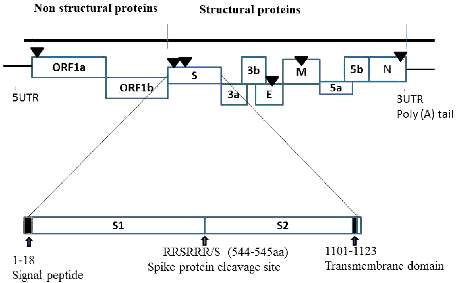
IBV belongs to genus Gammacoronavirus of family Coronaviridae within order Nidovirales (Jackwood, Reference JACKWOOD2012). This round, enveloped virus usually found in pleomorphic shape has size of approximately 120 nm in diameter (Cavanagh, Reference CAVANAGH2007). The positive sense RNA genome of this virus is 27.6 kb in size and encompasses 3′ and 5′ untranslated regions (UTRs) with a poly (A) tail (Figure 2) (Mo et al., Reference MO, HUANG, WEI, WEI, CHEN, WANG, LI and FAN2012). A major part of the genome is organised as overlapping 1a and 1b open reading frames (ORFs), which are translated into large polyproteins 1a and 1ab through a ribosomal frame shift mechanism. The remaining part of the genome consists of regions coding for the main structural proteins e.g. spike (S), envelope (E), membrane (M) and nucleocapsid (N). Two additional genes, ORF3 and ORF5, have been described coding for 3a & 3b and 5a & 5b accessory proteins respectively (Montassier, Reference MONTASSIER2010).
A schematic genome organisation of the infectious bronchitis virus, showing locations of 5′ and 3′ UTRs and coding regions (not drawn to scale). Black triangles (▼ ) indicate recombination hot spots within the genome.
The spike glycoprotein of all coronaviruses contains four domains that are involved in anchoring of the S protein into lipid bilayer of host cell. The S protein consisting of 1162 amino acids is sliced into two sub-units, the C-terminal S2 subunit (627 amino acids) and the N-terminal S1 subunit (535 amino acids). Attachment to host cell and neutralisation of immune response are the responsibilities of serotype-specific antigenic determinants and virus neutralisation functions which are comprised by S1 (Li et al., Reference LI, KANG, LIU, MAKKINJE, WILLIAMSON, LEIBOWITZ and GIEDROC2008). The variation in the nucleotide arrangement in spike genes among various serotypes may lead to minor cross protection, leading to altered defence against a vaccine or immunity (Abro et al., Reference ABRO, RENSTRÖM, ULLMAN, BELÁK and BAULE2012a). The C-terminal S2 sub-unit comprises of a fusion peptide-like area and two heptade areas, almost 10-13 nm long, which have involvement in protein oligomerisation and host cell entry (Guo et al., Reference GUO, TISONCIK, MCREYNOLDS, FARZAN, PRABHAKAR, GALLAGHER, RONG and CAFFREY2009). The S1 subunit has an amino acid level variation somewhere around 2 and 25%, however, the S2 subunit is more conserved (De Wit et al., Reference DE WIT, COOK and VAN DER HEIJDEN2011; Liu et al., Reference LIU, MA, XU, SUN, HAN, SUN, GUO, SHAO, KONG and LIU2013a). Gene 3 contains two ORFs (ORF3a and ORF3b) that are functionally tricistronic in nature (Lin et al., Reference LIN, LIN, CHIOU, HSU, LEE, CHANG, CHANG, SHIEN and HSU2012). ORF3a and ORF3b contain highly conserved nucleotide sequence regions (Mo et al., Reference MO, HUANG, WEI, WEI, CHEN, WANG, LI and FAN2012), not only in IBV but also in other gammacoronaviruses (Lin et al., Reference LIN, LIN, CHIOU, HSU, LEE, CHANG, CHANG, SHIEN and HSU2012). The envelope protein of virus (E) is known to have role in viral assembly (Almeida et al., Reference ALMEIDA, TORTELLY, NASCIMENTO, CHAGAS, KHAN and PEREIRA2012; Guo et al., Reference GUO, TISONCIK, MCREYNOLDS, FARZAN, PRABHAKAR, GALLAGHER, RONG and CAFFREY2009) and this function may be influenced by mutations in this protein (Valastro et al., Reference VALASTRO, MONNE, FASOLATO, CECCHETTIN, PARKER, TERREGINO and CATTOLI2010). The integral membrane protein (M) along with other structural proteins has the responsibility of arrangement and assembling the virus particle (Abro et al., Reference ABRO, RENSTRÖM, ULLMAN, ISAKSSON, ZOHARI, JANSSON, BELÁK and BAULE2012b). Gene 5 encompasses two ORFs (ORF5a and ORF5b) that encode the 5a and 5b proteins, respectively (Almeida et al., Reference ALMEIDA, TORTELLY, NASCIMENTO, CHAGAS, KHAN and PEREIRA2012; Kuo et al., Reference KUO, KAO, HOU, WANG, LIN and SU2013). It has been demonstrated that these proteins are functionally bicistronic (Valastro et al., Reference VALASTRO, MONNE, FASOLATO, CECCHETTIN, PARKER, TERREGINO and CATTOLI2010). The nucleocapsid protein (N) is a phosphoprotein consisting of 409 amino acids and has role in viral assembly, packing, core formation, signal transduction and modifying the processes of host cell (Krapez et al., Reference KRAPEZ, SLAVEC and ROJS2011). This protein is profoundly conserved; the amino acid arrangement varies from 2-6% percent among distinct IBV isolates (Mo et al., Reference MO, HUANG, WEI, WEI, CHEN, WANG, LI and FAN2012). A number of IBV serotypes, e.g. Arkansas, Connecticut (Conn), Massachusetts (Mass), 4/91 or 793B, D274, H120, Italy-02, Beaudette, California, Georgia 98 and QX have been identified and reported around the globe (De Wit et al., Reference DE WIT, COOK and VAN DER HEIJDEN2011). The appearance of a large number of IBV variants in different regions is a major constraint for the practical application of serotyping (De Wit et al., Reference DE WIT, COOK and VAN DER HEIJDEN2011). Genotyping of IBV is centred on the amplification and sequencing of the highly variable sequence region of the S1 gene by reverse transcriptase polymerase chain reaction (Abro et al., Reference ABRO, RENSTRÖM, ULLMAN, BELÁK and BAULE2012a). The molecular diagnostic assays revealed that different genotypes, such as Massachusetts, 4/91, D274, Italy-02 and European QX-like, are circulating in European countries (Krapez et al., Reference KRAPEZ, SLAVEC and ROJS2011; Abro et al., Reference ABRO, RENSTRÖM, ULLMAN, ISAKSSON, ZOHARI, JANSSON, BELÁK and BAULE2012b).
Evolution of infectious bronchitis virus
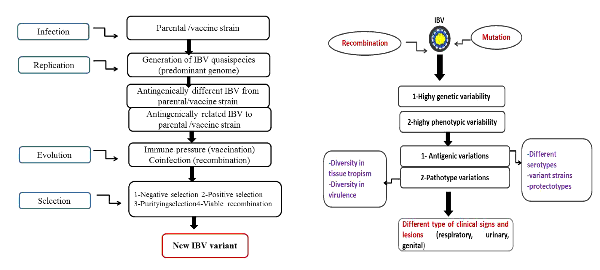
Avian coronavirus IBV creates genetic diversity through rapid replication and large population sizes coupled with a high mutation rate and recombination. Mutations include substitutions, which are the result of a high error rate and limited proof reading capability of the viral RdRp, and insertions and deletions, caused by recombination events or by RdRp stuttering or slippage. Broad genetic diversity facilitates survival of the virus in a constantly changing environment (Jackwood et al., Reference JACKWOOD, HALL and HANDEL2012; Marandino et al., Reference MARANDINO, PEREDA, TOMÁS, HERNÁNDEZ, IRAOLA, CRAIG, HERNÁNDEZ, BANDA, VILLEGAS, PANZERA and PÉREZ2015). Evolutionary progression of IBV is quite complex and inadequately understood. However, investigations carried out to date have highlighted the role of three factors: (1) lack of RNA polymerase proofreading, leading to replication errors in RNA genomes with mutation at the order of 10-4; (2) interference of continuous use of live and often multiple attenuated vaccines formulated with different IBV strains, and (3) immune pressure exerted on circulating viruses by the constant presence of partially immune bird populations. New variants of IBV have emerged due to spontaneous mutation and recombination during virus replication, followed by replication of those phenotypes favoured by selection (Awad et al., Reference AWAD, BAYLIS and GANAPATHY2014; He et al., Reference HE, LI, WEI, MO, WEI and LI2012; Toro et al., Reference TORO, PENNINGTON, GALLARDO, VAN SANTEN, van GINKEL, ZHANG and JOINER2012a, b; Liu et al., Reference LIU, SHAO, MA, SUN, ZHANG, LI, HAN, YAN, KONG and LIU2013b; Mo et al., Reference MO, HUANG, WEI, WEI, CHEN, WANG, LI and FAN2012). However, few variants are able to persist for extended time periods and spread in new territories to become of evolutionary and economic importance. The genetic forces such as mutations, recombination and selective pressure play significance role in the viral genome evolution as shown in Figure 3a and 3b (Marandino et al., Reference MARANDINO, PEREDA, TOMÁS, HERNÁNDEZ, IRAOLA, CRAIG, HERNÁNDEZ, BANDA, VILLEGAS, PANZERA and PÉREZ2015).
(A - left) Possible evolutionary pathway and emergence of new IBV variants from parental strain (B - right) Evolution of IBV giving rise to important antigenic and pathotype variability.
Recombination commonly occurs between two or more viruses infecting the same cell. It is believed that a high rate of recombination events occurs in the genomes of non-segmented RNA viruses, such as in IBV and in other coronaviruses (Jackwood et al., Reference JACKWOOD, HALL and HANDEL2012). Recombination can reduce mutational load, create genetic variants that may be very different from parental strains, and result in the emergence of new strains (Holmes, Reference HOLMES2009; Sumi et al., Reference SUMI, SINGH, DHAMA, GOWTHAMAN, BARATHIDASAN and SUKUMAR2012). Recombination has been reported in many coronaviruses (Thor et al., Reference THOR, HILT, KISSINGER, PATERSON and JACKWOOD2011). Recombination hot spots, or regions of the viral genome with higher incidences of recombination breakpoints, have been reported in IBV. Hot spots (Figure 2) tend to lie immediately upstream of the S glycoprotein gene, as well as in nsp 2, nsp 3, and nsp 16 (Thor et al., Reference THOR, HILT, KISSINGER, PATERSON and JACKWOOD2011; Jackwood et al., Reference JACKWOOD, HALL and HANDEL2012). Recombination in the non-structural proteins associated with the RdRp can alter replication efficiency of the virus, which can in turn affect pathogenicity.
Mutations and selective pressure in genes, especially in hypervariable regions, enable viruses to cross the species barrier and adapt to new host species, hence contributing to viral evolution (Lim et al., Reference LIM, LEE, LEE, LEE, PARK, YOUN, KIM, LEE, PARK, CHOI and SONG2011). The average rate of synonymous mutation in all coronaviruses including IBV is approximately 1.2 × 10⁻³ substitutions/site/year (Holmes, Reference HOLMES2009; Jackwood et al., Reference JACKWOOD, HALL and HANDEL2012). The rate of other RNA viruses with smaller genomes can be as high as 1 × 10⁻¹substitutions/site/year. The difference is presumably due to the presence of a 3′ to 5′ exoribonuclease (ExoN) domain in Nsp 14, which contains similarities to host proteins involved in proofreading and repair (Snijder et al., Reference SNIJDER, BREDENBEEK, DOBBE, THIEL, ZIEBUHR, POON, GUAN, ROZANOV, SPAAN and GORBALENYA2003). Using an nsp 14-ExoN mutant SARS-CoV, Eckerle et al. (Reference ECKERLE, BECKER, HALPIN, LI, VENTER, LU, SCHERBAKOVA, GRAHAM, BARIC, STOCKWELL, SPIRO and DENISON2010) showed impaired growth and a 21-fold increase in mutation rates for the mutant virus compared to the wild type. This data and previous work with a mouse hepatitis virus coronavirus (MHV-CoV) ExoN mutant (Eckerle et al., Reference ECKERLE, LU, SPERRY, CHOI and DENISON2007), confirmed that ExoN, which is conserved in all coronaviruses, contributes to the fidelity of the viral RdRp. High fidelity of polymerase results in a greater ‘error threshold’ and may permit the virus to maintain a large genome size (Holmes, Reference HOLMES2009; Jackwood et al., Reference JACKWOOD, HALL and HANDEL2012). The emergence of new strains and serotypes of IBV is largely due to the accumulation of mutations in the S gene over time as opposed to recombination events. This is thought to be the primary method of cross-species transmission and was shown to lead to the emergence of SARS-CoV (Hon et al., Reference HON, LAM, SHI, DRUMMOND, YIP, ZENG, LAM and LEUNG2008).
Different environmental determinants within the host i.e. immune responses, affinity for cell receptors, physical and biochemical conditions are implicated in the selection process (Toro et al., Reference TORO, VAN SANTEN and JACKWOOD2012b). Amino acid changes within the three S1 glycoprotein hypervariable regions determine the most relevant phenotypic changes, resulting in new serotypes and the induction of non-cross reacting VN antibodies (non-cross protecting).Thus, it is assumed that due to widespread vaccination, the immune selection pressure involving the S1 subunit of the S gene and the high mutation rate of the viral genome together can result in the emergence of many serotypes and variants (Abro et al., Reference ABRO, RENSTRÖM, ULLMAN, BELÁK and BAULE2012a).
Three hypervariable regions (HVRs) have been described in the first 395-amino acid region of the S1 subunit. Variants may attain increased virulence, efficient receptor binding, rapid transmission and persistence in host system causing significant disease in vaccinated flocks of all ages. A large number of variant viruses have been reported in America, North America, Australia, China, Africa, India and Pakistan (Ducatez et al., Reference DUCATEZ, MARTIN, OWOADE, OLATOYE, ALKALI, MAIKANO, SNOECK, SAUSY, CORDIOLI and MULLER2009; Han et al., Reference HAN, SUN, YAN, ZHANG, WANG, LI, ZHANG, MA, SHAO, LIU, KONG and LIU2011; Acevedo et al., Reference ACEVEDO, DE ARCE, BRANDAO, COLAS, OLIVEIRA and PEREZ2012; Cook et al., Reference COOK, JACKWOOD and JONES2012; Jackwood, Reference JACKWOOD2012; Dhama et al., Reference DHAMA, SINGH, BARATHIDASAN, DESINGU, CHAKRABORTY, TIWARI and KUMAR2014).
Vaccination and the evolution of IBV
Control of IBV in commercial poultry is normally via the use of live attenuated and killed vaccines. Since little or no cross-protection occurs between different serotypes or variants of the virus, a variety of different viral antigenic types are used in vaccines. Typically more than one immunisation is given and multivalent vaccines containing two or more antigenic types are used in an attempt to provide broad protection. It is not uncommon for vaccines to induce only partial protection against varied antigenic types of the virus, which allows some viruses to continue to replicate and persist in a flock. Presumably replication of the virus in a vaccinated host provides it with an opportunity to further adapt and evade the immune response (Jackwood et al., Reference JACKWOOD, HILT, MCCALL, POLIZZI, MCKINLEY and WILLIAMS2009; Jackwood, Reference JACKWOOD2012, Cook et al., Reference COOK, JACKWOOD and JONES2012; Jackwood et al., Reference JACKWOOD, HALL and HANDEL2012).
The use of modified live vaccine can result in faster evolutionary rates in IBV. Mutation rates for the 793/B IBV type where vaccines against that specific serotype were not used have been estimated to be 3 × 10⁻³ substitutions/site/year (Cavanagh, Reference CAVANAGH2007), whereas the GA98 virus, which emerged in the face of vaccination with the closely related DE072 type, was reported to have a mutation rate of 1.5 × 10⁻² substitutions/site/year (Jackwood, Reference JACKWOOD2012). However, caution must be exercised when estimating mutation rates of IBV antigenic types when live attenuated vaccines against that same virus type have been used. Mutation rates for Mass and Conn IBV types, where live vaccines against those types are commonly used, were estimated to be 1.9 × 10⁻³ and 1.5 × 10⁻⁴ nucleotide changes/site/year, respectively (Jones, Reference JONES2010; Cook et al., Reference COOK, JACKWOOD and JONES2012). Reisolation of vaccine viruses that are continuously introduced into the field and circulating in birds for a relatively short period of time likely contributed to the apparently low mutation rates. Emergence of new variants or serotypes of IBV is dependent on the ability of existing strains to be transmitted from one individual to another (Liu et al., Reference LIU, ZHANG, WANG, LI, HAN, SHAO, LI and KONG2009a).
Recent advances and future challenges in IBV vaccination
Vaccination has the strategic role in control and prevention of infection. IBV infection is extremely difficult to control because multiple serotypes and variants of the virus are continuously emerging and are not cross protective (Cavanagh, Reference CAVANAGH2007; Dhama et al., Reference DHAMA, SINGH, BARATHIDASAN, DESINGU, CHAKRABORTY, TIWARI and KUMAR2014). In most countries, day-old chicks are vaccinated in the hatchery with low virulence IBV vaccines. Thereafter, a booster immunisation is followed with virulent vaccines, usually in the drinking water. This low virulence is suitable for chicks with lower levels of maternal immunity as they do not cause the respiratory reactions which can occur with vaccines of higher virulence, a disadvantage being that low level immunity is not always enough to protect the respiratory tract (Kataria et al., Reference KATARIA, MOHAN, DEY, DASH and DHAMA2005).
The main vaccine types e.g. live attenuated and killed are usually formulated in an oil emulsion adjuvant. Live attenuated IBV vaccines have been administered to breeders and commercial egg layers for priming prior to oil-emulsion inactivated vaccines (OEIV). The OEIV is given prior to egg laying of breeders or layers, at about 17 weeks of age, and never during laying (Jackwood et al., Reference JACKWOOD, HILT, MCCALL, POLIZZI, MCKINLEY and WILLIAMS2009; Liu et al., Reference LIU, ZHANG, WANG, LI, LIU, HAN, ZHANG, KONG and TONG2009b). Nevertheless, for inactivated vaccines to be effective birds should have been previously ‘primed’ with a live vaccine. Preparation of live vaccines involves serial passages of IBV strains in embryonated chicken eggs (ECE) in order to achieve the required level of attenuation. With optimal conditions, vaccination may give immunity for many months and this may be life-long (Cook et al., Reference COOK, JACKWOOD and JONES2012; Dhama et al., Reference DHAMA, SINGH, BARATHIDASAN, DESINGU, CHAKRABORTY, TIWARI and KUMAR2014). The most common live vaccine strains are representative of the Massachusetts serotype, such as H120, which is a mild vaccine and is known for the typical level of attenuation due to the number of passages it has undergone. It is usually used for vaccinations for the first time without inducing a long lasting immunity in regions where there is increased level of field challenge, with the intention of maintaining local protection of the respiratory tract at a high level. Initial vaccination can be done individually by eye drop, intra tracheal or intranasal routes or by mass vaccination (e.g. coarse spray or drinking water). Such procedures are usually inexpensive and induce local as well as systemic immunity. Unfortunately it can cause reactions which are observed for a few days after vaccination (Dhama et al., Reference DHAMA, SINGH, BARATHIDASAN, DESINGU, CHAKRABORTY, TIWARI and KUMAR2014; Matthijs et al., Reference MATTHIJS, VAN ECK, LANDMAN and STEGEMAN2003). Ma5, a single component vaccine, is mild and used as a single component and can be included in first vaccination programs with IB 4/91 vaccines and inactivated vaccines for broad protection against different IBV serotypes. Live vaccines are generally used in breeders and layers as young birds to maintain a good level of local protection of the respiratory tract and are advisable in areas of increased level of field challenge.
The selection of vaccine must be based on strains prevalent in the local area/country. Different vaccine strains in SPF chickens provides cross-protection against homologous and reference strains and variant field isolates (Li et al., Reference LI, KANG, LIU, MAKKINJE, WILLIAMSON, LEIBOWITZ and GIEDROC2008; Marandino et al., Reference MARANDINO, PEREDA, TOMÁS, HERNÁNDEZ, IRAOLA, CRAIG, HERNÁNDEZ, BANDA, VILLEGAS, PANZERA and PÉREZ2015). Higher levels of cross-protection to some heterologous strains are given by a combination of Mass and Conn or Mass and JMK. The occurrence of multiple serotypes of the virus has complicated and increased the cost of disease prevention and warrants the use of local strains in vaccines for their effective control. The inactivated vaccines are used primarily at the point of lay to avoid stress and loss of production. The Massachusetts (Mass or M41) strain is the most popular as it is the representative of the initial isolates reported from many countries (De Wit et al., Reference DE WIT, COOK and VAN DER HEIJDEN2011). In order to give specific protection against the IBV type, IB 4/91 serotype (containing a strain of the 4/91serotype) or IB 274 vaccine virus (containing a strain of the D207(D274) serotype) are used; when combined with Ma5 and IB multi vaccines, they provide broad protection (Ma et al., Reference MA, SHAO, SUN, HAN, LIU, GUO, LIU, KONG and LIU2012). Inactivated vaccines induce long lasting immunity, show no vaccination reactions, usually cost more than live vaccines and a combination of different antigens apart from IBV can be achieved when given individually. Elevated levels of circulating antibodies were more stimulated by inactivated vaccines compared to live vaccines, which is useful in a breeder program where maternal antibody protection is needed. Still, due to induction of better T cell responses and higher local antibody (IgA) stimulation, modified live vaccines can play a significant role in protecting commercial layers.
Chickens must be properly primed with live vaccines, in order to exploit the potential of the inactivated vaccine and ensuring the highest titres will be obtained at an interval of at least four to six weeks (i.e. the period of vaccination between last live and inactivated vaccine) (Liu et al., Reference LIU, SHAO, MA, SUN, ZHANG, LI, HAN, YAN, KONG and LIU2013b). Furthermore, vaccination programs may be simplified by combining inactivated antigens against two or more serotypes (or two or more diseases) into one vaccine (Hong et al., Reference HONG, KWON, KIM, MO and KIM2012). Both live attenuated vaccines and inactivated water-in-oil emulsions are available for the Massachusetts, Connecticut and Arkansas serotypes in North America; California strains and Georgia 98 vaccines being used in USA. In Europe, ‘Holland variants’ commonly designated by number (e.g., D-274, D-1466) have been documented, with the IB H120 based vaccine being used in most parts of Europe. IB Ma5 and IB 4/91 (as mentioned above) are live, freeze-dried vaccine serotypes, giving long lasting protection. Infectious Bronchitis D274 is a live vaccine against strain D274 in poultry for vaccination of future breeding and layers stock. Vic S vaccine is used in most vaccination programs in Australia. The K2 vaccine might be useful for the control of newly evolving IBV recombinants (new cluster 1) and variants (new cluster 2) in Korea (Lim et al., Reference LIM, LEE, LEE, LEE, PARK, YOUN, KIM, LEE, PARK, CHOI and SONG2011). Massachusetts-serotype vaccine is the only live attenuated vaccine approved in Brazil, but the genotypes are greatly divergent and the most of them belongs to Non-Massachusetts types (Brandao, Reference BRANDAO2010).
Use of two heterologous IBV vaccines has been shown to be very successful, even though the mechanisms have not been elucidated to date. It is usual to first administer a Massachusetts-type vaccine followed by a variant (e.g. 793B-type in Europe) which offers wide protection against different types. Such a programme has been shown to be efficacious against, for example, Italy 02 and QX (Jones, Reference JONES2010).
In the EU, current control is centred on the use of Mass and major variant (793B)-type live and killed vaccines. The mass-type vaccines used are H120, MM (modified Mass) and Ma5. The 794B-type vaccines are denoted 4/91 and CR88. These are given to birds during the growing period as monovalent vaccines, with timing frequently involving Mass followed by 793B type, resulting in wide protection. One manufacturer produces bivalent live vaccines, an IB primer, comprising H120 and Dutch variant D274 and IBMM+Ark. Inactivated IB vaccines are given prior to point of lay often in combination with other inactivated viruses. Live vaccines are occasionally given to flocks in lay (Abro et al., Reference ABRO, RENSTRÖM, ULLMAN, ISAKSSON, ZOHARI, JANSSON, BELÁK and BAULE2012b).
With advances in molecular biology, novel solutions including DNA, sub-unit and vectored vaccines using S1 glycoprotein gene, along with reverse genetics vaccines have been tested in different laboratories (Dhama et al., Reference DHAMA, MAHENDRAN, GUPTA and RAI2008; Lin et al., Reference LIN, LIN, CHIOU, HSU, LEE, CHANG, CHANG, SHIEN and HSU2012). One of the most promising of the new generation vaccines appears to be the 'spike swapping' technology. Using reverse genetics, an infectious clone is produced into which specific S1 spike genes can be incorporated, appropriate to the new variant (Matthijs et al., Reference MATTHIJS, VAN ECK, LANDMAN and STEGEMAN2003; Brandao, Reference BRANDAO2010). Variations on this theme have included incorporation of nucleocapsid genes or specific cytokines to induce a broader immunity (Cavanagh, Reference CAVANAGH2007). Use of spike protein based DNA vaccines has revolutionised the concept of immunisation against IB (Matthijs et al., Reference MATTHIJS, VAN ECK, LANDMAN and STEGEMAN2003). Such tailor made-vaccines can be designed to suit the locally prevailing IBV strains and the problem of live attenuated strains reverting to virulence can be avoided. Recombinant or vector-based vaccines have been designed to introduce antigens from two or more viruses which act as multivalent vaccine giving protection against two or more diseases. DNA vaccine is another promising area as shown in initial clinical trials. These new generation vaccines can be administered safely in-ovo or to live chickens. The efficacy of these vaccines needs to be tested in large scale experiments before introduction for commercial purpose (Matthijs et al., Reference MATTHIJS, VAN ECK, LANDMAN and STEGEMAN2003; Brandao, Reference BRANDAO2010; Ullah et al., Reference ULLAH, RIAZ, UMAR and SHAH2013). The current need is to combat this economically important and notorious poultry pathogen by adapting and developing newer diagnostics, effective and safer vaccines, exploring novel therapeutics and following appropriate prevention and control strategies (Cook et al., Reference COOK, JACKWOOD and JONES2012; Jackwood et al., Reference JACKWOOD, HALL and HANDEL2012; Umar et al., Reference UMAR, YOUNUS, ARSHAD, SHAH, KHAN, NUMAN and TAHIR2015b).
Conclusions
Infectious bronchitis virus imposes a heavy welfare and economic burden on the poultry industry. There is considerable literature describing the continuous evolution of this virus due to mutations, recombination and strong selective pressure. These forces lead to a wide genetic diversity and generation of new variants of this virus. The disease possesses a continuous threat to poultry despite effective vaccines due to the emergence of new IBV variants from time to time, for which control by improved biosecurity, hygienic measures and vaccination programmes are required. However, effectiveness is very much dependent on challenge exclusively with a homologous serotype in the field. Little or partial cross-protection occurs between vaccine strains and new field strains which necessitates the development of new vaccines. Improved diagnostic and vaccination strategies are needed to tackle the growing threat of IBV infection in order to effectively prevent and control the disease in the future. It is critical to monitor IBV populations with continued nation-wide surveillance to map IBV genetic diversity, and to identify and prevent spreading of relevant genotypes.