Unwanted blood flow is one of the common problems in the field of paediatric cardiology. To date, vascular closure has been achieved through surgery and utilisation of various devices.Reference Gournay 1 – Reference Bass and Wilson 3 On the other hand, several authors have reported vascular narrowing as a potential complication of radiofrequency ablation,Reference Yan, Wang, Du, Yuan and Liang 4 – Reference Viles-Gonzalez, de Castro Miranda, Scanavacca, Sosa and d’Avila 6 with pulmonary venous stricture after pulmonary vein isolation and coronary narrowing after nearby radiofrequency ablation being two salient examples. Given this potential of radiofrequency energy, we successfully used radiofrequency for vascular closure and blood flow restriction in a sheep,Reference Dalili 7 and managed to confirm the procedure’s safety via pathological examination even after a prolonged high-powered use of the energy. Here, we present the closure of a conical patent ductus arteriosus with radiofrequency energy in a human.
Patient and method
The patient was a 3-year-old boy, who received a single-chamber pacemaker for his congenital complete heart block. He had also a small patent ductus arteriosus. Follow-up revealed an increase in the cardiac size; asynchrony owing to single-chamber pacing and patent ductus arteriosus were the likely culprits. The patient was admitted for patent ductus arteriosus closure. However, ductal closure from the venous side carried the risk of pacemaker lead dislodgment, and devices for ductal closure from the arterial side were unavailable at the time. Furthermore, the parents refused to consent to the implantation of any more devices. The parents, as well as the centre’s medico-legal committee, were provided with thorough explanations about our novel technique, and the patient was candidated for ductal closure with radiofrequency energy.
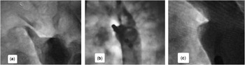
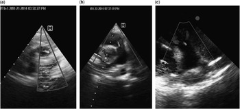
In the catheterisation room, an aortogram confirmed the presence of a small patent ductus arteriosus (Fig 1a). A 6-Fr ablation catheter (Blazer II HTD, Boston, Massachusetts, United States of America) was advanced from the left femoral artery into the patent ductus arteriosus. From the right femoral artery, a 4-Fr pigtail catheter was advanced adjacent to the ductus. Radiofrequency energy (Maestro, Boston, Massachusetts, United States of America) with a power of 45 W and a temperature level of 65°C were applied to the ductus orifice. For a detailed observation of the procedure and the progress, a small amount of contrast was injected via the pigtail catheter every 10 seconds. Radiofrequency application was continued for 2 minutes. After the procedure, the ablation catheter was pulled back and a standard amount of contrast material was injected, revealing the total occlusion of the ductus without any extravasation (Fig 1b and c). Post-procedural echocardiography showed a completely closed ductus without any complications (Fig 2b), and the pacemaker analysis demonstrated no adverse events. The patient was discharged from the hospital 2 days after the procedure with no complications. At 1 month’s follow-up, he was in full health. Echocardiography illustrated no residual ductal flow (Fig 2c). Serial echocardiographic pictures are depicted in Figure 2.
Aortogram before, during, and after ductal closure. ( a ) A conical ductus (arrow) with an ampulla is seen. ( b ) Radiofrequency catheter is seen in the ductal ampulla. ( c ) After radiofrequency energy application, the ductus is occluded. The pigtail tip is visible adjacent to the ductus; complete retrograde filling of the aortic arch confirms adequate contrast material injection. Tip of the ablation catheter is also observed.

Serial echocardiographic pictures of the patient. ( a ) Ductal flow is observed in hybrid view. ( b ) On the day after the procedure, no flow is seen in the closed ductus. ( c ) Echocardiography 1 month after the procedure shows no ductal flow. Trivial pulmonary regurgitation flow in ( b ) and ( c ) confirm that the frames have been recorded in diastole.

Discussion
This is the first report of a novel technique for patent ductus arteriosus closure. As is the case with other novel techniques, the future of the work depends on a larger number of case reports and clinical trials, the paramount concern of which would be safety. Use of radiofrequency energy is fully approved for the ablation of arrhythmia sources. In specific arrhythmias such as some outlet ventricular ectopies, the energy is applied to the vascular bed.Reference Dixit, Lin and Marchlinski 8 Apart from the treatment of arrhythmias, radiofrequency energy has been used for vascular sympathetic denervation to treat hypertension. A large study on 153 cases reported one vascular dissection; nevertheless, it was not clear whether radiofrequency had a direct role in the complication.Reference Krum, Barman and Schlaich 9
The technique for the application of radiofrequency energy to the ductus is not very complicated. A deflectable catheter can be easily applied and advanced to the ductus from the right or left femoral artery. The challenge may lie in the selection of appropriate catheters and sheet sizes. For our patient, we chose a 6-Fr catheter. Smaller ablation catheters may be available, but their smaller tips fail to provide sufficient effective areas for energy application. However, if the technique becomes widely accepted, specific catheters could be designed. For our first case, we used two arterial accesses. Use of a single access or a venous access will depend on the future of the work.
The main advantage of radiofrequency ductal closure is the exclusion of foreign body implantation. The novel method precludes the risk of device dislocation and spares the patient from the undesirable sensation of hosting a foreign body. Overall costs are reduced with the radiofrequency method, especially for centres in which the reuse of non-lumen catheters has been approved.Reference Josephson 10 Shortening of the procedural and fluoroscopy times are other predictable advantages.
First and foremost, among the possible disadvantages of radiofrequency ductal closure is the likelihood of vascular perforation. Needless to say, this catastrophic scenario could be the complication of the other currently used techniques too. The recurrent laryngeal nerve should be deemed a sensitive adjacent part, which could be damaged. Another drawback is that, although radiofrequency seems to be applicable for the closure of small vessels, the larger ducts may not be so amenable.
There are still some ethical issues to be solved before radiofrequency ductal closure can be fully approved. Further work on this novel technique is ongoing at our centre, and new findings will be published in due course.
Conclusion
Closure of undesirable vessels such as patent ductus arteriosus and collateral arteries could be achieved via radiofrequency energy. The principal advantage of this method is avoidance of foreign object implantation in the body. Further experience will elucidate the future scope of this novel technique.
Acknowledgements
Ramin Baghaei-Tehrani, cardiac surgeon, is highly appreciated for preparing an on-call operating room. The author thanks Professor Mohammad-yusef Aarabi-Moghaddam, paediatric cardiologist and head of the Pediatric Catheterization Laboratory, and Dr Hassan Birjandi, fellow in paediatric interventional cardiology, for arranging the accessory requirements, as well as to the Pediatric Catheterization and Electrophysiology Laboratory staff for their sincere collaborations. Special thanks to Dr Jayakeerthi Y. Rao for his contribution towards the preparation of the manuscript.
Financial Support
This work received no specific grant from any funding agency, commercial, or not-for-profit sectors. Rajaie Cardiovascular, Medical and Research Center provided the catheterisation laboratory and required equipment.
Conflicts of Interest
None.
Ethical Standards
The author asserts that all procedures contributing to this work comply with the ethical standards of the Iranian guideline on human experimentation of 1999, as revised in 2004, and with the Helsinki declaration of 1975, as revised in 2008, and has been approved by the ethics committee of Rajaie Cardiovascular, Medical and Research Center.