Cardiac rhabdomyomas are the most common cardiac tumours in children.Reference Beghetti, Gow, Haney, Mawson, Williams and Freedom 1 These benign hamartomas often demonstrate spontaneous regression over the first few years of life, and intervention is only indicated when there is sufficient mass effect to adversely affect cardiac output. Classically, surgical debulking has been the only means of alleviating obstructive symptoms.
Rhabdomyomas are one of the many manifestations of the tuberous sclerosis complex that is typically caused by mutations in the TSC1 and TSC2 tumour suppressor genes. Mutations in these genes result in hyperactivation of the mammalian target of rapamycin signalling pathway through the loss of tuberin or hamartin.Reference Crino, Nathanson and Henske 2 Mammalian target of rapamycin is a protein that regulates cell growth, proliferation, protein synthesis, and transcription.Reference Crino, Nathanson and Henske 2 Recently, mammalian target of rapamycin inhibitors have been successfully used to treat other tumours associated with tuberous sclerosis, including subependymal giant-cell astrocytomas, renal angiomyolipomas, and facial angiofibromas.Reference Bissler, McCormack and Young 3 – Reference Krueger, Care and Holland 5 In this report, we describe rapid regression of large cardiac rhabdomyomas in two neonates treated with mammalian target of rapamycin inhibitors.
Case 1
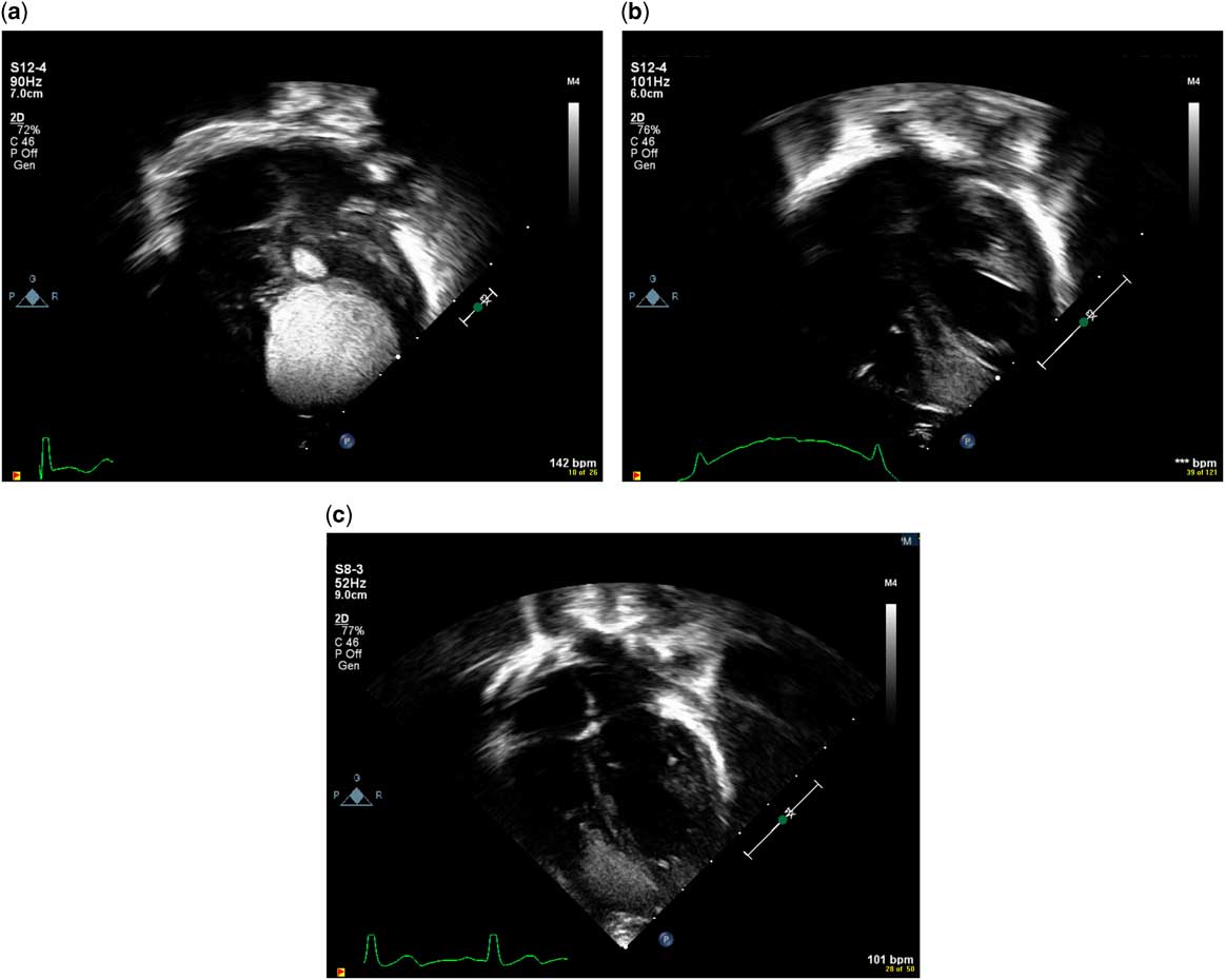
A 35-year-old woman was followed up by our high-risk fetal clinic owing to the prenatal finding of large cardiac tumours. Following an uncomplicated delivery, echocardiogram again showed a large, homogeneous and well-circumscribed cardiac tumour consistent with rhabdomyoma. The tumour was located at the apex of the heart, encroaching on the left and right ventricular cavities and measured 25×25×33 mm in orthogonal planes (Fig 1). There were also three smaller, non-obstructive tumours. Genetic testing showed a missense mutation in the TSC2 gene confirming a diagnosis of tuberous sclerosis.
Echocardiogram images demonstrating tumour progression in the patient described in case one. At birth, the largest tumour measured 25×25×33 mm in orthogonal planes ( a ). After 4 weeks of sirolimus therapy, the tumour size decreased to 9×9×9 mm ( b ). After being off of sirolimus for 9 months, there was some rebound in tumour size, which measured 16×12×10 mm ( c ).

Although the neonate was stable, the tumour was massive and we were concerned that it would impair diastolic and/or systolic ventricular function with an adverse impact on growth and development. The size and location of the large primary tumour made surgical intervention for debulking excessively risky. Sirolimus was started at a dose of 0.3 mg (0.1 mg/kg) daily, with a steady state target level of 5–15 ng/ml. Repeat echocardiogram at first outpatient follow-up 11 days after starting therapy showed a 74% reduction in the tumour volume. At follow-up 4 weeks after starting sirolimus, the tumour volume was further reduced to 12% of the baseline tumour volume (Fig 1). However, the sirolimus level at this follow-up was 22.5 ng/ml, well above the target range. Despite the supra-therapeutic level, no side effects were evident. As the rhabdomyoma was no longer considered large enough to adversely affect ventricular systolic or diastolic function, and because sirolimus is a narrow therapeutic index drug with significant potential for toxic adverse effects, sirolimus was discontinued. She was followed up over time, and at her most recent follow up – 9 months after sirolimus was stopped – the rhabdomyoma has shown interval size increase but remains less than one-third of the original calculated volume (Fig 1). The tumour remains unobstructive.
Case 2
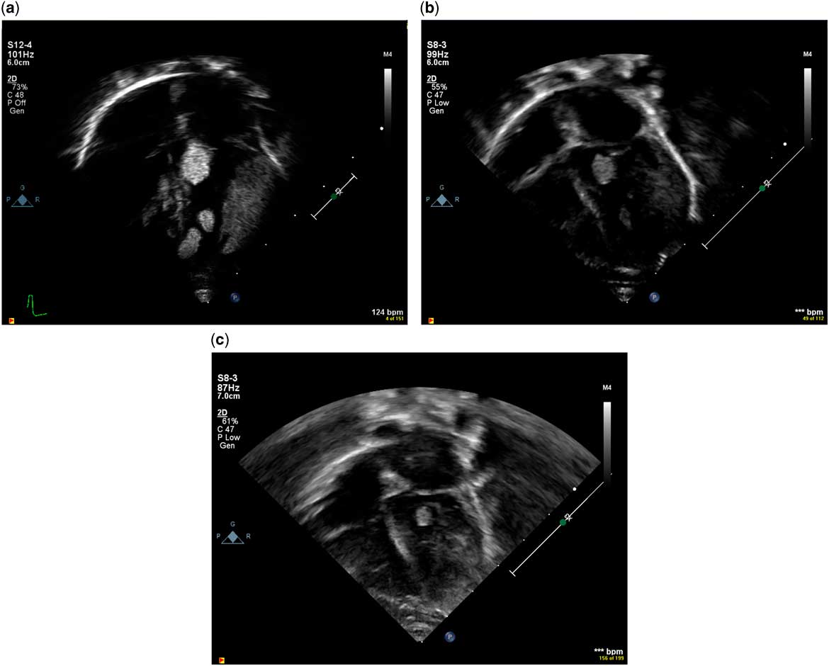
A 30-year-old woman was followed up by our fetal clinic owing to the presence of multiple cardiac masses. After an uncomplicated delivery, echocardiogram demonstrated multiple well-circumscribed, homogeneous tumours in the right and left ventricles that were felt to be consistent with cardiac rhabdomyomas. Notably, there was one large tumour involving most of the lateral wall of the left ventricle and a moderate-sized mobile tumour in the left ventricular outflow tract. The left ventricular lateral wall mass initially measured 22.1×14.5×8.0 mm and the left ventricular outflow tract mass initially measured 10.6×9.6×9.7 mm (Fig 2). By Doppler, there was mild obstruction through the left ventricular outflow tract with flow acceleration to 2.4 m/second. Genetic testing confirmed a diagnosis of tuberous sclerosis with a novel pathologic variant in the TSC2 gene.
Echocardiogram images demonstrating tumour progression in the patient described in case two. At birth, the left ventricular outflow tract mass measured 11×10×10 mm and the left ventricular lateral wall mass initially measured 22×15×8 mm ( a ). After 12 days of sirolimus therapy, the tumour in the left ventricular outflow tract measured 8×6×5 mm and the left ventricular free wall tumour measured 16×7×10 mm ( b ). At follow-up 1 month later, the left ventricular outflow tract tumour measured 6×5×5 mm and the lateral wall tumour measured 10×9×7 mm ( c ).
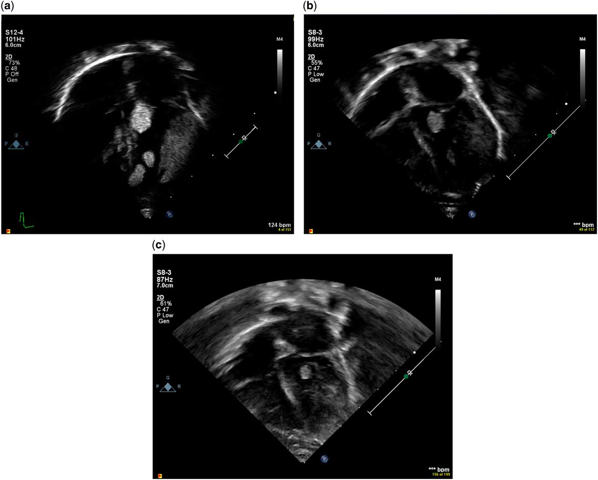
Sirolimus was initiated at 0.1 mg/kg every 12 hours with a target range of 5–15 ng/ml. We monitored the child in the hospital until steady state was reached. Repeat echocardiogram 12 days after treatment initiation showed a decrease in the size of the rhabdomyomas. The tumour volume in the left ventricular outflow tract was now approximately one quarter the original volume, and the left ventricular free wall tumour volume was decreased by >50% (Fig 2). There was no evidence of outflow tract obstruction. Sirolimus level was rechecked at that visit and was supratherapeutic at 24.3 ng/dl, so the dose was halved to 0.05 mg/kg. Follow-up 1 month later showed continued improvement in tumour size (Fig 2). Sirolimus level at that time was 12.1 ng/dl. The dose was decreased again to 0.03 mg/kg daily, with a resulting level of 9.1 ng/dl. No side effects were encountered during treatment.
Discussion
In this report, we describe two neonates with large cardiac rhabdomyomas that were considered at risk for haemodynamic complications but where surgical resection was considered prohibitively risky. Both were treated with the mammalian target of rapamycin inhibitor sirolimus with rapid regression over the course of about 4 weeks of therapy. After cessation of the drug in the first patient, a rebound increase in size of the rhabdomyoma was seen. Reinitiation of therapy was not required, as the patient continued to progress well clinically. The second patient, who stayed on sirolimus therapy, had a continued decrease in tumour size.
Primary cardiac tumours in children are rare, but the most common is the rhabdomyoma. More than 90% of rhabdomyomas are associated with tuberous sclerosis. The natural history is for spontaneous regression, which typically occurs during the first few years of life.Reference Beghetti, Gow, Haney, Mawson, Williams and Freedom 1 , Reference Crino, Nathanson and Henske 2 Consequently, most rhabdomyomas require no treatment. However, if there is potential for mass-related haemodynamic impairment, then surgical resection is indicated. Surgical intervention in a neonate is not a benign undertaking, and surgical debulking risks damage to surrounding cardiac structures including the atrioventricular or semilunar valve apparatus.
Tuberous sclerosis is associated with hyperactivation of the mammalian target of rapamycin signalling pathway, and mammalian target of rapamycin inhibitors have recently shown promise in the treatment of other tumours associated with tuberous sclerosis.Reference Bissler, McCormack and Young 3 – Reference Krueger, Care and Holland 5 Following two randomised, controlled trials,Reference Franz, Belousova and Sparagana 4 , Reference Krueger, Care and Holland 5 everolimus was recently approved by both the United States Food and Drug Agency and the European Medicines Agency for the treatment of subepyndomal giant-cell astrocytomas and renal angiolipomas. Although without a labelled indication for tuberous sclerosis-associated manifestations, trials of sirolimus have demonstrated benefits in the treatment of angiomyolipomas associated with tuberous sclerosis.Reference Bissler, McCormack and Young 3
Sirolimus and everolimus are currently the most studied mammalian target of rapamycin inhibitors used for the treatment of tumours associated with tuberous sclerosis. Sirolimus is readily available as an oral solution, whereas everolimus must be prepared from tablet form in water. Oral bioavailability of everolimus is slightly higher, at 20 versus 14% for sirolimus oral solution. The half-life of sirolimus is shorter in children than in adults, with a half-life of 20.5±11.4 hours in children as compared with 46–78 hours in adults.Reference Schubert, Venkataramanan and Holt 6 The half-life of everolimus is consistent in children and adults and is ~30 hours. The majority of pharmacokinetic information has been obtained from paediatric transplant patients, which may not be generalisable to our patients.
Four prior reports have described rhabdomyoma regression with mammalian target of rapamycin inhibitors in patients with tuberous sclerosis. The first report was in a 5-year-old receiving everolimus therapy for the treatment of a subepyndomal giant-cell astrocytoma. After 13 months of treatment, physicians noted clear regression of the rhabdomyoma.Reference Tiberio, Franz and Phillips 7 However, because the patient was older at the time of treatment initiation and received a relatively longer duration of therapy, the findings of this report were somewhat confounded by the natural history of spontaneous regression of rhabdomyomas. Three subsequent reports have described the use of mammalian target of rapamycin inhibitors in neonates with symptomatic, obstructive rhabdomyomas. Only one of these describes the use of sirolimus and, similar to our report, near complete tumour regression occurred within 1 month of treatment initiation.Reference Breathnach, Pears, Franklin, Webb and McMahon 8 In the two other reports, everolimus therapy was initiated, also with marked reductions in tumour burden but with a slower response to therapy in both patients – 2–3 months before the haemodynamic burden was considered resolved.Reference Demir, Ekici, Yazal Erdem, Emir and Tunc 9 , Reference Hoshal, Samuel, Schneider, Mammen and Vettukattil 10 It should be noted that both patients in these reports had significant clinical manifestations related to the rhabdomyomas, and the main end point for treatment success was clinical improvement rather than size reduction seen on serial echocardiography. The more rapid regression seen with sirolimus in our case and in the previously noted report from Breathnach et al may be purely coincidental but nonetheless warrants mention. This possibility, along with a more stable oral solution and otherwise similar profile to everolimus, may make sirolimus more advantageous than everolimus for administration. Further investigation in non-transplant children is needed.
Taken together, these reports highlight the potential therapeutic role of mammalian target of rapamycin inhibitors in children with haemodynamically significant cardiac rhabdomyomas. In all of the neonatal reports, there has been marked and rapid reduction of tumour bulk beyond what is typically expected based on the natural history of rhabdomyomas. Reassuringly, there was no substantial reoccurrence of the tumours with discontinuation of therapy, at least in the short term. This is an important finding, as published evidence indicates that treatment must be sustained, perhaps indefinitely, for durable treatment effect for other hamartomas associated with tuberous sclerosis.Reference MacKeigan and Krueger 11
Despite the success of mammalian target of rapamycin inhibition in these two patients, we only advocate for this therapeutic intervention in patients with large obstructive or potentially obstructive rhabdomyomas. Given the natural history of cardiac rhabdomyomas, treating with mammalian target of rapamycin inhibitors based only on size may lead to excessive use of the medication beyond therapeutic benefit. Furthermore, sirolimus and everolimus are both narrow therapeutic drugs with the potential for serious adverse events including mouth ulcers, hypercholesterolaemia, marrow suppression, infections, and metabolic effects.Reference MacKeigan and Krueger 11 There are limited data on the pharmacokinetics of either of these agents in neonates. Indeed in both of our patients doses that were initially associated with therapeutic drug levels (in the typical target range of 5–15 ng/ml) subsequently resulted in supra-therapeutic levels. We have no good explanation for this, but note that Breathnach et al had similar dosing issues in the only other reported experience with sirolimus for this indication.Reference Breathnach, Pears, Franklin, Webb and McMahon 8 Therefore, we recommend close monitoring of drug levels with discontinuation of therapy once tumours are sufficiently de-bulked.
In conclusion, our report adds to an emerging body of literature describing the potential therapeutic benefits of mammalian target of rapamycin inhibition for various tumours associated with the tuberous sclerosis complex. We suggest that mammalian target of rapamycin inhibition might be a reasonable alternative to surgical intervention in patients with haemodynamically significant rhabdomyomas, but we recommend close monitoring of drug levels with drug discontinuation once the tumour burden is sufficiently reduced to relieve haemodynamic concerns.
Acknowledgements
None.
Financial Support
This research received no specific grant from any funding agency, commercial or not-for-profit sectors.
Conflicts of Interest
None.