Introduction
The incidence of choroidal metastasis is increasing according to the available literature. The increase in the incidence is due to the longer survival rates of oncology patients and the improvement in the diagnostic studies that allow the identification of these metastases. Some studies report a prevalence of choroidal metastases from 2 to 7% of all oncology patients Reference Bloch and Gartner1–Reference Greear4 .
The most frequent primary tumour associated with choroidal metastasis is breast cancer, 47% of cases arising from breast cancer. Reference Demirci, Shields and Chao5–Reference Shields, Welch and Malik7 The second most frequent primary tumour is lung cancer, 20–29% of cases arising from lung cancer. Reference Shields, Welch and Malik7,Reference Shah, Mashayekhi and Shields8 Other primary tumours include prostate cancer, kidney cancer, skin cancer or gastrointestinal tumours.
The diagnosis of choroidal metastasis occurs in the progression of the primary tumour in 67% of cases. However, among the patients diagnosed with choroidal metastases from lung cancer, 44% present the choroidal metastasis as the first symptom. Reference Albert, Rubenstein and Scheie2
About 75% of choroidal metastases have other synchronous or metachronous extraocular metastases Reference Su, Chen and Perng3,Reference Jardel, Sauerwein and Olivier9 requiring multidisciplinary assessment with local and systemic therapies.
Overall survival is poor, with 57 and 23% at 1 and 5 years, respectively. Analysing the two most frequent cancers, overall survival after diagnosis of choroidal metastasis at 5 years for breast cancer is 25% and for lung cancer is 13%, worsening the prognosis of these patients. Reference Shields, Welch and Malik7,Reference Freedman and Folk10
In this study, we present a case of lung adenocarcinoma with choroidal metastasis.
Case
A 63-year-old male, non-smoker for a year, without comorbidities and without medical treatment.
In April 2018, the patient attends the emergency department for paresthesia in the left arm. The neurological examination showed no other findings. Chest X-ray showed a mass in the upper left lobe. A 52 × 51 mm hypodense mass in the left upper lobe with infiltration of the bronchus and the left pulmonary artery was observed on computerised tomography (CT). Brain CT showed two brain lesions, one in the right frontal lobe and the other in the left parietooccipital region. Magnetic resonance imaging (MRI) confirmed the presence of these two intraparenchymal lesions and one more in the left occipital lobe.
The diagnostic investigations were completed with a bone scan and positron emission tomography (PET) without new findings. Finally, the biopsy confirmed a non-small-cell lung adenocarcinoma, anaplastic lymphoma kinase (ALK) negative, Kirsten rat sarcoma 2 viral oncogene homolog (KRAS) mutated.
This case was presented to the Tumor Board and it was decided to treat the three brain lesions with radiosurgery (RS) with a single dose of 17 Gy and induction chemotherapy with cisplatino–pemetrexed to a subsequent surgical salvage of the primary tumour.
After RS, a complete response of the three brain metastases was observed. After four cycles of induction chemotherapy, a partial response of the primary tumour was observed.
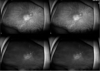
In August 2018, the patient presented with a loss of vision, ptosis, pain and oedema in the right eye. The patient was evaluated by the Ophthalmology Department. The fundoscopy showed a choroidal white mass, with intraretinal and subretinal flows, without bleeding and a normal papilla (Figures 1 and 2). The left eye was normal.
Right eye fundus with choroidal metastasis.

Pretreatment right eye fundus shows a whitish choroidal mass, with intraretinal and subretinal fluids.

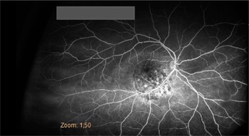
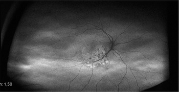
An ultrasound of the right eye showed a choroidal mass of 3–4 mm and angiography revealed a choroidal block (Figures 3 and 4). These characteristics established the diagnosis of choroidal metastasis.
Superficial normal angiography.

Choroidal block angiography.

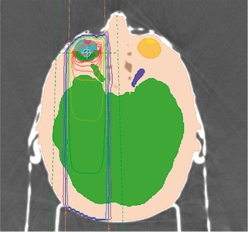
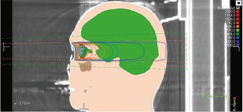
In August 2018, the patient received external beam radiotherapy for the choroidal metastasis. The patient was immobilised using a thermoplastic mask and underwent CT simulation, contouring and planning using 6 MV photons produced by a linear accelerator. The total prescribed dose was 20 Gy in five fractions (Figures 5 and 6).
Radiotherapy treatment plan (axial view).

Radiotherapy treatment plan (sagittal view).

The treatment was well tolerated, with no relevant toxicity and with improved pain and complete recovery of vision.
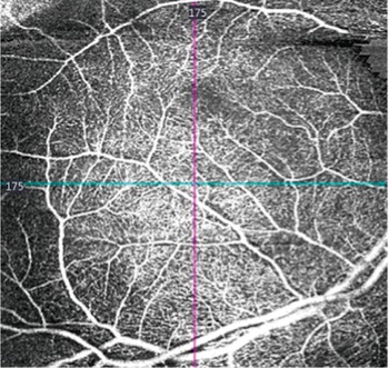
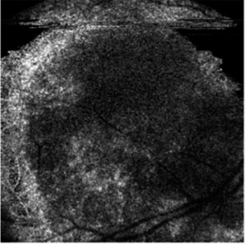
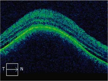
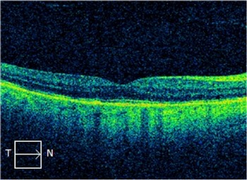
After treatment, the right fundus showed an area of non-elevated, mottled pigment alteration in the location of the previous lesion (Figure 7). Optical coherence tomography (OCT) showed a complete response of the choroidal metastasis (Figures 8 and 9).
Post-treatment right eye fundus.

Pre–treatment OCT.

Post–treatment OCT.

The patient continued treatment with pemetrexed for 3 months. In October 2018, he presented lung and mediastinal progression, and he started on a second chemotherapy agent, Nivolumab and a month later Docetaxel was added to treatment due to a progression of locoregional disease.
In July 2019, he was referred to Radiation Oncology for treatment for the pain of the primary tumour. He was treated with a total prescription dose of 37·5 Gy in 15 fractions, which resulted in an improvement of pain.
In November 2019, he presented progression with a right adrenal metastasis so the patient took part in a clinical trial that continued with systemic chemotherapy.
In July 2020, 22 months after treatment with radiotherapy, the patient had a complete response in the brain and choroidal metastases, with good vision and no late side effects associated with the treatment.
Discussion
We present the case of a 63-year-old male with a choroidal metastasis in the right eye of lung adenocarcinoma treated with external beam radiotherapy. After 22 months of follow-up, the patient continues with a complete choroidal response and good visual acuity.
The differential diagnosis of choroidal metastasis should be made in an oncology patient presenting with a loss of vision or intraocular pain. Intraocular pain has been described as the main symptom of choroidal metastasis specifically from lung cancer according to different studies. Reference Demirci, Shields and Chao5,Reference Shah, Mashayekhi and Shields8,Reference Konstantinidis, Rospond-Kubiak and Zeolite11 However, many of these patients are asymptomatic, which causes a delay in diagnosis. Among asymptomatic patients, the presence of choroidal metastasis in autopsy represents 12·6%. Reference Eliassi-Rad, Albert and Green12
Early diagnosis is essential and includes the assessment of visual acuity by the Ophthalmology Department. In the eye fundus, choroidal metastasis appears as a flat, yellowish-whitish (86%) orange (8%) or brown (4%) lesion, associated or not with intraretinal or subretinal fluid. Reference Shields, Welch and Malik7
These characteristics are not pathognomonic and are sometimes indistinguishable from choroidal melanoma, which is the second most frequent intraocular tumour. Reference Salah, Khader and Yousef13
Intraocular ultrasound shows differences between metastasis and choroidal melanoma due to reflectivity and the height/base ratio, which is the maximal prominence and the maximal base diameter of the tumour. In choroidal metastasis, there is greater reflectivity with a mean of 70% compared to 22% in melanoma. Also, the height/base ratio is lower in choroidal metastasis with a mean of 0·18 mm compared to 0·6 mm in melanoma. Reference Sobottka, Schlote and Krumpaszky14,Reference Sobottka and Kreissig15
Angiography in choroidal metastasis shows global hyperfluorescence that starts in the arterial phase and increases gradually, while fluorescence at choroidal area is obstructed during the complete test. OCT allows evaluation of the density and irregularity in the anterior part of the lesion; an elevated cupula is common. Reference Jorge, Labarrere and Rodrigues16–Reference Mathis, Jardel and Loria18 Other techniques such as OCT angiography or Enhanced Displacement Imaging (EDI-OCT) are also used in the diagnosis.
Biopsy is used in patients with unknown oncological diagnosis. Reference Zhang, Ma and Feng19
A diagnosis of choroidal metastasis presents a poor prognosis for patients, the mean survival is between 4 and 12 months. Reference Shields, Welch and Malik7,Reference Freedman and Folk10,Reference Wiegel, Bottke and Kreusel20–Reference Ferry and Font22
Among the different treatment options for choroidal metastasis, radiotherapy is the most commonly used in the literature. Reference Jardel, Sauerwein and Olivier9,Reference Mathis, Jardel and Loria18 Radiotherapy improves visual acuity between 81 and 90% and has good results with 64–80% complete responses in several studies. Reference Demirci, Shields and Chao5,Reference Shah, Mashayekhi and Shields8,Reference Jardel, Sauerwein and Olivier9,Reference Wiegel, Bottke and Kreusel20,Reference Rosset, Zografos and Coucke23,Reference Rudoler, Corn and Shields24
External beam radiation therapy (EBRT) is the most commonly used technique, allowing the treatment of single and multiple lesions. Hypofractionated schemes are the most used, included 30 Gy in 10 fractions (biologically effective dose (BED) = 39 Gy) and 20 Gy in 5 fractions (BED = 28 Gy). Reference Jardel, Sauerwein and Olivier9,Reference Eliassi-Rad, Albert and Green12,Reference Ratanatharathorn, Powers and Grimm25 In our case, we have performed a hypofractionated treatment with 20 Gy in five fractions. Hypofractionated schemes are effective with low acute complications and they provide a good treatment option due to the reduced life expectancy of the patients. Reference Jardel, Sauerwein and Olivier9,Reference Eliassi-Rad, Albert and Green12
In selected patients, who have a longer life expectancy, treatments with conventional dose schemes, 40 Gy in 20 fractions (BED = 48 Gy) are suitable to decrease the late side effects. Reference Wiegel, Bottke and Kreusel20,Reference Rudoler, Corn and Shields24,Reference Amer, Pe’er and Chowers26
EBRT achieves 80% complete responses and 57–89% clinical improvement. Reference Demirci, Shields and Chao5,Reference Shah, Mashayekhi and Shields8,Reference Jardel, Sauerwein and Olivier9,Reference Wiegel, Bottke and Kreusel20,Reference Rudoler, Corn and Shields24,Reference D’Abbadie, Arriagada and Spielmann27 Fifty percent of patients present with mild acute toxicities such as skin erythema and conjunctivitis. Late side effects are uncommon and include cataracts (6·5%), glaucoma (3%), neovascularisation of the iris (2%), vitreous haemorrhage (2%), retinopathy (<1%), papillopathy (<1%), macular oedema (1%), dry eye syndrome (<1%), eye pain (<1%). Reference Shah, Mashayekhi and Shields8,Reference Wiegel, Bottke and Kreusel20,Reference Rudoler, Corn and Shields24
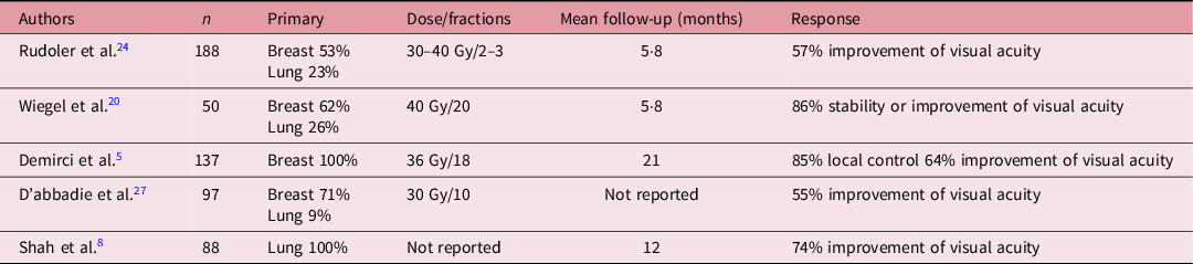
Some studies describe the use of lens-sparing technique with a contact lens mounted lead shield during the treatment to reduce complications. Reference Wiegel, Bottke and Kreusel20,Reference Rudoler, Corn and Shields24,Reference Mccormick, Ellsworth and Abramson28 Table 1 summarises the main studies of choroidal metastases treated with EBRT.
Main studies of choroidal metastases treated by external beam radiation therapy (EBRT)

Up to 38% of patients have bilateral choroidal metastases. For these cases, some studies recommend prophylactic bilateral treatment. Reference Rosset, Zografos and Coucke23 Others use a treatment plan with two anteroposterior and lateral fields, this may eliminate the microscopic disease in the contralateral eye due to the lateral field. Reference Jardel, Sauerwein and Olivier9,Reference Wiegel, Bottke and Kreusel20
Depending on the size, proximity to the macula, number of lesions, intention to treat, histology or prognosis of the patient, other radiotherapy techniques may be used.
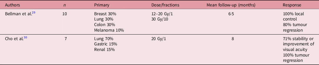
In the treatment of choroidal metastasis, high-dose radiotherapy techniques, such as stereotactic body radiation therapy (SBRT), may be used in one or more fractions. SBRT allows a high radiation dose to the tumour with low doses to the organs at risk, achieving better local control and lower side effects. SBRT is indicated in patients with a good performance status. The mean dose used is 20 Gy in a single fraction and local control is 80–100%. Reference Bellmann, Fuss and Holz29–Reference Wackernagel, Holl and Tarmann31 Table 2 summarises the main studies of choroidal metastases treated with SBRT.
Main studies of choroidal metastases treated by stereotactic body radiation therapy (SBRT)

Brachytherapy is indicated as initial or salvage treatment. Brachytherapy is used in defined metastasis with a diameter of <18 mm. The most commonly used radioactive isotope is Iodine-125 with a mean dose of 40–70 Gy applied over 3 days. Brachytherapy has very good results with 90–100% tumour response Reference Shah, Mashayekhi and Shields8,Reference Shields, Shields and De Potter32–Reference Sas-Korczyńska, Dixon and Skołyszewski34 and the duration of treatment is shorter compared to EBRT. However, it is an invasive technique that is done exclusively in specialised centres. Complications such as retinopathy, cataracts and atrophy of the optic nerve similar to EBRT. Reference Shields, Shields and De Potter32 Table 3 summarises the main studies of choroidal metastases treated with brachytherapy.
Main studies of choroidal metastases treated by brachytherapy

Proton beam therapy is indicated in radio-resistant histologies, including melanoma, sarcoma or renal cell carcinoma and in long-term survival patients. The mean total dose used is 20–28 Gy in two fractions, with local control rates between 94 and 98%. Reference Kamran, Collier and Lane35 The characteristics of protons and its energy liberation (Bragg peak) provide superior dose distributions and have a dosimetric advantage compared to photon beam therapy, with a better protection of the organs at risk and a shorter treatment duration. However, proton therapy is not always accessible. Reference Sas-Korczyńska, Dixon and Skołyszewski34,Reference Tsina, Lane and Zacks36 Table 4 summarises the main studies of choroidal metastases treated with proton beam therapy.
Main studies of choroidal metastases treated by proton beam therapy

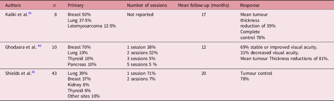
Photodynamic therapy associated with intravitreal verteporfin is another local treatment option for choroidal metastases. However, its effectiveness is variable (39–81%) depending on the results from different studies. Reference Yasui, Sato and Takeyama37–Reference Hua, Li and Wu41 It appears not to be useful in large tumours. Intravitreal injections of anti-vascular endothelial growth factor therapy (anti-VEGF) have also been used in combination with systemic therapies, but their benefit is not completely clear. Reference Yasui, Sato and Takeyama37 Table 5 summarises the main studies of choroidal metastases treated with photodynamic therapy.
Main studies of choroidal metastases treated by photodynamic therapy

This review has some limitations, as there are no clinical trials, most of the studies are cases with a limited number of patients. Besides these studies are heterogeneous, including different primary tumours, with different doses and fractions of radiotherapy and different endpoints. While acknowledging these limitations, radiotherapy is an effective and safe treatment option for these patients.
Conclusions
Choroidal metastasis treated with radiotherapy achieves good local control, with limited side effects, allowing an improvement in visual acuity and consequently, an improvement in the patient´s quality of life.
Acknowledgements
None.
Financial Support
None.
Conflicts of Interest
None.