Introduction
Angiostrongylus cantonensis, a metastrongyloid nematode widely found in the Asia-Pacific region among many others (Wang et al., Reference Wang, Lai, Zhu, Chen and Lun2008; Barratt et al., Reference Barratt, Chan, Sandaradura, Malik, Spielman, Lee, Marriott, Harkness, Ellis and Stark2016), causes human angiostrongyliasis, which can result in eosinophilic meningitis or meningoencephalitis (EOM) (Cross, Reference Cross1978; Pien and Pien, Reference Pien and Pien1999; Graeff-Teixeira et al., Reference Graeff-Teixeira, da Silva and Yoshimura2009). So far, at least 2800 cases of the disease have been recorded globally (Tsai et al., Reference Tsai, Liu, Kunin, Lee, Chen, Lin, Tsai, Lin, Huang, Yen and Yen2001, Reference Tsai, Chen and Yen2013; Eamsobhana et al., Reference Eamsobhana, Lim and Yong2013; Thiengo et al., Reference Thiengo, Simões Rde, Fernandez and Maldonado2013). Human infection comes mainly through the consumption of undercooked snails infected with the parasite (Pila or Pomacea snails) (Wang et al., Reference Wang, Chen and Lun2007; Tesana et al., Reference Tesana, Srisawangwong, Sithithaworn, Laha and Andrews2009) or vegetables contaminated with infective third-stage larvae (L3) (Slom et al., Reference Slom, Cortese, Gerber, Jones, Holtz, Lopez, Zambrano, Sufit, Sakolvaree, Chaicumpa, Herwaldt and Johnson2002; Tsai et al., Reference Tsai, Lee, Huang, Yen, Chen and Liu2004). A definitive diagnosis is confirmed by the discovery of the worms in cerebrospinal fluid after lumbar puncture or in the eyes during surgery, but direct detection of the parasite in patients is rare (Punyagupta, Reference Punyagupta and Cross1979; Cross and Chen, Reference Cross, Chen, Murrell and Fried2007). Diagnosis based on clinical presentation is difficult because the symptoms are often confused with those produced by other parasitic diseases such as paragonimiasis, gnathostomiasis and cysticercosis (Jaroonvesama, Reference Jaroonvesama1988). Antigenic proteins with molecular masses of around 29–31 kDa from A. cantonensis worms have been used as the specific markers in immunological diagnosis of angiostrongyliasis such as enzyme-linked immunosorbent assay (ELISA) and immunoblot tests (Nuamtanong, Reference Nuamtanong1996; Maleewong et al., Reference Maleewong, Sombatsawat, Intapan, Wongkham and Chotmongkol2001; Intapan et al., Reference Intapan, Maleewong, Sawanyawisuth and Chotmongkol2003; Eamsobhana and Yong, Reference Eamsobhana and Yong2009). However, these immunological tests are laborious to perform and none are commercially available. A 31 kDa antigen of A. cantonensis worms purified by electroelution from SDS-polyacrylamide gels was recently applied to a lateral-flow immunochromatographic test (ICT) assay for the simple and rapid diagnosis of angiostrongyliasis (Eamsobhana et al., Reference Eamsobhana, Tungtrongchitr, Wanachiwanawin and Yong2018). Nevertheless, no ICT kit using recombinant A. cantonensis antigen is available that would allow for unlimited mass production. A previous study identified a 31 kDa A. cantonensis antigen as a galectin-2 protein, and a recombinant A. cantonensis galectin-2 (rAcGal2) protein was produced as a result (Somboonpatarakun et al., Reference Somboonpatarakun, Intapan, Sanpool, Wongkham and Maleewong2019). The recombinant showed high specificity and sensitivity in immunoblotting assays. This study used rAcGal2 as an alternative antigen in the development of an ICT kit and compared its effectiveness with an ELISA.
Materials and methods
Parasite antigens
An antigenic protein with a mass of approximately 31 kDa from young adult female A. cantonensis worms was identified as galectin-2, and a recombinant A. cantonensis galectin-2 (rAcGal2) was produced as previously described (Somboonpatarakun et al., Reference Somboonpatarakun, Intapan, Sanpool, Wongkham and Maleewong2019). Briefly, young adult female worms were obtained from the brains of A. cantonensis-infected Sprague-Dawley rats (Rattus norvegicus) using a method described by Maleewong et al. (Reference Maleewong, Sombatsawat, Intapan, Wongkham and Chotmongkol2001). Total RNA obtained from the worms was used to amplify the full-length cDNA of A. cantonensis galectin-2 (AcGal2). The cDNA insert of AcGal2 was subcloned into a pET43.1b(+) expression vector (Novagen, Darmstadt, Germany) and transformed into Escherichia coli Rosetta-gami 2(DE3) expression host (Novagen). The soluble rAcGal2 was purified using Ni-NTA His Bind Resin (Novagen), dialysed against distilled water, and stored at −80 °C until being used in the ELISA and ICT.
Serum samples
A total of 244 serum samples were obtained from the serum bank at the Khon Kaen University Faculty of Medicine. These included 25 samples from normal healthy volunteers who were not found intestinal parasitic infection [stool samples were negative based on the formalin ethyl acetate concentration (FEC) method; Elkins et al., Reference Elkins, Haswell-Elkins and Anderson1986] at the time of blood collection, 100 samples of angiostrongyliasis sera (six from patients with proven angiostrongyliasis confirmed by the recovery of the worms from the patients' eyes and 94 from the patients with EOM and clinical suspected angiostrongyliasis diagnosed based on immunoblotting reacted with antigenic component at approximate molecular mass of 29 kDa; Maleewong et al., Reference Maleewong, Sombatsawat, Intapan, Wongkham and Chotmongkol2001), and 119 samples from patients with other parasitic diseases, i.e. gnathostomiasis (N = 10), capillariasis (N = 10), strongyloidiasis (N = 9), trichinellosis (N = 10), trichuriasis (N = 7), ascariasis (N = 9), hookworm infections (N = 9), cysticercosis (N = 10), paragonimiasis (N = 10), fascioliasis (N = 10), opisthorchiasis (N = 10), taeniasis (N = 10) and sparganosis (N = 5). The infections were diagnosed parasitologically, except in cysticercosis patients, whose infections were diagnosed using a computerized tomography scan and immunological methods (Intapan et al., Reference Intapan, Khotsri, Kanpittaya, Chotmongkol, Maleewong and Morakote2008). Pooled positive and negative reference sera were produced by mixing equal volumes of sera from 10 EOM and clinically suspected angiostrongyliasis patients and 10 normal healthy volunteers, respectively. These pooled positive and negative reference sera were further used as control sera for within-day and between-day precisions in ELISA plate and ICT kit.
The rAcGal2 antigen-based enzyme-linked immunosorbent assay
The rAcGal2 at a concentration of 5 µg mL−1 in carbonate buffer, pH 9.6 was incubated in the wells of a microtiter plate overnight at 4 °C, followed by washing five times with 0.05% Tween 20 in 0.1 M phosphate buffered saline, pH 7.4 (PBST). After blocking non-specific binding sites with 300 µL of PBST-3% bovine serum albumin (BSA) for 1 h at room temperature, the plate was then washed with PBST, incubated with 1:800 diluted serum in PBST-1% BSA for 1 h at 37 °C, and washed again with PBST. This was followed by incubation in horseradish peroxidase conjugated monoclonal anti-human IgG (Invitrogen, Frederick, MD, USA) at a dilution of 1:50 000 for 1 h at 37 °C. After washing thoroughly, o-phenylene-diamine dihydrochloride was used as a substrate and incubated for 30 min at room temperature in the dark. The reaction was stopped by adding 50 µL of 8 N sulphuric acid. Optical density (OD) was read spectrophotometrically at 492 nm with an ELISA reader (Tecan, Salzburg, Austria). Antigen, serum, conjugate and substrate were used at volumes of 100 µL per well. Optimal concentration of the antigen and optimal dilutions of serum and conjugate were pre-determined using titration experiments. Positive, negative and PBST controls were included in all test runs. For each serum sample, the experiment was performed in duplicate in the same plate to ensure the reproducibility of the tests. The result was considered positive if the OD value exceeded the mean plus three standard deviations (mean + 3s.d.) of the values obtained from the 25 negative sera. The diagnostic sensitivity, specificity and positive and negative predictive values were calculated as previously described (Galen, Reference Galen1980).
The rAcGal2 antigen-based immunochromatographic test kit
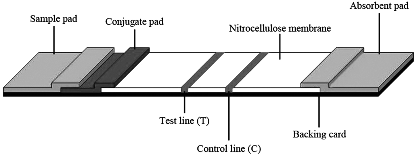
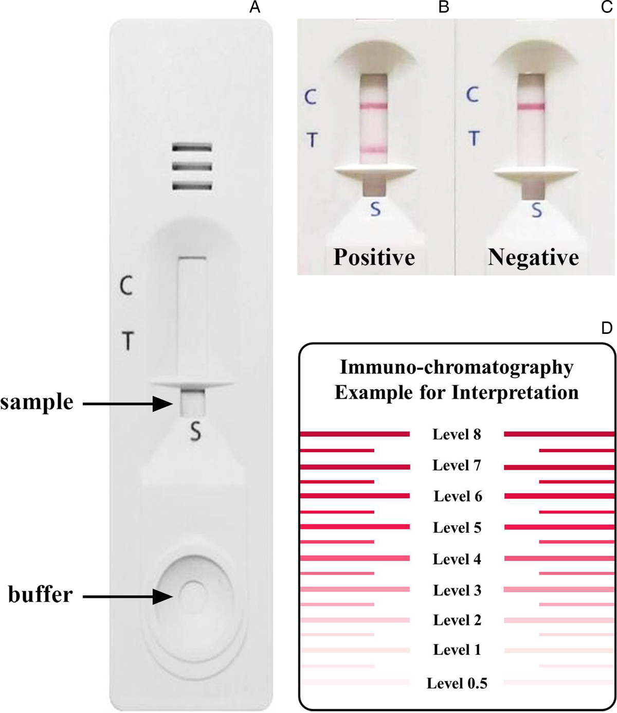
The ICT device was developed using the rAcGal2 antigen. Schematic diagram of the ICT strip accessories was shown (Fig. 1). The process was as follows: rAcGal2 antigen at a concentration of 2 mg mL−1 was immobilized onto nitrocellulose membrane (Sartorius Stedim Biotech, Goettingen, Germany) at the test line (T), and 1 mg mL−1 of goat anti-mouse IgG antibodies (Lampire Biological Laboratories, Pipersville, PA, USA) were also immobilized on the control line (C) of the nitrocellulose membrane (Fig. 2A). The colloidal gold particles (40 nm diameter in solution) were conjugated with mouse anti-human IgG antibodies (Kestrel BioSciences Co., Pathumthani, Thailand) to OD10 at 540 nm. Gold conjugated antibody was then sprayed at a flow rate of 1 µL mm−1 onto the conjugate pad (a piece of glass microfiber filter GF33; Whatman Schleicher & Schuell, Dassel, Germany) as a detector reagent. The strip was housed in a plastic cassette and sealed with a desiccant in an aluminium foil pouch. The test kit was composed of an immunochromatographic device, sample buffer for diluting the serum sample and running buffer for chromatography.
Schematic diagram of the immunochromatographic test strip. An absorbent pad, immobilized nitrocellulose membrane, conjugate pad and sample pad were glued together on a plastic adhesive backing card. The recombinant Angiostrongylus cantonensis galectin-2 and goat anti-mouse IgG antibodies were immobilized at the test line (T) and control line (C), respectively.

The immunochromatographic test for human angiostrongyliasis. An immunochromatographic device (A) and representative images of positive (B) and negative (C) results are presented. Each serum sample was diluted with a buffer at 1:30 dilution ratio. A criterion of the diagnostic result is whether or not a red band appears at the test line within 15 min. In the positive serum sample, the test band and control band turned red (B), whereas only the control band turned red (C) in the negative serum sample. The intensity of the bands was estimated visually according to the interpretation card (D). The cut-off level for a positive result was >0.5.

For testing, 5 µL of diluted serum samples (1:30) was added in a sample well, followed by 90 µL of running buffer. The result was confirmed through visual examination within 15 min after the addition of the running buffer. If red bands appeared at both T and C lines, the result was positive (Fig. 2B) and if a red band appeared only at the C line, the result was negative (Fig. 2C). The band intensity was evaluated visually according to the reference board (Fig. 2D, with 0.5 as the cut-off level). Strip tests that showed T line intensities greater than the the cut-off point were regarded as positive results. The diagnostic parameters of the ICT kit were computed with those of the ELISA, as described above, and the percentage of total concordance was computed [per cent concordance (%) = no. of positives and negatives by both the ICT kit and ELISA × 100/the number of tested samples].
Results
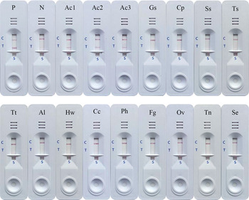
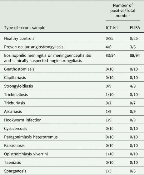
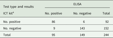
The rAcGal2 protein was used as an antigen in both the ICT kit and the ELISA to evaluate the diagnostic values for human angiostrongyliasis using individual sera from normal healthy subjects who were not found intestinal parasites by FEC stool examination, angiostrongyliasis patients and patients with other parasitoses (Table 1, Fig. 3). Most of the serum samples from patients with EOM and clinical suspected angiostrongyliasis were positive using both ICT (83/94) and ELISA (88/94), while four out of six and three out of six confirmed ocular angiostrongyliasis sera were positive by the ICT kit and ELISA, respectively. Neither method yielded positive results for any of the 25 healthy control sera. Cross-reactivity was detected in serum samples of trichinellosis (1 of 10), ascariasis (1 of 9), hookworm infection (1 of 9), opisthorchiasis (1 of 10) and sparganosis (1 of 5) for the ICT kit, and of strongyloidiasis (4 of 9) for the ELISA. The calculated diagnostic sensitivity, specificity and positive and negative predictive values of the ICT kit were 87.0, 96.5, 94.6 and 91.4%, respectively, and of the ELISA were 91.0, 97.2, 95.8 and 94.0%, respectively. Results attained from the sera of 244 patients using the ICT kit and ELISA are compared in Table 2. The concordance of the ICT kit was determined to be 93.9% (229/244 sera).
Representative images of anti-A. cantonensis antibody detection by the immunochromatographic test. The sensitivity and specificity were evaluated using human sera from P, pooled positive angiostrongyliasis; N, pooled healthy control; Ac1-3, three individuals angiostrongyliasis; Gs, gnathostomiasis; Cp, capillariasis; Ss, strongyloidiasis; Ts, trichinellosis; Tt, trichuriasis; Al, ascariasis; Hw, hookworm infection; Cc, cysticercosis; Ph, paragonimiasis; Fg, fascioliasis; Ov, opisthorchiasis; Tn, taeniasis; and Se, sparganosis.

Reactive frequencies of the ICT kit and ELISA using different serum samples against the recombinant Angiostrongylus cantonensis galectin-2 fusion-tagged protein

Comparison of the ICT kit and ELISA using recombinant Angiostrongylus cantonensis galectin-2 antigen

a There was no significant difference with ELISA (P > 0.05; Exact McNemar's test).
Discussion
Central nervous system manifestations of A. cantonensis have become an important problem affecting human health for people both living in and visiting endemic regions (Barratt et al., Reference Barratt, Chan, Sandaradura, Malik, Spielman, Lee, Marriott, Harkness, Ellis and Stark2016). Serological methods of diagnosing angiostrongyliasis (e.g. immunoblotting and ELISA) are time-consuming, complicated and require specialized equipment and medical personnel (Maleewong et al., Reference Maleewong, Sombatsawat, Intapan, Wongkham and Chotmongkol2001; Podwall et al., Reference Podwall, Gupta, Furuya, Sevigny and Resor2004; Kirsch et al., Reference Kirsch, Dekumyoy, Loscher and Haberl2008). This makes the diagnosis of human angiostrongyliasis problematic, as none of the serological tests that have been developed is commercially available. Recently, a purified 31 kDa A. cantonensis antigen from native worm extract was used in a rapid lateral-flow immunochromatographic assay for angiostrongyliasis (Eamsobhana et al., Reference Eamsobhana, Tungtrongchitr, Wanachiwanawin and Yong2018). However, the limitations of using a native antigen were the low yield of material extracted from parasites and the need to maintain the parasite's life cycle in the laboratory. Thus, a recombinant protein is necessary for the mass production of antigens for use in a serodiagnostic assay.
We developed a novel ICT kit for detecting anti-A. cantonensis antibodies in human serum using rAcGal2 as an antigen. The test is rapid, simple, and sensitive and specific. In addition, the results of this ICT kit were not significantly different (P > 0.05) from those obtained by the ELISA (Table 2). Hence, the kit can be used in place of the previous serodiagnostic methods for human angiostrongyliasis, such as ELISA and immunoblotting, which are time consuming. In addition, antigens for use in these methods are generally crude or partially purified 31 kDa components of A. cantonensis which have limitations due to the low yield of material for serodiagnostic assay and the need to maintain parasite life cycles in the laboratory. However, the 87.0% sensitivity and 96.5% specificity of the recombinant protein-based ICT developed in this study are lower than those of the test format developed based on an antigen from native worm extract (100 and 98.7%; Eamsobhana et al., Reference Eamsobhana, Tungtrongchitr, Wanachiwanawin and Yong2018). Possible reasons for these are the different types and number of sera used, single rAcGal2 antigenic protein that was not correctly folded and possible lack conformational epitopes, the different phrases of the infection when samples were collected. The cross-reactivity among opisthorchiasis, trichinellosis, ascariasis, hookworm infection and sparganosis may be attributable to concomitant or previous subclinical infection with A. cantonensis or one limitation in the use of single rAcGal2 antigenic protein for detection is the possibility of shared epitopes with other parasites. However, this should not cause difficulties in diagnosis because clinical symptoms of these parasitic infections usually differ from those of angiostrongyliasis (Cross, Reference Cross1978). The ICT kit and ELISA yielded false-negative results in two and three cases of ocular angiostrongyliasis, respectively. These negative results may be due to the parasite's location in the eye where the immune response is limited (immunologically privileged site; Zhou and Caspi, Reference Zhou and Caspi2010), which may fail to induce the production of antibodies against the worm.
In conclusion, we have successfully developed an ICT kit with rAcGal2 as a massive production of antigen source for the detection of anti-Angiostrongylus antibodies in human sera that can be employed quickly and easily. The kit can be used to support the clinical diagnosis and for large-scale sero-epidemiological investigation in endemic areas without additional facilities or ancillary supplies.
Acknowledgements
We wish to acknowledge the support of the English Consultation Clinic at the Khon Kaen University Faculty of Medicine Research Affairs Division and the Khon Kaen University Publication Clinic at the Research and Technology Transfer Affairs Division for their assistance. We also extend our gratitude to Jitaporn Harasan from the Mekong Health Science Research Institute for providing such an effective lateral flow immunochromatographic technique.
Financial support
This study was supported by the Thailand Research Fund's Distinguished Research Professor Grant (P.M.I. and W.M., grant number DPG 6280002) and Khon Kaen University. C.S was supported by a scholarship under the Post-Doctoral Training Program at the Khon Kaen University Research Affairs and Graduate School (grant number 59146).
Conflict of interest
None.
Ethical standards
All procedures performed involving human participants were in accordance with the Khon Kaen University Ethics Committee for Human Research (HE 611507). Experiments involving the use of animals were approved by the Animal Ethics Committee of Khon Kaen University, according to the Ethics of Animal Experimentation guidelines from the National Research Council of Thailand (AEMDKKU 002/2018).