Introduction
Sarcopenia is defined as the loss of skeletal muscle mass and strength with age.Reference Morley, Baumgartner and Roubenoff 1 There is increasing recognition of the serious health consequences of sarcopenia in terms of disability,Reference Sayer, Syddall and Martin 2 morbidityReference Sayer, Dennison and Syddall 3 mortalityReference Gale, Martyn, Cooper and Sayer 4 and major healthcare costs.Reference Janssen, Shepard, Katzmarzyk and Roubenoff 5 Adult determinants of sarcopenia including age, gender, size and level of physical activity have been well described.Reference Morley, Baumgartner and Roubenoff 1 , Reference Marcell 6 – Reference Arden and Spector 8 In particular, the importance of physical activity and resistance exercise training as interventions to slow the age-related loss of muscle is widely accepted.Reference Vincent, Braith and Feldman 9 , Reference Skelton, Young, Greig and Malbut 10 Twin studies suggest a heritable componentReference Arden and Spector 8 and a recent genome-wide association study identified TRHR as an important gene for lean body mass.Reference Liu, Tan and Lei 11 Nevertheless, there remains considerable unexplained variation in muscle mass and strength between older adults, which may reflect not only the current rate of loss but also the peak attained earlier in life. To date most observational and interventional epidemiological studies of sarcopenia have focused on factors modifying decline in later life, however, a life course approach to understanding sarcopenia, additionally, focuses on factors operating earlier in life including developmental influences.
A series of epidemiological studies demonstrating the relationship between small size at birth and an increased risk of age-related disease in later life has led to the developmental origins of health and disease hypothesis. This proposes that disease risk in adult humans is influenced by environmental exposures during critical periods of prenatal and postnatal life.Reference Barker, Osmond, Forsen, Kajantie and Eriksson 12 Findings from the Dutch Famine Winter (1944–1945) support the influence of early nutritional and hormonal environments on long-term optimal health demonstrating the importance of both the type and timing of prenatal nutrient deprivation on subsequent adverse health outcomes. For example, exposure to famine during any stage of gestation was associated with glucose intolerance and exposure to famine in early gestation was related to coronary heart disease and obesityReference Roseboom, de Rooij and Painter 13 although there have been some inconsistencies in the findings.Reference Lumey, Stein and Kahn 14 This environmentally driven phenomenon is called programming, a process whereby a stimulus or insult acting at a critical period of development has lasting or even lifelong significance.Reference Lucas 15 Clues to the mechanisms have come from evolutionary biology where Darwin described two adaptational strategies in animals: natural selection based on genetic variation acting over many generations and developmental plasticity acting within the lifetime of an individual.Reference Gluckman and Hanson 16
Developmental plasticity is defined as ‘the ability of a single genotype to produce more than one alternative form of structure, physiological state or behaviour in response to environmental conditions’.Reference West-Eberhard 17 This enables production of a range of phenotypes better suited to the prevailing environmental conditions than would otherwise be possible.Reference Bateson, Barker and Clutton-Brock 18 These early environmentally driven adaptations in phenotype may also occur in preparation for the ‘predicted’ postnatal environment, giving rise to the concept of the predictive adaptive response. When the prediction is correct, an advantageous health benefit may prevail, however, when a mismatch occurs between the predicted and the actual environment, the chosen phenotype may be associated with an increased disease risk over the longer term.Reference Gluckman, Hanson and Bateson 19
The induction of persistent changes to tissue structure and function, as a consequence of early life environmental conditions, is likely to involve life-long alterations to the regulation of gene transcription and mechanisms are starting to be identified. For example, there is an evidence for changes in the epigenetic regulation of transcription, specifically, DNA methylation and covalent modification of histones in the induction of an altered phenotype by nutritional constraint in early life.Reference Burdge, Hanson, Slater-Jefferies and Lillycrop 20 The contribution of such mechanisms to adult muscle mass and strength has yet to be described although studies to identify the effect on neonatal body composition are underway.Reference Bergmann, Bergmann and Dudenhausen 21 This area is of particular interest because of the central role of muscle in metabolic as well as mechanical function.
Epidemiological evidence
Evidence for developmental influences on human muscle has come from a series of observational epidemiological studies linking low birth weight, as a marker of adverse early environmental conditions, with reduced adult muscle strength, mass and size. The association between low birth weight and reduced muscle strength was first reported a decade ago in the Hertfordshire Ageing Study, a retrospective birth cohort study of men and women born in Hertfordshire, UK between 1920 and 1930 and still living there, 60–70 years later.Reference Sayer, Cooper and Evans 22 They had historical health visitor records of weight at birth and 1 year and were traced through the National Health Service Central Registry in Southport, UK. Following a home interview, 717 men and women attended a local clinic for measurement of current size and markers of ageing in different body systems including grip strength. Lower birth weight and weight at 1 year were significantly associated with lower grip strength in later life. These relationships remained significant although attenuated after allowing for adult height, suggesting that an adverse early environment may have an effect on muscle quality and quantity. Ten-year follow-up data including muscle strength have recently been collected in this cohort and will enable the investigation of early influences on the age-related loss of muscle strength.
The association between birth weight and adult grip strength has been replicated using a similar study design in a younger Hertfordshire cohort born 1931–1939Reference Sayer, Syddall, Gilbody, Dennison and Cooper 23 and in a national birth cohort of middle-aged men and women born in 1946 and participating in the National Survey of Health and Development.Reference Kuh, Hardy and Butterworth 24 More recent work has demonstrated a similar effect size of birth weight on adult muscle strength in young women aged 20–34 years taking part in the prospective Southampton Women’s Survey suggesting an association between birth weight and peak muscle strength rather than decline in this younger cohort.Reference Inskip, Godfrey and Martin 25
It has been possible to add to these findings with a more detailed life course approach using longitudinal data collected in the National Survey of Health and Development, which has a prospective cohort study design.Reference Wadsworth, Kuh, Richards and Hardy 26 Grip strength, weight and height were measured in a representative British sample of 1406 men and 1444 women who were 53 years old and had prospective childhood data on weight, height, motor milestones, cognitive ability and information on lifetime social class, current physical activity and health status. Birth weight and prepubertal height gain were associated with midlife grip strength, independently of later weight and height gain. Pubertal growth was also independently associated with midlife grip strength. In men, weight gain during puberty was beneficial, whereas in women height gain was found to be beneficial. Those participants with earlier infant motor development had better midlife grip strength, which was partly confounded by the growth trajectory. This suggests that components of prenatal, prepubertal and pubertal growth have long-term effects on midlife grip strength.Reference Kuh, Hardy and Butterworth 27
Using a meta-analysis approach, it has been possible to compare the findings from published epidemiological studies investigating the relationship between lower birth weight and reduced grip strength in later life. These show remarkable homogeneity of association with a pooled effect size estimate of a 2.06 kg increase in grip strength per kilogram increase in birth weight. Adult height typically attenuated, but did not remove, the birth weight v. grip strength relationship.Reference Sayer, Syddall and Martin 28
Studies investigating the relationship between growth in early life and muscle mass have demonstrated consistent findings linking low birth weight with reduced muscle mass. A study of older men participating in the Hertfordshire Cohort Study showed that birth weight was strongly positively associated with fat-free mass but showed only weak positive associations with measures of adult fat mass. In contrast, weight at the age of 1 year was strongly associated with fat-free mass and adult fat mass estimated using anthropometry.Reference Sayer, Syddall and Dennison 29 Similar findings were observed in studies of men and women using urinary creatinine excretionReference Phillips 30 and dual X-ray absorptiometryReference Gale, Martyn, Kellingray, Eastell and Cooper 31 to estimate muscle mass.
A study from Helsinki has replicated the previously observed relationships between small size at birth, lower muscle mass and reduced grip strength in older adults.Reference Yliharsila, Kajantie and Osmond 32 Other research investigating the link between birth weight and muscle mass in earlier stages of life demonstrate similar findings for children,Reference Hediger, Overpeck and Kuczmarski 33 teenagersReference Singhal, Wells, Cole, Fewtrell and Lucas 34 and young adults.Reference Kahn, Narayan, Williamson and Valdez 35 More recent work using peripheral quantitative computed tomography to measure muscle size directly has shown that birth weight is positively associated with forearm and calf muscle area in older men and women suggesting tracking of relative muscle size throughout life.Reference Sayer, Dennison and Syddall 36
There is considerable interest in which aspects of the early environment underlie these epidemiological associations. Few retrospective cohort studies have sufficiently detailed data on prenatal and postnatal environmental influences to assess specific effects on long-term muscle mass and strength. However, these questions are being addressed in the prospective Southampton Women’s Survey.Reference Inskip, Godfrey and Robinson 37 This study has recruited over 12,500 women living in the city of Southampton, UK and interviewed them to assess health, body composition, lifestyle and diet. They have been followed in subsequent pregnancies and their offspring followed through childhood with the aim of identifying prospectively the influence of the preconceptional and antenatal environment on the growth and development of the foetus, infant and child.
Using data from 448 mother-offspring pairs in this cohort it has been possible to examine maternal influences on neonatal body composition ascertained by dual X-ray absorptiometry.Reference Harvey, Poole and Javaid 38 Taller women and those with higher parity had offspring with increased birth weight, fat and lean mass, whereas women who smoked during pregnancy had lower birth weight babies, with reduced fat and lean mass. Maternal walking speed as a marker of maternal physical activity was negatively associated with birth weight, and maternal fat mass positively predicted neonatal relative fat but was negatively correlated with proportionate lean mass. Current analyses are focusing on the influence of maternal diet on neonatal body composition and childhood grip strength. These findings have the potential to inform the development of beneficial interventions in young women to maximise the growth and development of their children.
Underlying mechanisms
Cellular mechanisms and adaptation
Muscle fibre number is a critical determinant of muscle mass and strength and a number of studies have described the regulators of myofibre number, type and size.Reference Dauncey and Gilmour 39 Limited data derived from children suggest that an increase in muscle fibre size (hypertrophy) but not number (hyperplasia) accounts for muscle growth.Reference Lexell, Sjostrom, Nordlund and Taylor 40 It is believed that fibre number is set at 24 weeks of human gestation, and fibres are incapable of replication, hence any growth, maintenance and repair is believed to be achieved through recruitment of myoblasts, derived from embryonic stem cells at approximately 6 weeks of gestation in the human embryo.Reference Elder and Kakulas 41 Most myoblasts fuse to form primary myotubes attached via tendons to skeleton. Some remain as single cells, which are intrinsic to the successful growth, maintenance and repair of muscle.Reference Alessandri, Pagano and Bez 42 These observations suggest that key periods during gestation influence the initiation of fibre formation, or the recruitment/turn-over of myoblasts, but an association between low birth weight and myoblast function in later life remains to be identified.
Despite this paucity of in vivo information pertaining to low birth weight and myoblast function, there is good evidence from animal models to support the concept of developmental plasticity of muscle as for example exploited in the meat industry. Here, early nutritional manipulation is routinely used to optimise muscle growth and development with evidence for a permanent effect of early nutrition on muscle fibre number, type and quality.Reference Maltin, Delday, Sinclair, Steven and Sneddon 43 Indeed a recent study linking these phenotypes showed that peri-implantation and late gestation maternal undernutrition differentially affected skeletal muscle development in foetal sheep.Reference Costello, Rowlerson and Astaman 44 There was evidence of reduced slow-twitch myofibre and capillary density in the foetal triceps but not the soleus with both modes of undernutrition. This reduction in fibre density, which may have been driven by decreased capillary density, was associated with higher insulin receptor, GLUT4 and type 1 insulin growth factor receptor mRNA levels, suggesting a redistribution of resources at the expense of specific peripheral tissues as a consequence of late gestational undernutrition. Other studies involving experimental manipulation of early nutrition have shown that prenatal maternal diet restriction is associated with reduced neonatal muscle weight in sheep,Reference Greenwood, Hunt, Hermanson and Bell 45 and a reduction in postnatal muscle fibre number in the pig,Reference Dwyer, Stickland and Fletcher 46 guinea pigReference Dwyer, Madgwick, Ward and Stickland 47 and rat.Reference Wilson, Ross and Harris 48 There is some evidence that these adverse effects persist.Reference Prakash, Fournier and Sieck 49 , Reference Pond, Yen, Mersmann and Maurer 50 Interestingly, these detrimental effects of early undernutrition, contrast with the beneficial effects of dietary restriction instituted later in life on both ageing and longevity.Reference Aihie Sayer and Cooper 51
Hormonal and metabolic influences
There appear to be several stages in early life where specific nutritional and hormonal environmental influences become particularly important and this may vary between tissues and organs. There is evidence for the role of the growth hormone (GH)/insulin-like growth factor (IGF) axis as well as altered insulin signalling and glucose metabolism linking poor early growth and adult health.Reference Petry, Ozanne and Hales 52 , Reference Ozanne, Wang, Dorling and Petry 53 Specifically, poor foetal nutrition has been linked to altered insulin sensitivity in both muscle and adipose tissue of male offspring of protein malnourished animals.Reference Ozanne, Smith, Tikerpae and Hales 54 There is also evidence for the importance of the IGF-I axis in the control of cellular mechanisms of growth.Reference Brameld, Mostyn and Dandrea 55 Ovine maternal undernutrition, particularly in mid-gestation culminates in altered associations between IGF-I expression and term body weight,Reference Heasman, Brameld and Mostvn 56 indeed maternal nutrition alters IGF expression in the skeletal muscle and liver of sheepReference Brameld, Mostyn and Dandrea 55 and normal birth weight at term is related to levels of IGF and IGF binding proteins (IGFBPs) in second trimester amniotic fluid.Reference Tisi, Liu, Wykes, Skinner and Koski 57 These data suggest that not only is birth weight influenced by diet and circulating IGFs, but that the subsequent expression of these components in the offspring is also altered. These studies suggest a shift in the set point of the IGF-I axis with reduced IGF-I occurring in utero and postnatally in small for gestational age babies.Reference Ogilvy-Stuart, Hands and Adcock 58
The major influence on primary fibre number appears to be genetic, whereas environmental factors such as maternal undernutrition have a predominant effect on the growth and development of secondary fibres.Reference Maltin, Delday, Sinclair, Steven and Sneddon 43 This is demonstrated in polytocous species such as the pig, which display large intra-litter variation not only in birth weight but also in fibre number. Indeed, low birth weight in piglets correlates with low post-natal growth, reduced catch up growth and increased mortality. Reduced fibre number in these animals is associated with increased fibre size, the lowest lean percentage at slaughter and poor meat quality,Reference Rehfeldt and Kuhn 59 all indicators of the lifelong impact of reduced birth weight. Successful prenatal development depends on nutritional supply, utilisation and hormonal regulation. Indeed, the maternal somatotropic axis plays a significant role in the control of myogenesis, with the infusion of GH into pregnant sows (until mid-gestation), culminating in overall increases in birth weight and increased fibre numbers in the smallest littermates. This suggests a key role for cross talk between nutrient availability, the GH-IGF axis and regulation of fibre number with ultimately consequences for muscle form and function in later life.
Other studies have taken these investigations further using the spontaneous differential foetal growth of pigs to investigate the mechanisms underpinning the link between foetal size and muscle development.Reference Tilley, McNeil, Ashworth, Page and McArdle 60 The development of the Longissimus muscle (m. longissimus) in small foetuses and their average sized littermates was investigated on days 45, 65 and 100 of gestation (term is 113–116 days). Small foetuses were reported to have significantly lower body weight (P < 0.05) at all time points and significantly reduced secondary to primary muscle fibre ratio on day 100 compared with littermates. The expression of the IGF-I receptor and IGFBP three were significantly higher at day 65 (P < 0.05) in m. longissimus of the small compared with average sized foetuses. IGF-I receptor expression remained elevated (P = 0.001) at day 100 and was accompanied by significantly increased expression (P < 0.05) of the IGF-II receptor and of IGFBP-5. These data suggest that reduced foetal muscle development is associated with increased expression of several genes of the IGF system in mid-to-late gestation, which may be a compensatory response to poor growth.
Interestingly, rodent studies from almost two decades ago suggest that hindlimb skeletal muscle biopsies from small neonatal rats secrete significantly more IGFBPs (P < 0.05) and significantly less IGF (P < 0.05) than average sized littermates.Reference Frampton, Jonas and Larkins 61 This differential expression, if maintained postnatally, may account for the retention of small muscle size throughout the lifecourse and may also influence metabolic function. Indeed, recent research findings suggest that mechanical properties of human muscle (strength, loading and unloading) may be directly linked with metabolic function and cell matrix interactions, which may ultimately influence the mitogenic, differentiation and anti-apoptotic roles of the IGFs. IGFs are key to skeletal muscle structure and function and may therefore be associated with a wider systemic effect. These studies suggest that early nutrition may influence future muscle growth (which is dependant on muscle stem cell recruitment) and thus determine muscle function and insulin resistance, which is important for understanding the pathogenesis of age-related disorders such as sarcopenia and impaired glucose tolerance but also for developing and assessing effective preventive and therapeutic interventions.
The postnatal consequences of reduced birth weight in lambs are associated with hypoglycaemia and slowed postnatal engagement of the GH/IGF system. Critically, however, when small lambs are reared in an optimum environment, they grow at rates comparable with average-weight lambs, however, they lay down more fat as a consequence of high energy intakes, low maintenance energy requirements and limited capacity for bone and muscle growth.Reference Greenwood and Bell 62 These growth characteristics are accompanied by higher plasma concentrations of GH and leptin, and lower concentrations of IGF-I during the first 2 weeks of postnatal life, and higher concentrations of insulin during subsequent growth up to 20 kg body weight. These data support the notion that foetal exposure does indeed impact on later life metabolic dysfunction, however, the underlying molecular processes of impaired muscle function remain poorly understood, with only limited data available to address this issue.
Molecular pathways
Efficiency of nutrient utilisation is high in neonates with normal birth weights but is reduced in those with intrauterine growth restriction, suggesting altered function of the major organs involved in the digestion, absorption and metabolism of dietary nutrients.Reference Wang, Chen and Li 63 Indeed, proteomic analyses of the gastrocnemeus from small and normal weight newborn piglets, illustrated major adaptations in 13 proteins, which were associated with reduced muscle size at birth. Annexin A1 (phospholipase A2-inhibitory protein), apolipoprotein AI, catechol O-methyltransferase, β-actin, eukaryotic translation initiation factor-3 subunit 2β, keratin 10, peroxiredoxin 1, phosphoglycerate kinase, and serum albumin precursor were all decreased (P < 0.05) by 22–66% and the levels of mitochondrial F1-ATPase, proteasome α-1 subunit, and tubulin β-chain were all increased (P < 0.05) by 31–52% in the muscle of small compared with normal weight piglets, respectively.Reference Wang, Chen and Li 63 Importantly, these are proteins related primarily to macronutrient metabolism (particularly protein synthesis and degradation), immune response, cellular structure, extracellular matrix and antioxidant function. Importantly, in the presence of increased protein degradation, increased concentrations of mitochondrial F1-ATPase would accelerate ATP hydrolysis, reducing its availability for example for nutrient transport and protein synthesis, which may culminate in amino acids being directed to oxidation and fatty acid synthesis, thus possibly decreasing protein accretion and increasing fat deposition and impaired metabolism in skeletal muscle of small birth weight piglets.Reference Wang, Chen and Li 63
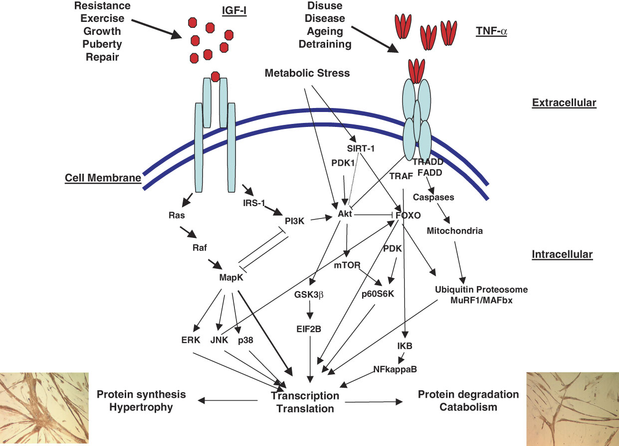
Additional studies using rodent models of maternal protein restriction to induce insulin resistance and diabetes in adult male offspring have begun to investigate the altered signalling pathways affected in association with reduced size at birth. Basal glucose uptake was not different between normal and reduced weight animals. However, the ability of insulin to stimulate glucose uptake in the soleus was blocked in the smaller animals, in the face of normal insulin receptor and GLUT4 expression levels. Further analyses revealed that protein kinase C ξ, which is positively involved in GLUT4 regulated glucose transport, was reduced (P < 0.001) compared with controls.Reference Ozanne, Olsen and Hansen 64 Akt, which is a serine/threonine kinase essential for metabolism and survival acts downstream of both the IGF-I and insulin receptors.Reference Haddad and Adams 65 , Reference Kandel and Hay 66 Furthermore, signalling through Akt following the binding of IGF-I to its receptor is associated positively with muscle protein synthesis. Myofibre contraction has also recently been demonstrated to stimulate Akt signalling and importantly also GLUT4 translocation, both central regulators of glucose homoeostasis. Among the intracellular mechanisms controlling protein synthesis, signalling via Akt appears to regulate gene transcription through inactivation of Fork-head box Os (FOXOs), which are associated with E3 ubiquitin ligase expression.Reference Saini, Al-Shanti and Stewart 67
Like Akt, mTOR is a protein kinase, which regulates translation and whose expression is regulated by hypertrophy (increases) and atrophy (downregulates), suggesting an important role in muscle mass.Reference Bodine, Stitt and Gonzalez 68 Given that nutrition, insulin and IGF-I activate the Akt/mTOR pathway, it is possible to envisage a situation in older adults with reduced birth weights and displaying insulin resistance where mTOR concentrations or activation may be downregulated. Furthermore, the ubiquination and degradation of muscle proteins in association with the tissue and substrate specific MuRF1 and MAFbx culminate in muscle atrophy.Reference Saini, Al-Shanti and Stewart 67 Therefore, since Akt activation influences not only protein synthesis but also the expression of MuRF1 and MaFBx and their regulation by FOXOs,Reference Saini, Al-Shanti and Stewart 67 downregulation of Akt, may not only be associated with altered metabolism as a consequence of small birth weight but also with reduced muscle mass as a consequence of increased protein degradation in the absence of Akt control. The balance between synthesis/degradation appears therefore to converge at these kinases and ligases. Therefore, it is important that studies are performed to ascertain the impact of small birth weight and subsequent reductions in muscle mass and type on Akt/mTOR/FOXO/MuRF1 and MAFbx signal transduction pathways, to determine their roles in controlling muscle mass and function in later life, as is the case, for PKCξ above.
In addition to reduced shuttling of GLUT4 as a consequence of reduced Akt activity, studies investigating the mechanisms controlling reduced GLUT4 expression have also focussed on epigenetic regulation and demonstrated that epigenetic mechanism(s) underpin not only diminished skeletal muscle GLUT4 mRNA in intrauterine growth-restricted female rat offspringReference Raychaudhuri, Raychaudhuri, Thamotharan and Devaskar 69 but also underpin reduced myofibre size in glucose restricted murine C2C12 skeletal muscle cell lines.Reference Fulco, Cen and Zhao 70 In the former study, an increase in MEF2D (inhibitor) in the face of decreased MEF2A (activator) and MyoD (co-activator) binding to the GLUT4 promoter was evident in hind limb skeletal muscle from small v. control rats. Methylation of the binding sites within the GLUT4 promotor did not appear to be involved in altered gene transcription; however, histone code modification did appear to be key in repressing GLUT4 transcription, not only in the postnatal period but also into adulthood of small birth weight rodents.Reference Raychaudhuri, Raychaudhuri, Thamotharan and Devaskar 69 In the latter study, investigators ascertained using cell culture models that glucose restriction impaired the differentiation of myoblasts and that this was associated with increased activation of the AMP-activated protein kinase (AMPK), augmented Nampt activity and consequently modified intracellular [NAD+]:[NADH] ratios and nicotinamide levels, which culminated in the inhibition of myogenesis.
Critically, myoblasts derived from SIRT1+/− heterozygous mice were resistant to glucose restriction or to AMPK activation, suggesting that AMPK, Nampt and SIRT1 are central to the ability of skeletal muscle cells to sense and react to nutrient availability.Reference Fulco, Cen and Zhao 70 Importantly, SIRT1 overexpression in mice culminates in increased insulin expression by pancreatic β cells in response to glucoseReference Moynihan, Grimm and Plueger 71 and SIRT1 knock out mice show blunted insulin secretion by islet cells.Reference Bordone, Motta and Picard 72 SIRT1 also alters the activities of peroxisome proliferator-activated receptor (PPAR)-γ and PPAR-γ coactivator 1α (PGC-α), transcription factors critical for the regulation of differentiation of skeletal muscle, adipogenesis and liver metabolism,Reference Fulco, Schiltz and Iezzi 73 perhaps therefore linking nutrition, metabolism and responsiveness of peripheral metabolic tissues. Finally, SIRT1 knock out mice show reduced activity compared with controls when on calorie restricted diets, worsening potentially compromised metabolic pathways.Reference Chen, Steele, Lindquist and Guarente 74 As a consequence of this potential link with the sirtuins, it leads to the question of whether sirtuin expression is also important when nutrient restriction is encountered in utero or in early childhood and the implications for interactions with Akt, FOXO and cellular adaptations (Fig. 1).
(Colour online) Potential pathways regulating muscle hypertrophy and atrophy.

Human studies
There has been little work linking early environmental influences with changes in human muscle at a molecular or cellular level. One small study involving 27 adult women looked at the relationship between size at birth and skeletal muscle morphology but did not find significant associations.Reference Thompson, Sanderson and Sandeman 75 In contrast, a more recent study focused on a group of 40 young men and showed altered skeletal muscle fibre composition and size in those with low birth weight.Reference Jensen, Storgaard, Madsbad, Richter and Vaag 76 More recently further studies carried out in the vastus lateralis of low birth weight (⩽10th centile) young men revealed reduced skeletal muscle expression of the PI3K p85α regulatory subunit and the p110β catalytic subunit, PKCξ and GLUT4 in the fasting state.
When biopsies were obtained in an insulin stimulated state (following a 4-hr hyperinsulinemic euglycemic clamp), expression of aPKCξ (P < 0.001) and Akt1 (P < 0.001) as well as Akt phosphorylation (ser 473; P < 0.01) were decreased, while IRS1 (P < 0.05), PI3K p85α (P < 0.001) and p110β (P < 0.05) subunits were stimulated in muscle of low birth weight men when compared with matched controls. Expression and phosphorylation of p38 MAPK, phospho-p38 MAPK (Thr180/Tyr182), phospho-ERK (Thr 202/Tyr204), JNK1, JNK2 and phospho-JNK (Thr 183/Tyr185) were not different between groups, suggesting that altered insulin activation of the PI3K/Akt but not the MAPK pathway may contribute to development of insulin resistance and type 2 diabetes in low birth weight men.Reference Jensen, Martin-Gronert and Storgaard 77 This work needs to be taken forward in studies of older adults to investigate mechanisms underlying the developmental origins of sarcopenia.Reference Aihie Sayer and Cooper 78
Conclusions
The epidemiological evidence linking small size at birth with reduced muscle mass and strength is strong and consistent with replication in a number of different groups including children, young and older adults. However, most of the evidence for the cellular, hormonal, metabolic and molecular mechanisms underlying these associations comes from animal models. The next stage is to translate the understanding of mechanisms from animal muscle to human muscle enabling progress to be made not only in earlier identification of individuals at risk of sarcopenia but also in the development of beneficial interventions across the life course.
Acknowledgements
This work was supported by the Medical Research Council, the University of Southampton and Manchester Metropolitan University, UK.
Statement of Interest
None.