Introduction
Malaria is one of the most common fatal syndromes, affecting more than >200 million people and causing 0.5 million deaths per year (World Health Organization, 2018). Cerebral malaria (CM) is a major complication of Plasmodium falciparum infection, usually occurring in children and non-immune travellers visiting endemic countries (Osuntokun, Reference Osuntokun1983) causing mortality of 30–50% of the total CM patients (Brewster et al., Reference Brewster, Kwiatkowski and White1990). It is the most severe neurological complication causing acute non-traumatic encephalopathy in tropical countries. Boivin et al had reported that approximately 20–22% of infants <5 years aged with CM have cognitive deficits even after 6 months of discharge from the hospital (Boivin et al., Reference Boivin, Bangirana, Byarugaba, Opoka, Idro, Jurek and John2007). They showed increased seizure frequency and prolonged coma duration with persistent cognitive deficits. Current views support the idea that CM arise due to high avidity of adherence of parasite-infected erythrocytes, and, thereafter pathogenic effects controlled by inflammatory cytokines and effector cells, leading to morphological and functional alterations of brain microvascular endothelial cells (Franke-Fayard et al., Reference Franke-Fayard, Janse, Cunha-Rodrigues, Ramesar, Buscher, Que, Lowik, Voshol, den Boer, van Duinen, Febbraio, Mota and Waters2005). These clinical findings could be explained by an excitotoxic mechanism in which a large amount of release of glutamate neurotransmitter in intersynaptic spaces causes the death of neuronal cell and neurodegeneration (Wang and Qin, Reference Wang and Qin2010). It has been reported that during CM the release of glutamate is increased in the brain and increased levels of this neurotransmitter in the cerebrospinal fluid (CSF) of Plasmodium berghei (P. berghei) ANKA infected mice are associated with behavioural changes (Miranda et al., Reference Miranda, Vieira, Lacerda-Queiroz, Souza, Rodrigues, Vilela, Gomez, Machado, Rachid and Teixeira2010). Glutamate, a type of acidic amino acid, is the major excitatory neurotransmitter in the central nervous system (CNS) of class Mammalia of the vertebrate groups, playing an essential role in the development of neuronal cells, synaptic plasticity, learning and memory processes under normal physiological conditions (Choi, Reference Choi1988; Constantine-Paton et al., Reference Constantine-Paton, Cline and Debski1990; Bliss and Collingridge, Reference Bliss and Collingridge1993; Tsien et al., Reference Tsien, Huerta and Tonegawa1996). The excitotoxicity process which is mediated by glutamate neurotransmitter and other factors like quinolinic acid and aspartic acid that act via glutamate receptors have been involved in the occurrence of neurological and cognitive symptoms in CM (Dobbie et al., Reference Dobbie, Crawley, Waruiru, Marsh and Surtees2000). Subtypes of ionotropic glutamate receptor are N-methyl-D-aspartate receptors (NMDARs) which are involved in synaptic plasticity, mechanisms of learning and memory and mediates excitotoxic neuronal damage. Quinolinic acid is an endogenous excitotoxin that is a selective agonist of N-methyl-D-aspartate (NMDA) glutamate receptors. High level of the local concentration of quinolinic acid which is a neuroactive metabolite of the kynurenine pathway in the brain could lead to damage of neuronal cells (Clark et al., Reference Clark, Mackay, Smythe, Bustamante, Stone and Phillips2005).
It is well established that during inflammation, either central or peripheral nervous system of the brain, is associated with an increased sensitivity of NMDAR, which are a type of membrane-bound ionotropic glutamate receptors that function as ligand-gated ion channels and non-selective cation channels. Stone (Reference Stone and Perkins2001) had reviewed the role for quinolinic acid in neurodegenerative disorders (Stone, Reference Stone2001) and we postulate that the activation of NMDAR might be increased during murine CM leading to damage of neuronal cells and contributing to the neurological symptoms and mortality associated with the disease.
NMDAR are assembled as heterologous tetramers, composed of the functional NR1 subunit and modulatory NR2 subunits. Four NR2 subunits, each encoded by a separate gene, have been identified: NR2A, NR2B, NR2C, NR2D (Moriyoshi et al., Reference Moriyoshi, Masu, Ishii, Shigemoto, Mizuno and Nakanishi1991; Kutsuwada et al., Reference Kutsuwada, Kashiwabuchi, Mori, Sakimura, Kushiya, Araki, Meguro, Masaki, Kumanishi, Arakawa and Mishina1992; Ishii et al., Reference Ishii, Moriyoshi, Sugihara, Sakurada, Kadotani, Yokoi, Akazawa, Shigemoto, Mizuno, Masu and Nakanishie1993; Monyer et al., Reference Monyer, Burnashev, Laurie, Sakmann and Seeburg1994). The existence of NR3 subunits in the brains of rat has also been reported as NR3A, and NR3B (Ciabarra et al., Reference Ciabarra, Sullivan, Gahn, Pecht, Heinemann and Sevarino1995; Sucher et al., Reference Sucher, Akbarian, Chi, Leclerc, Awobuluyi, Deitcher, Wu, Yuan, Jones and Lipton1995; Nishi et al., Reference Nishi, Hinds, Lu, Kawata and Hayashi2001; Matsuda et al., Reference Matsuda, Kamiya, Matsuda and Yuzaki2002). NR3B isoforms have been proving to modulate NMDA receptor-mediated intracellular calcium [Ca2+] influx (Domingues et al., Reference Domingues, Neugebauer and Fern2011).
In spite of having several reports, the mechanisms underlying the fatal cerebral complications are still not fully understood. It also remains unclear that why only a limited proportion (1–3%) of P. falciparum infections progress to severe or fatal episodes of CM (Malaria Genomic Epidemiology Network, 2008) while others remain non-symptomatic or develop an uncomplicated illness is still not fully understood. In the current study, we have used P. berghei (Pb) ANKA infection in C57BL/6 mice as a murine model of experimental CM (ECM) to investigate the role of different NMDAR expression during CM. We have also assessed the effect of one of the widely used artemisinin derivative α/β arteether on NMDAR expression as well as on survival of the CM mice.
Materials and methods
Mice and experimental infections
Female C57BL/6 mice (6–7 weeks old) weighing 17–18 g were used for all experiments. Animals were housed in polypropylene cages under standard conditions of temperature (24 ± 1 °C) and humidity (55–68%) and fed standard pellet diet with water ad libitum. Plasmodium berghei ANKA, (MRA-311) strain was obtained from Malaria Research and Reference Reagent Resource Centre (MR4), ATCC Manassas, Virginia. All experimental mice were infected intraperitoneally with 3 × 10⁶ PbA parasitized red blood cells (RBCs), which were withdrawn from a previously infected mouse of the same strain. Parasitaemia was monitored daily by counting the number of pRBCs per 10 000 RBCs by microscopic examination of Giemsa-stained thin smears from mice tail blood. Mice were monitored daily for survival and neurologic signs of CM such as deviation of head, ruffled hair, hind limb paralysis, ataxia, convulsions and coma (Engwerda et al., Reference Engwerda, Belnoue, Gruner and Renia2005). Approximately 80% of the PbA-infected animals showed typical CM symptoms between days 6 and 8 post-infection.
Treatment
Treatment was started on day 6 after the development of CM symptoms. Three consecutive doses of arteether i.e. 15 mg kg−1 × 3 days, 25 mg kg−1 × 3 days and 50 mg kg−1 on the first day followed by 25 mg kg−1 × 2 days were given by intramuscular route, while MK801 at 2 mg kg−1 × 3 days was given by intraperitoneal route.
Brain tissue collection
Mice were sacrificed by ether anaesthesia and their brains were excised after intra-cardiac perfusion with chilled normal saline according to the method of Glowinski and Iversen (Reference Glowinski and Iversen1966) and kept on an ice-cold plate immediately. After washing from Diethyl pyrocarbonate treated water, brain samples were stored in TRIzol (Thermo-scientific) for further studies.
Glutamate assay in brain synaptosomes
Brain synaptosomal preparation was done as described earlier (McGovern et al., Reference McGovern, Maguire, Gurd, Mahler and Moore1973) with some modifications. Brain tissues were homogenized in 10 vol 0.32 M sucrose in a Potter-Elvehjem homogenizer, and the homogenate was centrifuged at 4–8 °C for 10 min at 600 g. The supernatant was then diluted 1:1 with 1.3 M sucrose, to yield a suspension at a final concentration of 0.8 M sucrose. This suspension was further centrifuged at 20 000 g for 30 min at 4 °C. The supernatant was discarded, and the pellet consisting of synaptosomes and free (extrasynaptosomal) mitochondria was resuspended in HEPES buffer pH 7.4 (adjusted by NaOH). The resulting synaptosomal preparation was kept at −80 °C till the experiments were performed to estimate neurotransmitters glutamate. Glutamate levels in the brain synaptosomes were measured using a colorimetric (optical density at 450 nm [OD450]) glutamate assay kit (MAK004; Sigma Aldrich, USA) according to the manufacturer's instructions.
Assessment of blood brain barrier (BBB) permeability
To analyse the permeability of the BBB, C57BL/6 mice infected with Pb ANKA and showing neurological symptoms (usually between days 6 and 10 after infection) were intravenously injected with a 200 µL volume of a 2% (wt/vol) solution of Evans Blue (Sigma) in PBS. After 2 hours, mice were anaesthetized with ether and the brain was harvested, photographed and incubated in 1 mL formamide (Sigma) for 48 h to extract dye. Absorbance was measured at 620 nm in an Enzyme-Linked Immunosorbent Assay (ELISA) reader and Evans blue concentration was assessed by linear regression.
Measurement of mRNA expression using quantitative real-time polymerase chain reaction (qRT-PCR)
Total RNA was extracted from isolated brains by TRIzol reagent (Thermo-scientific) and 1 µg of RNA was reverse transcribed using oligo (dT) primer with the Revert aid H Minus first strand cDNA synthesis kit (Thermo-scientific). Real-time PCR was carried out using ABI StepOnePlusTM system. The applied thermal profile was: 1 cycle at 95 °C for 10 min, 40 cycles at 95 °C for 10 s, 62 °C for 30 s and 72 °C for 20 s. Housekeeping gene glyceraldehyde 3-phosphate dehydrogenase (GAPDH) was used as an endogenous control. The relative gene expression levels were calculated by the 2–ΔΔCT method (Nunes et al., Reference Nunes, Pandey, Yadav, Goel and Ateeq2017). Supplementary Table S1 lists the primers used in the current study.
Immunohistochemistry assay
Mice were deeply anaesthetized with diethyl ether and perfused intracardially with physiological 0.9% saline solution. At the end of perfusion, brain samples were removed and fixed overnight in 4% paraformaldehyde at 4–8 °C. The tissues were rinsed with water, dehydrated through an ascending series of alcohol (100, 95, 70, 50 and 30%) and embedded in paraffin through xylene. Sections (5 µm) were cut using a Leica paraffin wax slicing machine. Microwave-assisted antigen retrieval was performed in a citrate antigen retrieval solution and sections were cooled to room temperature and transferred to 2–3% hydrogen peroxide for 8–10 min to block endogenous peroxidase. Sections were washed with 0.01 M phosphate buffered saline (PBS; 5 min each wash) for three times and blocked with bovine serum albumins for 1 h at room temperature. Slices were incubated with primary antibodies with dilution 1:100 (anti-NR1 and NR2A antibodies from Santacruz Biotechnology) for overnight at 4–8 °C. Subsequently, the sections were equilibrated to room temperature for 1 hour, washed with PBS (pH 7.4) for 5 min, and incubated with appropriate secondary antibodies (Sigma) for 30 min at room temperature. This step was followed by 3 washes in PBS (5 min each). The sections were then treated with 3,3′-diaminobenzidine (DAB) solution according to the manufacturer's instructions and then washed with distilled water to remove the reagent. Haemotoxyline staining was done for 1 min and then washed with distilled water, mounted with DPX mounting and were microscopically examined. Images were captured using the Olympus DP25 photomicrography system. For the specificity of the staining, parallel slices were stained identically but without primary antibodies.
Immunoblotting: Western blot analysis
Brains were removed from perfused mice and homogenized in ice-cold lysate buffer (200 mm HEPES (pH 7.5), 250 mm sucrose, 1 mm dithiothreitol, 1.5 mm MgCl2, 10 mm KCl, 1 mm EDTA, 1 mm EGTA) freshly supplemented with phosphatase and protease inhibitors [0.1 mm PMSF, 2 mm Na3VO4 and 1 × protease inhibitor cocktail (Sigma)]. The final protein concentration in each sample was determined using the Bradford reagent. Aliquots containing 50 µg of protein were re-suspended in SDS-PAGE (sodium dodecyl sulfate–polyacrylamide gel electrophoresis) loading buffer, resolved on 10% SDS acrylamide gels and transferred onto polyvinylidene difluoride membranes. Membranes were blocked in non-fat dry milk (5%) for 1 h followed by incubation with primary antibodies (1:1000) for overnight at 4 °C. Then, membranes were incubated with horseradish peroxidase-labelled secondary antibodies. Rabbit polyclonal anti-NR1, NR2A, NR2B, anti-actin and horseradish peroxidase-labelled goat monoclonal anti-rabbit antibodies were obtained from Santa Cruz Biotechnologies. Signal was developed using an enhanced chemiluminescence kit (GE Healthcare, Little Chalfont, UK) using Image Quant LAS 4000 (GE Healthcare). The probed membranes were stripped and re-probed with rabbit polyclonal β-actin antibody to detect total levels of protein.
Statistical analysis
Data are presented as the mean ± s.e.m. (standard error of the mean). Survival analysis was performed using the Kaplan–Meier log-rank test (Mantel-Cox) test. The statistical significance of the differences was analysed by the one-way analysis of variance (ANOVA) followed by the Tukey's multiple comparison post-test (Graphpad Prism5 software). A p value of <0.05 was considered significant. p value summary is mentioned as *p < 0.05; **p < 0.001; ***p < 0.0001 and ns as not significant.
Results
Pb ANKA infection causes BBB breakdown in ECM model
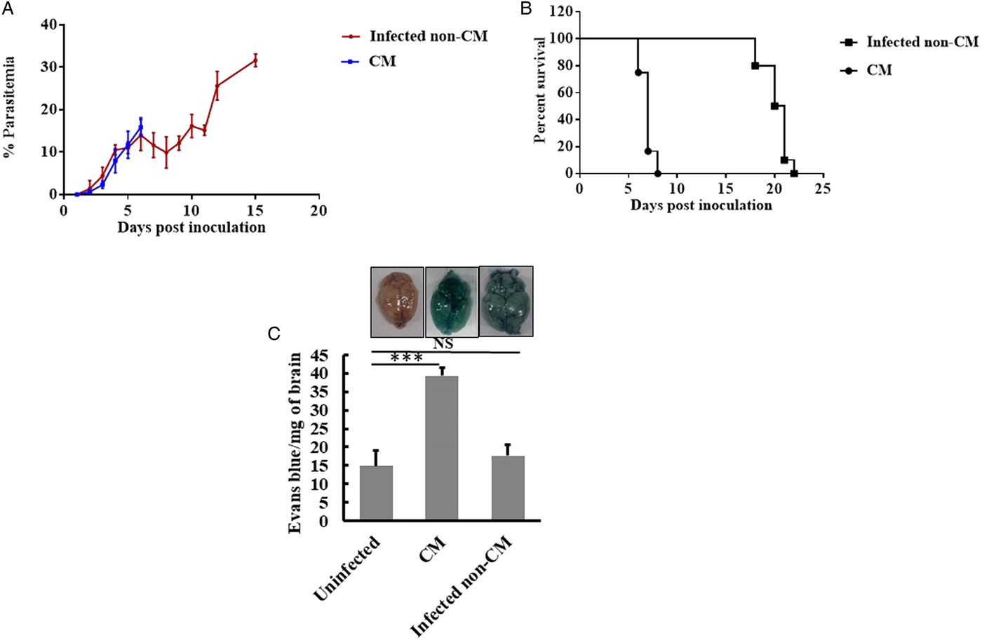
C57BL/6 mice were infected with 3 × 10⁶ Pb ANKA infected RBCs and the parasitaemia and symptoms were monitored daily. Mice infected with Pb ANKA showed a very low level of parasitaemia up to day 3 and then gradually increased in all the infected mice. Approximately 80% of the mice infected with Pb ANKA developed CM with neurological signs (data not shown) and became moribund between 7 and 10 days post infection (dpi) (Fig. 1A). Parasitaemia in CM mice was almost similar as compared to non-CM (infected mice without cerebral symptoms) mice on days 7–10 post-infection. Parasite load reached 15–20% at the time of death in CM mice while it was between 35 and 56% in the non-CM mice (Fig. 1A). Mice showing neurological signs (CM) usually died within a few hours after the appearance of CM-symptoms while the mice did not exhibit symptoms of CM died between day 14 and 22 (Fig. 1B) mainly due to other malaria-related pathologies such as hyper parasitaemia or severe anaemia without showing obvious neurological signs.
Time course of (A) infection (B) survival of C57BL/6 mice infected with Pb ANKA parasitized RBCs and (C) blood brain barrier destruction shown by Evans blue staining and quantification by spectrophotometry. C57BL/6 mice were infected with 3 × 10⁶ Pb ANKA-parasitized RBCs. Data are presented as Mean ± s.e.m. (n = 6) and are representative of two experiments. Significant differences were determined by using Tukey's multiple comparison tests.

BBB breakdown is a characteristic feature of ECM. Evans blue, a vascular marker has the ability to bind to albumin so it can detect plasma protein leakage across the BBB into the cerebral parenchyma. The infected C57BL/6 mouse showed dye leakage indicating a widespread increase in vascular permeability and accelerated loss of BBB integrity in the brain of mice whereas the brain of the uninfected mice showed no dye leakage (Fig. 1C). The earlier loss of BBB integrity in infected mice is consistent with the accelerated onset of mortality.
NMDAR are highly expressed in CM
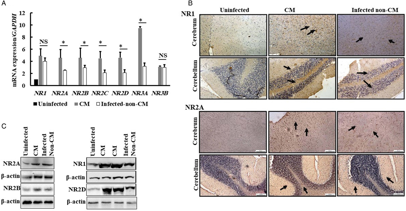
The mRNA expression levels of NMDAR were estimated in the brain samples. All the NMDAR were found to be overexpressed in CM mice compared to the uninfected and infected non-CM group. mRNA of NR1, NR2A, NR2B, NR2C, NR2D were overexpressed by ~4–5-fold while NR3A was overexpressed by 9-fold whereas ~3-fold increase in NR3B was observed when compared to uninfected mice (Fig. 2A). In infected non-CM group NR2A, NR2B, NR2C, NR2D, NR3A mRNA were found to be overexpressed when compared to the uninfected group. However, the expression of NR2A, NR2B, NR2C, NR2D, NR3A were found to be significantly higher in CM mice compared to infected non-CM mice group (Fig. 2A).
(A) NMDA (NR1, NR2A-D, and NR3A-B) receptors mRNA expressions in brains of C57BL/6 mice were analysed by using real-time quantitative polymerase chain reaction. The values were shown as a fold change to the non-infected controls. There were five to six mice per group and data are representative of three separate experiments. Values are means from triplicate measurements. *p < 0.05 and **p < 0.01. (B) Photomicrographs of NMDA receptor subunit NR1 and NR2A immunoreactivity from control and Pb ANKA infected CM mice brains. In the frontal cortex of CM mice, neuronal cell bodies were intensely stained for NR1 and NR2A as compared to controls. The pyramidal cell layer of the cerebellum region of CM mice brain was more immunopositive for NR1 and NR2A. (C) Immunoblot showing expression of NR1, NR2A, NR2B, NR2C and NR2D in uninfected, CM and infected non-CM mice.
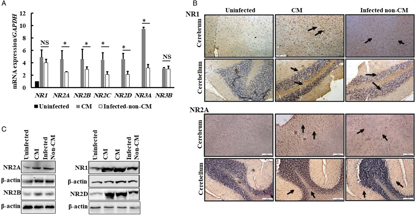
We further confirmed the expression of subunits of NMDAR (NR1 and NR2A) by Immunohistochemistry. Photomicrograph of NMDA receptor subunits after infection with Pb ANKA and control uninfected mice brains revealed marked differences in immunoreactivity. Our studies show that immunoreactivity of NR1 subunit was significantly increased in cerebrum and cerebellum regions of symptomatic mice brain as compared to uninfected and infected non-CM mice (Fig. 2B). Similarly, NR2A subunit expression was also notably elevated in Pb ANKA infected symptomatic mice (Fig. 2B). Immunoblot analysis also showed overexpression NR1, NR2A, NR2B and NR2D in infected mice and it is higher in CM group (Fig. 2C).
CM increases glutamate in nerve synaptosomes
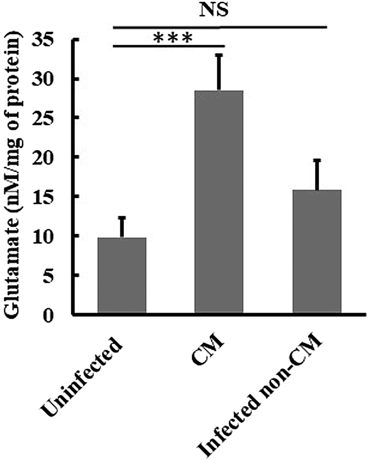
Glutamate, one of the two acidic proteinogenic amino acids is also a key molecule in cellular metabolism. Within the mammalian nervous system, glutamate is the most abundant excitatory neurotransmitter. Since abnormal glutamatergic neurotransmission has been implicated in a wide range of neurological diseases, we measured glutamate release from isolated brain cortical nerve terminals (synaptosomes) in the CM model. Glutamate concentration is determined by an enzymatic assay which results in a colorimetric (450 nm) product, proportional to the glutamate present. Glutamate level was significantly increased in CM mice on day 7 post-infection as compared to uninfected and infected non-CM mice (Fig. 3).
Glutamate level in synaptosomes of brain of uninfected Pb ANKA infected symptomatic and non-symptomatic C57BL/6 mice (n = 6). Data are represented as means ± s.e.m. from two independent experiments. **p ⩽ 0.01 (ANOVA followed by the Tukey's multiple comparison post-test).

Arteether increases survival of CM infected mice and inhibits NMDAR expression.
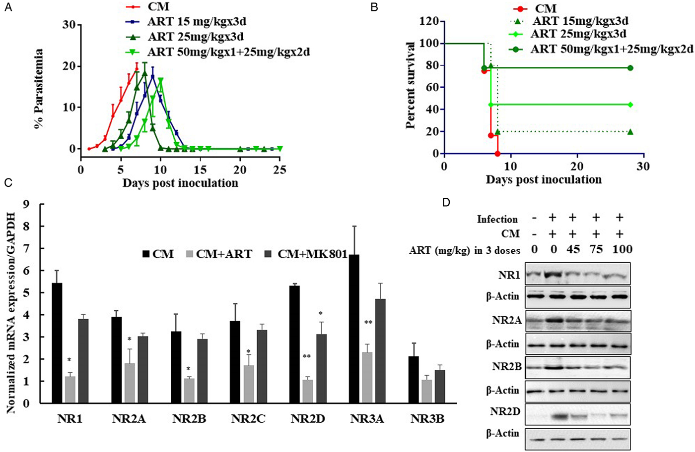
Artemisinin derivatives are first-line treatment for malaria, however, the action mechanism of artemisinin derivatives are still debatable. We have evaluated the antimalarial effect of arteether (one of the widely used artemisinin derivative) on the mice having cerebral symptoms due to Pb-ANKA infection. Interestingly, we found that treatment with arteether at different concentrations could decrease the parasite load and increase the overall survival of the mice and some of the mice survived beyond the observation period of 28 days. The data show that arteether treatment at 25 mg kg−1 × 3 days and 50 mg kg−1 × 1 day followed by 25 mg kg−1 × 2 days could save 50 and 80% of the CM mice, respectively (Fig. 4A and B).
Effect of arteether on (A) Pb ANKA infection (B) survival of CM mice (C-D) NMDA receptor expression. (C) mRNA expressions in brains of C57BL/6 mice treated with 25 mg kg−1 × 3 days were analysed using real-time quantitative polymerase chain reaction (MK801 2 mg kg−1 × 3 days was used as positive control and the data were compared with non-infected mice). The values were shown as a fold change ± s.e.m. to the non-infected controls. (n = 6). *p < 0.05 and **p < 0.01 were considered significant.

Further, to evaluate the effect of arteether treatment on the expression of NMDAR, we performed qRT PCR for NMDAR on the brain samples of CM mice with or without treatment of arteether and MK801 (a known NMDA receptor inhibitor). Results showed that 25 mg kg−1 × 3 days arteether treatment significantly reduced the expression of all the NMDAR and it was comparable with the uninfected mice. Furthermore, the effect of arteether treatment on NMDAR expression was found to be more pronounced than MK801 (Fig. 4B). Immunoblot data show a significant decrease in NR1, NR2A, NR2B, NR2D after arteether treatment in infected CM mice compared to untreated CM mice (Fig. 4D)
Discussion
CM is likely the outcome of interrelated events, including mechanical obstruction due to the adherence of the parasite-infected RBCs to the microvessels of the vital host organs and dysregulated immune responses. In parallel, BBB breakdown and neurological damage also occur. Survival of CM patients is very difficult and if they survive, they may show neurological sequelae. A minority have neurological deficits persisting beyond 6 months with memory impairment being the most common (Oluwayemi et al., Reference Oluwayemi, Brown, Oyedeji and Oluwayemi2013). A major role of astrocytes cells in the normal CNS is to induce and maintain BBB properties in the vascular endothelium (Brightman, Reference Brightman and Neuwelt1989). Astrocytes also make close contact with the synapses of neuronal cells and are thought to be intimately involved in controlling acid-base, electrolyte and neurotransmitter balance (Sykova et al., Reference Sykova, Svoboda, Simonova and Jendelova1992; Walz, Reference Walz1992). In addition, astrocytes regulate the local concentration of neurotransmitters, such as glutamate, in the extracellular fluid (Medana et al., Reference Medana, Hien, Day, Phu, Mai, Chu'ong, Chau, Taylor, Salahifar, Stocker, Smythe, Turner, Farrar, White and Hunt2002). The clinical picture of CM is likely to be due to some combination of the effects of sequestration, metabolic changes such as hypoglycaemia and metabolic acidosis, and the effects of systemic and CNS pro-inflammatory cytokine production (John et al., Reference John, Kutamba, Mugarura and Opoka2010).
For the assessment of neurochemical marker of CM, we investigated the involvement of glutamate during the development of CM. The amount of glutamate present in the cerebro-cortical synaptosomes of CM mice was significantly increased after the onset of symptoms i.e. on 7 dpi. Since there is a parallel increase of glutamate levels in synaptosomes of symptomatic brain with the neurological symptoms of CM, this may suggest a role of glutamate in CM pathogenesis and its association with their behavioural changes. Several studies also reported that the level of glutamate increases in the brain, CSF and particularly the synaptic clefts in ECM infection (Rae et al., Reference Rae, McQuillan, Parekh, Bubb, Weiser, Balcar, Hansen, Ball and Hunt2004; Miranda et al., Reference Miranda, Vieira, Lacerda-Queiroz, Souza, Rodrigues, Vilela, Gomez, Machado, Rachid and Teixeira2010). One group also showed that glutamate was significantly high in the plasma of children, who died of malaria due to cerebral complications (Planche et al., Reference Planche, Dzeing, Emmerson, Onanga, Kremsner, Engel, Kombila, Ngou-Milama and Krishna2002). Taken together, these studies suggest that an imbalance in metabolism of glutamate neurotransmitter may be relevant to pathogenesis of CM and also suggest a role for glutamate in the central nervous dysfunction found in CM disease.
The high amounts of glutamate release in intersynaptic spaces are reported to cause neuronal cell death and neurodegeneration via excitotoxicity processes (Ankarcrona et al., Reference Ankarcrona, Dypbukt, Bonfoco, Zhivotovsky, Orrenius, Lipton and Nicotera1995; Wang and Qin, Reference Wang and Qin2010). This process mediated by glutamate and factors such as quinolinic acid and aspartic acid that act via glutamate receptors has been implicated in the occurrence of neurological and cognitive symptoms in CM (Medana et al., Reference Medana, Hien, Day, Phu, Mai, Chu'ong, Chau, Taylor, Salahifar, Stocker, Smythe, Turner, Farrar, White and Hunt2002; Rae et al., Reference Rae, McQuillan, Parekh, Bubb, Weiser, Balcar, Hansen, Ball and Hunt2004; Parekh et al., Reference Parekh, Bubb, Hunt and Rae2006). CSF, sampled in 97 Kenyan children with CM, depicted a graded increment in quinolinic acid concentration across outcome groups of increasing severity suggesting that excitotoxic mechanisms may contribute to the pathogenesis of CM (Dobbie et al., Reference Dobbie, Crawley, Waruiru, Marsh and Surtees2000). Quinolinic acid can activate a subpopulation of neuronal glutamate receptors specifically sensitive to NMDARs producing depolarization (Stone and Perkins, Reference Stone and Perkins1981; Guillemin, Reference Guillemin2012) and excitotoxicity (Schwarcz and Kohler, Reference Schwarcz and Kohler1983) as well as increased CNS proinflammatory cytokine production and oxidative stress (Guillemin et al., Reference Guillemin, Smith, Smythe, Armati and Brew2003).
While almost every glutamate receptor subtype has been implicated in mediating excitotoxic cell death, NMDARs have received particular attention because of their crucial roles in excitatory synaptic transmission, plasticity and neurodegeneration in the CNS (Petrenko et al., Reference Petrenko, Yamakura, Baba and Shimoji2003). NMDAR-mediated signals are critical for the survival of developing neurons and for several forms of synaptic plasticity (Sheng and Kim, Reference Sheng and Kim2002). The overactivation of NMDAR by increased extracellular glutamate acting in synergy with high brain quinolinic acid and TNF-α concentration can lead to excitotoxic damage of NMDA receptor containing neurons. Furthermore, NMDA receptor antagonists were found to be neuroprotective in CM and such a mechanism may be particularly important in the development of long-term learning impairment and increased susceptibility to epilepsy later on in life among paediatric CM survivors (Stafstrom and Sasaki-Adams, Reference Stafstrom and Sasaki-Adams2003; de Miranda et al., Reference de Miranda, Brant, Vieira, Rocha, Vieira, Rezende, de Oliveira Pimentel, Moraes, Ribeiro, Ransohoff, Teixeira, Machado, Rachid and Teixeira2017).
Here, we demonstrated that NR1, NR2A-D and NR3A-B in brains of Pb ANKA infected mice were overexpressed in CM mice on 7 dpi as compared with infected non-CM and uninfected controls. Increased subtypes of NMDA receptor expression in the early infective stage may explain the expansion of excitotoxicity mediated neuropathogenesis in murine CM. Therefore, in the presented study, as increased levels of glutamate and increased expression of NMDAR in CM mice model exerted more aggravating effects on the progression of disease, it might be possible that these mice had toxic effects of glutamate and NMDAR to cause worse neurological symptoms. With prolonged glutamate exposure, NMDA receptor activation might have created a sustained increase in intracellular calcium that overexcite the neurons, often leading to cell death. Also, NMDA receptor stimulation can lead to mitochondria-mediated cell death (Waxman and Lynch, Reference Waxman and Lynch2005; Alberdi et al., Reference Alberdi, Sanchez-Gomez, Cavaliere, Perez-Samartin, Zugaza, Trullas, Domercq and Matute2010).
Artemisinin derivatives, a group of antimalarials having a fast therapeutic response and are effective against all stages of malaria parasite as well as drug-resistant P. falciparum malaria. For CM patients having severe complications, there are limited therapeutic options. WHO recommends quinine or artemisinin derivatives for severe CM patients (McIntosh and Olliaro, Reference McIntosh and Olliaro2000). However, quinine is not a perfect choice over artemisinin derivatives due to severe undesirable side effect and shorter half-life (WHO, 1998, 2015). Oil soluble artemisinin derivative; arteether co-developed in our institute, has a long elimination half-life, and is more stable than other derivatives and has been proved to have a potent effect on cerebral complications and survival of CM patients (Afolabi and Okoromah, Reference Afolabi and Okoromah2004). However, the exact mechanism of arteether on CM is still unknown.
Here, we have shown that arteether treatment decreases the expression of NMDAR and increases the survival of CM mice. However, arteether is a potent antimalarial agent and its effects on CM could be a function of its antimalarial property only. A deeper understanding and experiments are needed to confirm the underlying mechanism.
Supplementary material
The supplementary material for this article can be found at https://doi.org/10.1017/S0031182019000878.
Author ORCIDs
Renu Tripathi, 0000-0002-3719-8802.
Acknowledgements
Authors are thankful to the Director, C.D.R.I. for his continuous support throughout the study.
Financial support
This work is supported by grants provided under CSIR-Network project ‘New approaches towards understanding of Disease dynamics and to accelerate drug discovery (UNDO)’ and ‘Evaluation of rescue treatment for cerebral malaria in vitro / in vivo model (GAP0147)’ to RT. SKS is thankful to ICMR, HD and SG and SKP to CSIR and BSC to UGC for Senior Research Fellowship.
Conflict of interest
We have no competing interests to declare. CSIR-CDRI communication number: 9855.
Ethical standards
The study received ethical approvals vide permit number: IAEC/2010/142 from CSIR-Central Drug Research Institute's ‘Institutional Animal Care and Use Committee’ recognized by ‘Committee for the Purpose of Control and Supervision of Experiments on Animals (CPCSEA)’, Government of India. All animals were sacrificed using deep ether anesthesia during or after the study and all efforts were made to minimize suffering.