Introduction
Post-operative adhesion formation is a common and serious problem in ENT and other fields of surgery. For instance, intestinal adhesions can cause severe manifestations, such as ileus; in obstetrics and gynaecology, ovarian or intra-tubal adhesions lead to infertility and ectopic pregnancies, while in cardiovascular surgery, pericardial damage can lead to potentially fatal tamponade secondary to post-operative adhesions.Reference Yener, Bardakçı, Iriz, Özoğul, Yener and Bayram1 Inflammation occurring due to mucosal trauma in the middle ear can result in the formation of post-operative adhesions and related complications, such as ossicular fixation, retraction of graft and residual tympanic membrane.
Therefore, numerous haemostatic and anti-adhesive agents have been developed to prevent adhesions. An ideal anti-adhesive material has to be a non-adhesive product of adequate dimensions and without any potential risk of infection or adverse effects on the healing process.Reference Hellebrekers, Trimbos-Kemper, van Blitterswijk, Bakkum and Trimbos2 Many products, such as blood clots, lyophilised plasma, Sepragel™ Sinus, Seprafilm® and Gelfoam® have been used to coat the middle ear to prevent adhesions.Reference Hellström, Salén and Stenfors3 In animal experiments, Sepragel and Seprafilm have been reported to reduce adhesions with good tolerability and without ototoxic effects.Reference Jang, Park, Cho and Choi4 The application of Gelfoam, which had been used widely in middle-ear surgery since the 1950s, has been abandoned because of its inflammatory and adhesion-increasing effects.Reference Dogru, Haholu, Gungor, Kucukodaci, Cincik and Ozdemir5
The objective of our study was to evaluate the anti-inflammatory and anti-adhesive effects of two anti-adhesive products, Arista™ AH (a plant-based powder containing microporous polysaccharide hemospheres; Medafor Inc, Minneapolis, Minnesota, USA) and Ankaferd Blood Stopper (Ankaferd İlaç Kozmetik A.Ş., Istanbul, Turkey); both have well-known haemostatic and anti-adhesive properties on mucosal layers. To our knowledge, this is the first study reporting the use and effects of these products in otological practice.
Materials and methods
Approval for the study was obtained from the Animal Ethics Committee of Dokuz Eylül University, Turkey. The study protocol was also approved by the Animal Ethics Committee of Dokuz Eylül University.
The study was completed in two stages: a surgical and a histopathological stage. The surgical stage was performed at the Dokuz Eylül University Animal Laboratory. Fourteen 6-week-old guinea pigs weighing between 350 and 900 g were used in the study. Before surgery, the external auditory canals and eardrums of all guinea pigs were intact and not showing any middle-ear pathology. Anaesthesia was administered with intra-peritoneal xylazine (Basilazin, 5 mg/kg; aniMedica GmbH, Senden, Germany) and ketamine (Ketalar, 50 mg/kg; Pfizer Inc, New York, New York, USA) injections. The incision site was numbed with local lidocaine hydrochloride 20 mg/ml and adrenaline 0.0125 mg/ml (this is a mixed drug containing both lidocaine and adrenaline and its market name is “jetobain”) (Adeka, Samsun, Turkey) injections.
Surgical procedure
The posterior wall of the right external auditory canal was identified microscopically and the skin of the posterior wall of the external auditory canal was elevated. Following elevation of the inferior wall of the external auditory canal, the bony structure located inferiorly (i.e. the tympanic bulla) was reached. Then, the postero-inferior aspect of the tympanic bulla was perforated with a sharp pick and the hole was dilated. The middle-ear cavity was reached with a pick to create mucosal trauma. Thus, the middle-ear mucosa and the outer mucosal aspect of the cochlea facing the middle ear were traumatised. The middle ear was penetrated through the bone window in the tympanic bulla of the right ear. Then, Arista™ AH was injected into the middle ear of seven guinea pigs using the manufacturer's applicator. Ankaferd Blood Stopper was injected into the middle ear of the other seven guinea pigs. The left ears of the 14 guinea pigs were left untreated and they formed the control group. The incision site was sutured and covered with a dressing.
At post-operative week 4 (28–30 days post-surgery), the guinea pigs were euthanised using higher doses of the anaesthetising agents previously described, and then decapitated. The temporal bones were carefully excised without harming the cochlear bones and then fixed in 10 per cent formalin solution (Figure 1).
(a) Lateral and (b) medial views of the temporal bone following resection. Care was taken to leave the skin of the cochlea and external auditory canal intact. EAC = external auditory canal; TB = tympanic bulla; C = cochlea

Tissue specimens that had been fixed in formalin solution for 24–48 hours were dehydrated in calibrated ethanol solution, washed with xylene and embedded in paraffin. Paraffin-embedded tissue blocks from the middle ear, promontory of the tympanic cavity and cochlea were cut into 5-mm-thick sections. The sections were stained with haematoxylin and eosin and histological examination was performed under a light microscope (BX40; Olympus, Tokyo, Japan). Images were saved as digital media in a computerised system (Windows XP Pro/Image Pro Plus, version 3.0; MediaCybernetics Inc., Rockville, Maryland, USA). Two independent observers classified the intensity of the inflammation observed in the middle- and inner-ear sections as mild (+), moderate (++) or severe (+++).
Statistical analysis
Data were analysed using the SPSS software program, version 16.0 for Windows (SPSS Inc., Chicago, Illinois, USA). All differences associated with a p value of 0.05 or less were considered statistically significant. Continuous variables were presented as mean ± standard deviation. Parametric tests were applied to data with normal distributions and non-parametric tests were applied to data with questionable normal distributions. The Shapiro–Wilk test was used to assess the distribution of the data. The three groups were compared using the non-parametric Kruskal–Wallis test. Significant data (p < 0.08) were subjected to a post-hoc Bonferroni correction with a 95 per cent confidence interval.
Results
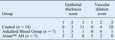
For each group, epithelial thickness (scoring: 1 = normal thickness; 2 = mild thickening; 3 = marked thickening) and capillary dilation (scoring: 1 = absent; 2 = mild increase; 3 = marked increase) in the middle and inner ears were quantitatively evaluated and tabulated (see Table I). In each of the three groups, consolidation of the lamina propria and epithelium mucosae, increments in the number of active fibroblasts, collagen fibrils and inflammatory cells, and increased vascular dilation were seen on haematoxylin and eosin-stained sections, and were more prominent in the control and Ankaferd Blood Stopper groups (Figures 2–4). Epithelial thickness and capillary vasodilation were significantly lower in the Arista™ AH group, when compared to the control and Ankaferd Blood Stopper groups (p < 0.008). In the Ankaferd Blood Stopper group, these changes were lower when compared to the control group, but the difference was not statistically significant (p > 0.008) (Table II).
Photomicrographs of the control group samples, showing staining of the middle ear at (a) × 100 and (b) × 400 (H&E) and cochlea at (c) × 100 and (d) × 400 (H&E). These demonstrate thickening of the epithelium and lamina propria, and capillary dilation. Irregularities, inflammation, vascularisation in the organ of Corti situated on the cochlear basal membrane and increased vacuolisation of the stria vascularis are also seen. C = capillary; E = epithelium; LP = lamina propria; SV = scala vestibuli; BM = basilar membrane; ST = scala tympani; RM = Reissner's membrane; SM = scala media (cochlear duct); Sv = stria vascularis (of cochlear duct); SL = spiral limbus
Photomicrographs of the Arista™ AH group samples, showing staining of the middle ear at (a) × 40 and (b) × 100 (H&E) and cochlea at (c) × 40 and (d) × 100 (H&E). These photomicrographs demonstrate minimal vascular dilation, reduced epithelial thickening and a lower number of inflammatory cells in this group. OC = organ of Corti; BM = basilar membrane; Sv = stria vascularis (of cochlear duct); SG = spiral ganglion
Photomicrographs of the Ankaferd group samples, showing staining of the middle ear at (a) × 40 and (b) × 100 (H&E) and cochlea at (c) × 40 and (d) × 100 (H&E); panels (e) and (f) show staining of the spiral (cochlear) ganglion at × 100 and × 400 (H&E), respectively. These photomicrographs demonstrate reduced inflammation in the Ankaferd group relative to the control group, though this difference was not statistically significant. LP = lamina propria; E = epithelium; MEC = middle-ear cavity; OC = organ of Corti; BM = basilar membrane; SL = spiral limbus; Sv = stria vascularis (of cochlear duct); SG = spiral ganglion
Epithelial thickness and vascular dilation scores for the middle and inner ears of the three groups
Epithelial thickness scores: 1 = normal thickness; 2 = mild thickening; 3 = marked thickening.
Vascular dilation scores: 1 = absent; 2 = mild increase; 3 = marked increase.
p = 0.04 for epithelial thickness, p = 0.038 for vascular dilation, p < 0.008 for Arista™ AH.
Number of inflammatory cells in the three groups
p < 0.05 for Arista™ AH and Ankaferd Blood Stopper groups. In the inter-group comparison: Arista™ AH, p < 0.008; Ankaferd Blood Stopper, p > 0.008.
Discussion
Various methods and agents have been used to prevent the formation of post-operative adhesions. Peri-operative preventive measures, such as minimisation of surgical trauma, precise tissue dissection and avoidance of any remaining foreign substances in the middle-ear cavity, desiccation of mucosal surfaces, excessive hot irrigation (warm irrigations usually use to clean intrabdominal spaces and also could be use in middle ear so excessive hot irrigation without paying attention could be a reason for postoperative fybrosis and syneschias), redundant use of retractors, cautery and laser application are notable factors which may help decrease the formation of adhesions.Reference diZerega and Tulandi6 Up to the present time, pharmacological agents, such as corticosteroids, non-steroidal anti-inflammatory drugs and fibrinolytic agents have been administered locally without any distinct beneficial effects. As a surgical barrier, blood clots, lyophilised plasma, Sepragel, Seprafilm, Gelfoam and Silastic® sheet packing have been used to coat the middle ear; however, owing to extensive inflammation and adhesion, the use of Silastic sheet packing and Gelfoam in clinical practice remains controversial.Reference Dogru, Haholu, Gungor, Kucukodaci, Cincik and Ozdemir5
Gelfoam had been widely used in middle-ear surgery since the 1950s, but its use has now been abandoned.Reference Dogru, Haholu, Gungor, Kucukodaci, Cincik and Ozdemir5 Reports about Sepragel and Seprafilm have stated that, in animal experiments, they were well tolerated and had favourable effects on adhesions without any ototoxic effect in the middle ear.Reference Dogru, Haholu, Gungor, Kucukodaci, Cincik and Ozdemir5, Reference diZerega and Tulandi6 GYNECARE INTERCEED®, which is commonly used in daily practice in obstetrics and gynaecology, is a promising agent for use in middle-ear procedures.Reference diZerega and Tulandi6 In recent years, cellular linkage proteins, such as intercellular adhesion molecule-1 and vascular cell adhesion molecule-1, which have been held responsible for the development of adhesions, and substance P and neurokinin 1 (tachykinin receptor 1) receptor antagonists, which reduce intra-abdominal adhesion formation and play a role in the formation of free oxygen radicals, have steadily become the focus of research interest.Reference Reed, Heydrick, Aarons, Prushik, Gower and Stucchi7
In this study, we aimed to assess and compare the anti-adhesive properties of Arista™ AH and Ankaferd Blood Stopper, which are well known for their haemostatic capabilities, though there are few studies on their anti-adhesive role in ENT practice.
In many in vivo and in vitro studies performed so far, Ankaferd Blood Stopper, which is a standard mixture of Thymus vulgaris (thyme), Glycyrrhiza glabra (liquorice), Vitis vinifera (grape vine), Alpinia officinarum (lesser galangal) and Urtica dioica (nettle), has been shown to be an effective haemostatic agent.Reference Firat, Ozdemir, Kosar, Goker and Haznedaroglu8 In addition, reports have shown that, in ENT practice, Ankaferd Blood Stopper has decreased peri-operative bleeding and operative times in children undergoing tonsillectomies.Reference Teker, Korkut, Gedikli and Kahya9 Many publications have shown its safe use for haemostatic control after thyroidectomy.Reference Guler, Maralcan, Kul, Baskonus and Yılmaz10 Only a few studies have questioned the anti-adhesive properties of Ankaferd Blood Stopper. In a relevant investigation, Cömert et al. reported that the intra-peritoneal application of Ankaferd Blood Stopper decreased intra-abdominal adhesions. They also added that the intra-peritoneal administration of Ankaferd Blood Stopper led to some minor changes in the lungs and serosal surfaces of the intestines, as well as minor architectural changes in the liver that were not considered toxic.Reference Cömert, Karakaya, Barut, Karadeniz Çakmak, Uçan and Gültekin11 Odabaş et al. reported the concentration-dependent cytotoxic effects of Ankaferd Blood Stopper on cell cultures and showed that it was cytotoxic to human pulp fibroblasts.Reference Odabaş, Ertük, Çinar, Tüzüner and Tulunoğlu12
Arista™ AH is a simple, safe and effective haemostatic agent that was recently approved by Food and Drug Administration (FDA) for use in cardiac, orthopaedic, spinal and general surgery, though not for use in cranial and ophthalmic interventions. It is a plant-based powder derived from purified plant starch that contains microporous polysaccharide hemospheres, a patented blood clotting technology; the powder rapidly exerts haemostatic effects and forms clots on contact with surfaces.Reference Emmez, Tonge, Tokgoz, Durdag, Gonul and Ceviker13 In animal studies, it has been shown that microporous polysaccharide hemospheres do not harm the intact epithelium, do not cause foreign substance reactions and do not increase the infection rate at the wound site.Reference Antisdel, Janney, Long and Sindwani14 In addition to its haemostatic and antibacterial properties, in controlled studies performed with several haemostatic anti-adhesive agents, various reports have shown that Arista™ AH alleviated peritoneal adhesions after intra-abdominal surgery.Reference Hoffmann, Siddiqui, Agarwal, McKellar, Kurtz and Gettman15 In 2005, Arista™ AH was approved by the FDA for use in ENT surgery. Since then, it has been widely used in post-nasal packing after endoscopic sinus surgery, haemostatic control after adenoidectomy and tonsillectomy, and local haemostasis following neck dissections and tonsillectomy.Reference Sindwani16 To the best of our knowledge, there have been no experimental animal studies or clinical experiences in the use of Arista™ AH in middle-ear procedures.
• Post-operative adhesions can lead to ossicular fixation, retraction of graft and residual tympanic membrane in middle-ear surgery
• The anti-adhesive haemostatic agent Arista™ AH can be used to prevent the formation of adhesions in middle-ear surgery, but its ototoxic potential should be further investigated
We designed this experimental animal study to examine the anti-adhesive properties of two agents with known haemostatic activities, to be used in the field of otology and especially in daily ENT practice. As an outcome of our study, histopathologically, we noted a statistically significant decrease in epithelial thickness and capillary vasodilation in the Arista™ AH group when compared to the control and Ankaferd Blood Stopper groups (p < 0.008). These reductions were also present in the Ankaferd Blood Stopper group, but the difference, when compared to the control group, was not statistically significant (p > 0.008). The number of inflammatory cells increased in the control and Ankaferd Blood Stopper groups, while they were close to normal values in the Arista™ AH group.
Conclusion
Arista™ AH can reduce the effects of surgical trauma in middle-ear surgery. Our results have shown that it is a promising agent to prevent adhesions in middle-ear surgery. Further experimental studies are needed to determine its ototoxic potential.